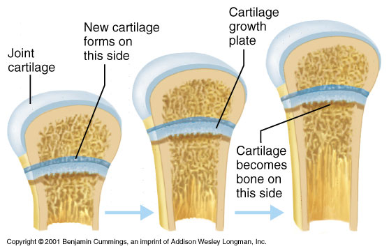
 I guess now would be a good time to explain why almost all forms of height increase ideas and product don’t work. In any process, there is usually a series of steps that has to happen for it to work out. In those steps, there is usually one step that is the slowest and that step is called the rate limiting part. A process or “reaction” can only be as fast or work as much as the slowest part can be. When it comes to achieving height increase, the part that really limits whether one can grow is the fact that after one passes puberty and reaches”physical maturity” the growth plates also known as epiphyseal plates which was originally cartilage has started to thin out and after a certain amount of estrogen is released into the system, the cartilage is slowly ossified until the epiphyseal plates disappear and fuse with the bones surrounding it leaving only a line known as the epiphyseal line. When we were still growing the growth plates are located on both sides of our long bones, like the femur and humerus.
From the Wikipedia Article on Epiphyseal plates, click HERE
Role in bone elongation
Endochondral ossification is responsible for the initial bone development from cartilage in utero and infants and the longitudinal growth of long bones in the epiphyseal plate. The plate’s chondrocytes are under constant division by mitosis. These daughter cells stack facing the epiphysis while the older cells are pushed towards the diaphysis. As the older chondrocytes degenerate, osteoblasts ossify the remains to form new bone. In puberty increasing levels of estrogen, in both females and males, leads to increased apoptosis of chondrocytes in the epiphyseal plate. Depletion of chondrocytes due to apoptosis leads to less ossificationand growth slows down and later stops when the entire cartilage have become replaced by bone, leaving only a thin epiphyseal scar which later disappears. Once the adult stage is reached, the only way to manipulate height is modifying bone length via distraction osteogenesis.
The growth plate has a very specific morphology in having a zonal arrangement. The growth plate includes a relatively inactive reserve zone at the epiphyseal end, moving distally into a proliferative and then hypertrophic zone and ending with a band of ossifying cartilage (the metaphysis). A mnemonic for remembering the names of the epiphyseal plate growth zones is ” Real People Have Career Options,” standing for: Resting zone, Proliferative zone, Hypertrophic cartilage zone, Calcified cartilage zone, Ossification zone. The growth plate is clinically relevant in that it is often the primary site for infection, metastasis, fractures and the effects of endocrine bone disorders.
Me: So let’s look at HOW EXACTLY does ossification and bone growth even happen. Let’s move to the Wiki article on ossification found HERE
Ossification (or osteogenesis) is the process of laying down new bone material by cells called osteoblasts. It is synonymous with bone tissue formation. There are two processes resulting in the formation of normal, healthy bone tissue: Intramembranous ossification is the direct laying down of bone into the primitive connective tissue (mesenchyme), while endochondral ossification involves cartilage as a precursor. In fracture healing, endochondral osteogenesis is the most commonly occurring process, for example in fractures of long bones treated by plaster of Paris, whereas fractures treated by open reduction and stabilization by metal plate and screws may heal by intramembranous osteogenesis.
Heterotropic ossification is a process resulting in the formation of bone tissue that is often atypical, at an extraskeletal location. Calcification is often confused with ossification. Calcification is synonymous with the formation of calcium-based salts and crystals within cells and tissue. It is a process that occurs during ossification, but not vice versa.
The exact mechanisms by which bone development is triggered remains unclear, but it involves growth factors and cytokines in some way.
Time Table For Human Ossification
| Time period | Bones affected |
|---|---|
| Third month of embryonic development | Ossification in long bones beginning |
| Fourth month | Most primary ossification centers have appeared in the diaphyses of bone. |
| Birth to 5 years | Secondary ossification centers appear in the epiphyses |
| 5 years to 12 years in females, 5 to 14 years in males | Ossification is spreading rapidly from the ossifcation centers and various bones are becoming ossified |
| 17 to 20 years | Bone of upper limbs and scapulae becoming completely ossified |
| 18 to 23 years | Bone of the lower limbs and os coxae become completely ossified |
| 23 to 25 years | Bone of the sternum, clavicles, and vertebrae become completely ossified |
| By 25 years | Nearly all bones are completely ossified |
Me: It would appear that once the estrogen starts getting released, the rate at which condrocytes die increases, thus leading to the number of chondrocytes that can multiply through mitosis decrease until all the condrocytes that can possibly multiply to form more stacking from endochondral ossification dies out. As stated there are two main forms of ossification.
What is interesting from the second article that talks about ossification is that in the chart, they say that the age range when bones of the lower limbs become ossified is 18-23, which I wonder whether refers to both male and female, or just male. If it is for a general average of both genders, then it is possible that the long bones can be stretched even if the bones have fused, since there is supposed to be still an epiphyseal line that is created.
In the time chart, it is not until the age f 23-25 before the vertebrate become completely ossified so I would state that even for people (girls and guys) who has past puberty and have have not grown in years within the 15-22 age range group they can still increase in height through the vertebrate and sternum expansion since those bones have not ossified.
What we notice is that the long bones seem to ossify before the torso bones like the vertebrate and sternum. When doctors like orthopedics and endocrinologists look at X-Rays of teenagers to check if they are growing, I doubt they also check the vertebrate and sternum since they don’t ossify for what appears to be 2 more years after the long bones. That means that the doctors can be wrong in many cases when they state to teenager height increase hopefuls that they can’t grow anymore since they did not look at the X-rays of the back. They only probably take X-rays of the wrists and knees !!
So Even if you are say 23 and your long bones are fused, there is still at least 3 ways to gain height, one of which is to make the rate of chondrocytes apoptosis decrease, the other is to pull at the line of epiphyseal since that line indicates the cartilage and bones are not completely sealed together, and the best indication is that the vertebrate has not ossified yet!
Tyler’s Notes:
I found this great paper on epiphyseal plates.
Does the epiphyseal cartilage of the long bones have one or two ossification fronts?
“Epiphyseal cartilage is hyaline cartilage tissue with a gelatinous texture, and it is responsible for the longitudinal growth of the long bones in birds and mammals. It is located between the epiphysis and the diaphysis. Epiphyseal cartilage also is called a growth plate or physis. It is protected by three bone components: the epiphysis, the bone bar of the perichondrial ring and the metaphysis. The epiphysis, which lies over the epiphyseal cartilage in the form a cupola, contains a juxtaposed bone plate that is near the epiphyseal cartilage and is in direct contact with the epiphyseal side of the epiphyseal cartilage. The germinal zone corresponds to a group of cells called chondrocytes. These chondrocytes belong to a group of chondral cells, which are distributed in rows and columns; this architecture is commonly known as a growth plate. The growth plate is responsible for endochondral bone growth. The aim of this study was to elucidate the causal relationship between the juxtaposed bone plate and epiphyseal cartilage in mammals. Our hypothesis is that cells from the germinal zone of the epiphyseal side of the epiphyseal cartilage are involved in forming a second ossification front that is responsible for the origin of the juxtaposed bone plate. We report the following: (a) The juxtaposed bone plate has a morphology and function that differs from that of the epiphyseal trabeculae; (b) on the epiphyseal edge of the epiphyseal cartilage, a new ossification front starts on the chondrocytes of the germinal area, which forms the juxtaposed bone plate. This ossification front is formed by chondrocytes from the germinal zone through a process of mineralisation and ossification, and (c) the process of mineralisation and ossification has a certain morphological analogy to the process of ossification in the metaphyseal cartilage of amphibians and differs from the endochondral ossification process in the metaphyseal side of the growth plate. The close relationship between the juxtaposed bone plate and the epiphyseal cartilage, in which the chondrocytes that migrate from the germinal area play an important role in the mineralisation and ossification process of the juxtaposed bone plate, supports the hypothesis of a new ossification front in the epiphyseal layer of the epiphyseal plate. This hypothesis has several implications: (a) epiphyseal cartilage is a morphological entity with two different ossification fronts and two different functions, (b) epiphyseal cartilage may be a morphological structure with three parts: perichondrial ring, metaphyseal ossification front or growth plate, and epiphyseal ossification front, (c) all disease (traumatic or dysplastic) that affects some of these parts can have an impact on the morphology of the epiphyseal region of the bone, (d) there is a certain analogy between metaphyseal cartilage in amphibians and mammalian epiphyseal cartilage, although the former is not responsible for bone growth.”
“[The growth plate] is nourished by three vascular systems: epiphyseal, metaphyseal and perichondrial vessels, which do not reach the interior of the growth plate.”
“[The] structures [that regulate the forces that reach the growth plate] are the epiphyseal bone structure, the bone bar of the perichondrial ring and the metaphyseal bone. The perichondrial ring is a structure that is considered part of the epiphyseal cartilage. ”
There is a small zone above epiphyseal cartilage.
“the mineralisation and neo-formation process of the juxtaposed bone plate were produced by a number of tissue and cell mechanisms that were very different from the endochondral ossification process that occurs in the metaphyseal side of the epiphyseal cartilage.”<-maybe the formation of this juxtabosed bone plate could be key to neo growth plate formation?
Differences were “columns of cells were not produced, cartilage septa did not appear, and the osteocytic cells did not have the typical polyhedral morphology described in the metaphyseal area. The mineralisation (manifested in the appearance of a tide-line) and ossification occured on an area of chondrocytes that have separated from the germinal area, changed their cell phenotype by acquiring a hypertrophic phenotype, and then died by a process of karyorrhexis followed by karyolysis (in contrast to the process of apoptosis that is described in the hypertrophic chondrocyte on the metaphyseal side). A new ossification front formed on these cells, which later formed the juxtaposed bone plate. However, when the germinal zone was affected by trauma or by ovariectomy (ovx) the juxtaposed bone plate did not appear or was delayed. Thus, we found an ossification front in the epiphyseal cartilage on the epiphyseal side and an endochondral ossification front on the metaphyseal side.”
“As an initial conclusion, on the epiphyseal side of epiphyseal cartilage a new ossification front can be observed. At the onset of ossification front, chondrocytes coming from germinal zone chondrocytes of the growth plate are involved. Some germinal zone chondrocytes of the growth plate migrate toward the epiphyseal edge of epiphyseal cartilage. These chondrocytes hypertrophy and die. An ossification front occurs on the epiphyseal side of the epiphyseal cartilage on the edge of the hypertrophic chondrocytes, originating from osteocytic cells from the epiphysis, which participate in the new formation of rough bone. Finally, a rough bone is deposited over the upper edge of the epiphyseal cartilage. Later, a few lamellar bones are deposited, and they both form the juxtaposed bone plate.“<-maybe we can form a juxtaposed bone plate to form a new growth plate. The composition of cells in this lateral germinal area(likely the zone of Ranvier) could be key to de novo growth plate formation.
The authors describe a paper where ” germinal zone cells in cultures of postnatal rat epiphyseal cartilage [could] regenerate the chondro-epifisis”
The scientists intend to research this germinal zone. The lead researcher is Emilio Delgado-Baeza. We should keep a lookout on his studies.
The paper:
Growth of the Perichondrium and the Chondroepiphysis: Experimental Approach in the Rat Proximal Tibial Epiphysis
The perichondrium is a fibrous capsule that covers the chondroepiphysis. It has two zones: an outer fibrous zone and an inner cellular zone. The growth plate without the perichondrium grew almost in a normal fashion. The cells that participated in epiphyseal growth plate came from the lateral region of the growth plate.
There are likely more insights from this study but I could not copy and paste. But the important thing is that the lateral zone could regenerate growth plates. How much that lateral region is maintained could affect growth plate regeneration.

Pingback: Complete List Of Posts - |
I hv consulted doctor 4 incrsng my height he tld md 2 gt d xray of my lft hnd afta seeing dt he sd my bones r ossified I am 18 years old gal n my height z jst 4.11 plzz help me out 🙁
Could you elaborate on
“So Even if you are say 23 and your long bones are fused, there is still at least 3 ways to gain height, one of which is to make the rate of chondrocytes apoptosis decrease, the other is to pull at the line of epiphyseal since that line indicates the cartilage and bones are not completely sealed together, and the best indication is that the vertebrate has not ossified yet!”
The wording is a bit unclear. So basically, if X Rays of my knees show that my growth plates have sealed and left a small epiphyseal line/scar, it means my vertebrate has not sealed yet? Also, how does on pull at the line of epiphyseal?
Thank you so much.
Hey I would also really appreciate any elaboration: i.e.
1. How to reduce the rate of chondrocyte apostosis?
2. How to pull at the epiphyseal line (vertebrae epiphysis I presume?)
Thanks