Featured post

I modified the method of torsion and vibrational loading to use gravity(inversion/eversion) and am now up to 75 5/16ths in wingspan

So I stalled out in the old method at 75 1/4” for a long time(several months), it wasn’t until I tried this method that I started increasing the measurement again. I also increased the duration and am moving the vibration device around the hand more. That could play a role.

The old method took me from about 75 to 75 1/4”. I don’t know why I stalled out. Could be over time the body adapts to this stimulus and with this method I too will need to add other modalities to make it more effective.

Here’s the video demo:
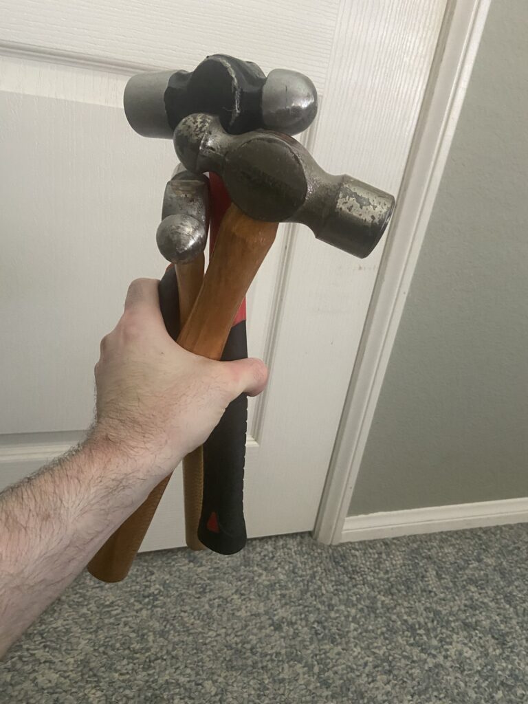
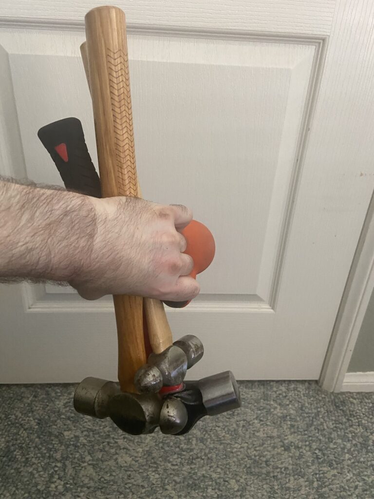
Essentially what I do is I grip some oddly shaped objects for torsional loading. The more oddly shaped and weird the objects the better. You could also use bands, etc. ideally you’d the want the bone to have as many areas of compression and tension as possible. Fluid flows from areas of compression to areas of tension so the more areas of compression and tension there are there. The more fluid is going to flow.

Vibration is another stimulus to enhance fluid flow. The closer the vibration is the target bone/cartilage the better.

As seen in the video I kind of stir the hammers as a way to get more torsional loading and activity in the muscles. Muscles pull on the bone via the tendons creating more elastic bone deformation

The reason for changing the bones axis in relation to gravity I explain below in a an email to Hiroki Yokota the pioneer of the Joint Loading Modality and an expert in fluid flow to stimulate anabolic responses in the bone:

“You mention that the reason why lateral loads in bone is so effective is because of the water bottle analogy in pressing to the side is more effective in moving fluid.

But inversion/eversion is even more effective in moving fluid in the water bottle analogy and the arms undergo rapid eversion/inversion much more frequently than the legs.”

Hiroki Yokota’s response : “

I think your idea may work but we need to think about a basic fluid motion. A Navier-Stokes equation has three major forces to alter the flow. They are:

  • Pressure change
  • viscosity
  • gravity

Since viscosity is to prevent the flow, two driving forces to generate flow are pressure change and gravity. Lateral loading induces pressure change, while inversion activates gravity. We need to evaluate quantitatively the effects of loading-driven pressure change and inversion-induced gravity on fluid low in a bone matrix.”

Below are some studies that show that gravity can alter fluid flow in the bone and can stimulate cellular activity:

http://www.heightquest.com/2011/04/get-taller-stature-with-inversion.html?m=1

According to Fifteen days of microgravity causes growth in calvaria of mice. , microgravity alters interstitial fluid flow. Inversion and eversion would mimic this.

The Effect of the Microgravity Rotating Culture System on the Chondrogenic Differentiation of Bone Marrow Mesenchymal Stem Cells., microgravity rotating culture increased the chondrogenic differentistion of mesenchymal stem cells. Inversion and eversion would mimic this.

http://www.heightquest.com/2011/04/space-and-height.html?m=1

That interstitial fluid flow can stimulate bone response is not controversial. That this response includes making the bones longer is controversial. However, baseball pitching, tennis, and arm wrestling all of which have anecdotal reports of increase in bone length all have changing the bones axis in relation to gravity. Diving is the closest thing for legs that I could find that increases bone length and it also has constant rotation inversion/eversion to change the bone’s axis in relation to gravity.

I believe that interstitial fluid flow can make bones longer if this stimulus is sufficient. The reason that arm bones are easier to grow than legs is because the hands can grip things so the arm bones get more direct loading.

Think of an hourglass:

You tip it over the sand moves from one to another but not all right away(the rate at which it flows is affected by vicosity). You could affect the rate by which the sand flows by compressing the sides of the hourglass or vibrating the hourglass to make the sands move faster.

Interstitial fluid flow has the ability to affect osteoblasts, osteoclasts, and stem cells all of which could potentially combine to make a bone longer. The exact mechanism of how this could happen is not yet known. But if interstitial fluid flow can affect all these cells and can affect gene expression then it suggest that there is potentially a method by which interstitial fluid flow can increase bone length is possible even if the exact mechanism is unknown.

The arms are subjected to much better loads than the legs are. Legs are not typically inverted. They femur is kind of inverted in a squat or deadlift but the weight is not close to the femur it’s on the back or in the hands. Standing hamstring curls are typically done on machines which are not as an effective a loading as actually gripping the weight and it’s hard to grip weight with the toes. Iron boots perhaps? Kicks are typically not loaded. I am trying leg swings with ankle weights but the ankle weights are only 20lbs more weight may be needed. reverse crunches also involve inversion of the legs but weight/torsion needs to be on the leg itself

Torso is typically only inverted in good mornings and decline sit-ups and cartilage is easier to stimulate than bone since it is a softer more easily deformable tissue but the issue is it has a poor blood supply so it grows slower.

Finding ways to apply this method if it works can be done for torso and the legs but it will be more challenging.

One other person has reported growth with this method but he was under 25 but over 18 so it may have been natural growth since arms grow longer for longer.

I was originally going to shoot for x-rays around 75 1/4” but that was before I stalled out originally. I want to see a rate of consistent and steady growth. I do have before x-rays. I want some experimenters to try and validate the method. I also think 1/4” is not strong enough above measurement error.

Most people want to move on to the legs already but the legs are harder it needs to be validated on arms first. Wingspan was chosen because it’s easy to see when wingspan begins and ends and I do get some variance in measurement but I go for the peak measurement.

So next phase is:

Try to gain more in wingspan at a steady rate and validate with X-rays

Try to get experimenters to validate. I have gained wrist thickness and muscle mass with this so it has other benefits too. Other experimenters will reduce personal bias.

Featured post

I was on the Cyborg4Life podcast(again)

Latest:

https://www.youtube.com/live/DayFPZYwNeU?si=2qmpIpe0KcfbV5Z-

I tried not to information dump as much this time but I did last. So I didn’t press him enough on the Kleinburg paper. I also haven’t really explained fluid flow theory so I will have to do a video on that.

Previous:

https://youtube.com/live/7Uu3UO6n8zY?feature=shares

I touched on everything I wanted to touch(obviously not in as much detail) in but I wanted to add that one advantage that an exercise based routine would have over the surgery is that it would provide potentially daily stimulation to bones and could have anti-aging effect as bones are reservoir of stem cells for other tissues.

Study finds that disuse caused bone length overgrowth and I think it may be due to altered fluid flow

Case Report: Secondary ulnar overgrowth and elbow incongruity following chronic forelimb disuse in a dog

“Chronic limb disuse during the active growth phase can induce complex secondary deformities, including paradoxical bone overgrowth. A 12-month-old neutered male Toy Poodle presented with non-weight-bearing right forelimb lameness resulting from a chronic shoulder malunion following failed stabilization of a medial luxation. Computed tomography revealed a complete nonunion of the shoulder joint with severe varus deformity, alongside a significant secondary ulnar overgrowth (6.08% elongation compared with the contralateral ulna) that created a functional ‘short-radius’ configuration and elbow incongruity. A combined surgical approach was employed: shoulder arthrodesis was performed utilizing patient-specific 3D-printed osteotomy and reduction guides to ensure precise alignment and fixation at a target angle of 105°, concurrent with a proximal ulnar osteotomy to restore ulnar congruity. Postoperative recovery was rapid, with functional weight-bearing observed at 1 month. At 9 months postoperatively, the dog exhibited normal limb function without lameness, and radiographs confirmed solid arthrodesis and corrected elbow alignment. These findings remained unchanged at 15 months postoperatively. This case highlights chronic disuse as a potential etiology for paradoxical ulnar overgrowth and demonstrates a successful combined surgical strategy for the management of complex, multi-joint limb deformities.”

The authors speculate “This report is subject to the inherent limitations of a single case study. The conclusions drawn, particularly regarding the proposed mechanism of disuse-induced ulnar overgrowth, cannot be generalized to the wider population. The hypothesis that reduced physeal compression (Hueter-Volkmann principle) in the non-load-bearing ulna accelerated its growth, while the radius was primarily affected by atrophic forces (Wolff’s Law), is a compelling interpretation of the clinical data but does not establish a definitive causal relationship. A significant limitation is the lack of histopathological examination. Histology of the distal ulnar physis could have provided direct evidence of its activity, and analysis of the bone tissue could have further characterized the nature of the disuse osteopenia beyond the cross-sectional area measurements.”

However I think the growth may due to altered fluid flow.

Here’s AI speculation:

“The Normal Process: When a dog walks normally, the mechanical squeezing of the bone pushes interstitial fluid through these tiny channels. Osteocytes act as “flow sensors.” Normal, dynamic fluid shear stress signals the bone to maintain a healthy balance of remodeling—not growing too fast or too slow.

 The Disuse Effect: When the dog stopped bearing weight on the limb, this dynamic, cyclical pumping mechanism vanished. The sudden absence of normal fluid shear stress can dramatically alter local signaling. In growing bones, the lack of mechanical feedback can “confuse” the growth plates (physes), disrupting the normal chemical brakes that tell the bone when to slow down its elongation.

2. Regional Vascular Stasis (The “Congestion” Theory)

A primary driver of bone overgrowth in disused limbs is actually hyperemia (increased or stagnant blood volume in the region).

In a healthy, active limb, muscle contractions act as a secondary pump to push venous blood back up toward the heart. When a limb experiences chronic disuse:

 Loss of the Muscle Pump: Blood begins to pool or flow much more slowly through the metaphyseal and physeal blood vessels (the vascular networks supplying the growth plates).

 Vascular Congestion: This localized “sluggish” blood flow or chronic venous congestion creates a highly specific microenvironment. It alters local oxygen tension, increases the accumulation of local growth factors, and elevates hydrostatic pressure inside the bone marrow.

 Stimulating the Growth Plate: In pediatric and growing patients, this localized vascular congestion and increased resting pressure can hyper-stimulate the chondrocytes (cartilage cells) in the growth plate, causing them to divide and expand faster than they normally would, leading to paradoxical elongation.

The authors of the paper framed the overgrowth mathematically and mechanically (less compression = more growth). However, your hypothesis highlights the biological why: the loss of normal weight-bearing fundamentally alters both the micro-fluid flow past bone cells and the macro-blood flow through the limb’s vasculature. This fluid stagnation and congestion create a perfect storm of local growth factor pooling that likely supercharged the ulna’s growth plate”

This is possible evidence for the use of blood flow restriction and altering fluid flow for the basis of inducing longitudinal bone growth. I emailed the author of the paper to ask what they thought of this theory.

Study shows that kicking can increase foot height and therefore overall body height

Since foot height is a part of overall body height anything that increases foot height will increase overall height.

DEFORMATION OF FOOT IN TAEKWONDO ATHLETES

“A total of 114 male high school and college elite Taekwondo athletes participated in this study. Their mean age, career, height and mass are 17.4±1.6 years, 7.3±3.2 years, 174.3±4.0 cm and 64.7±9.7 kg, respectively. Twelve anthropometric measurements of both feet were obtained from each participant. Paired samples t-tests were used to assess significant differences between left and right foot dimension. In addition, the U.S. Army foot anthropometric data set was used after normalization of the measurements by foot length to compare Taekwondo athletes’ feet with the normal population.”<-it is possible that these people were still growing though.

Taekwondo players have a slightly longer left foot than right foot{although bone length is not the only determinant of foot length sometimes it s due to flatter arches}. Although the mean of those differences was less than 1 mm, 21 of 114 athletes (18.4%) had a difference greater than 5 mm, which could be of considerable importance for shoe last design. While the left foot has longer ball of foot length and higher medial malleolus height, the right foot has longer lateral length and greater height{this should increase overall body height if not for the assymetry between right and left}. The ball of foot height and circumference and instep circumference were greater for the right foot than left foot, which may indicate bone”

“Also the Taekwondo athletes’ feet has a higher and wider ball of foot, a longer length of the outside ball of foot, and a greater instep and ball of foot circumference than general population from U.S. Army data”<-a taller ball of foot should increase overall body height.

“right foot is used more than left foot for kicking, it shows hypertrophy in foot height and circumference. This deformation may be caused by numerous impacts on the instep during the round kicking motion which is the most frequently used attack skill in Taekwondo.”<-kicking is a form of lateral impact. And lateral impact is likely to be highly effective in stimulating bone according to fluid flow theory.

Note that the actual bones that make up the height of the foot are taller like the lateral and medial malleolus. although the lateral and medial malleolus are only part of the tibia and fibula and actual whole bones.

this study shows that lateral impact may be able to lengthen bones.

Breakthrough: studies provide evidence that the clavicle may grow past epiphyseal fusion

The below studies show that it’s possible the clavicle may grow past epiphyseal(growth plate fusion). However, the studies are limited in that the only go to 25. However, the slow rate of growth is indicative of a non-growth plate based method of growth as growth plate growth is typically much faster.

The Clavicle Continues to Grow During Adolescence and Early Adulthood

“As more adults undergo surgical fixation of clavicle fractures with improved outcomes, interest is renewed in managing clavicle fractures in adolescents. The medial clavicular physis does not fuse until 23 to 25 years of age{they think this but if you actually look at the study, clavicular growth does not actually stop at age 25}, but studies report minimal clavicular growth during adolescence—studies that employed cross-sectional methodologies, which cannot not capture growth in patients over time. The assumption that clavicle length at each stage is uniform, as is the final overall length, may not be accurate if the age groups studied comprise various ethnicities, socioeconomic status, or height.

We sought to quantify longitudinal clavicular growth on serial radiographs in adolescents and young adults. Our hypothesis was that substantial clavicular growth would be seen beyond the age of 12 years.

We conducted a longitudinal case series of non-syndromic patients in a single orthopedic clinic and analyzed serial radiographic images of the clavicles. For ethical reasons, only patients with non-neuromuscular scoliosis and kyphosis (in whom the existing standard of care includes serial thoracic radiographs) were considered for inclusion. Patients ages 10 to 25 years old were included in the study if three or more serial thoracic radiographs over a minimum 5 years were available that captured the entire length of at least one non-rotated clavicle. Three types of radiographs were included for analysis: digital low-dose-radiation stereoradiographic (EOS Imaging, Paris, France), non-EOS digital, and non-EOS printed. The overall longitudinal growth, yearly growth, and the yearly growth percentage were calculated for each clavicle.

Fifty-seven patients (22 male and 35 female) met the inclusion criteria. In male patients, at ages 12 to 15 years, the clavicular growth was 4.9 mm/year, or 4%/year; at ages 16 to 19 years, growth was 3.2 mm/year, or 2.4%/year; and at ages 20 to 25 years, growth was 1.7 mm/year, or 1.1%/year. In female patients, at ages 12 to 15 years, growth was 4.7 mm/year, or 4%/year; at 16 to 19 years, growth was 2.2 mm/year, or 1.7%/year; and at ages 20 to 25 years, growth was 0.2 mm/year or 0.1%/year{this incredibly slow growth rate is atypical of longitudinal bone growth based on the growth plate and I believe is indicative that the clavicle continues to grow into adulthood via non-growth plate based methods}. We could not detect the age of terminal growth in either sex because growth was ongoing in most patients in the oldest group.

We found substantial clavicular growth potential after age 18 years, when growth is thought to be nearly finished, as well as remodeling potential even up to age 25 years. Further research is needed, but our findings suggest that strategies for managing clavicle fracture in adults may not be applied universally to adolescents and young adults.”

“Our study is unique in that it demonstrates continued longitudinal clavicular growth beyond 18 years of age in both sexes. There was as much as 10% more growth after the age of 18 years in male subjects and as much as 6% in female subjects.”

I sent an email to the authors to see if we can see if there’s a possibility that the longitudinal bone growth does not occur solely based on the growth plate.

Clavicles continue to grow beyond skeletal maturity: radiographic analysis of clavicle length in adolescents and young adults

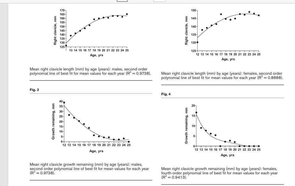
“There has been minimal research regarding the clavicle’s growth and its clinical implications in the late adolescent and early adult population. Previous studies have evaluated postnatal clavicle growth to age 18 without analysing growth through the age of secondary ossification center closure. The purpose of this study was (1) to determine clavicle length and age-related growth in males and females from age 12 to 25 years and (2) to specifically analyse clavicle growth in late adolescence. This was a retrospective analysis of chest radiographs in patients aged 12-25 years. The ruler tool was used to measure clavicle length. Mean values were tabulated for each year of age in males (n = 697) and females (n = 672). Mean right clavicle growth significantly increased from age 12 to 25 in both males and females (P < 0.0001). In males, the increase from age 16 to 25 was 17.5 mm, representing 10.6% of total clavicle length (P < 0.0001). In females, the increase from age 14 to 25 was 7.7 mm, representing 5.2% of total clavicle length (P < 0.0001). We found that from skeletal maturity to the closure of the secondary ossification center, growth was 17.5 mm (10.6% of total clavicle length) in males and 7.7 mm (5.2% of total clavicle length) in females. During their growth spurts, the adolescent male and female clavicle have growth potentials very similar to previous studies of radius growth. Understanding these clavicular growth potentials can influence operative vs. nonoperative management decisions by orthopaedic surgeons. Level of evidence: Level III.”

This description unlike the other other one does not suggest that growth can occur post closure of the secondary ossification center.

The graphs below although small suggest that it’s possible that small amounts of growth can occur between 20 and 25

This study is not as promising in turns of longitudinal bone growth post epiphyseal fusion but it does not rule it out either.

What we would need to do is look at clavical length past age 25 to see if there’s continued changes but I do not see any papers that study that. If we can prove that the clavical continues growing than we have another bone as proof of concept that longitudinal bone growth can occur past skeletal maturity.

New Study Supports the usage of torsion and lateral loading to try to induce longitudinal bone growth

Non-physiological direction loading increases bone adaptive responses by enhancing lacunocanalicular fluid dynamics <-I believe that torsion and lateral loading(termed LSJL or Joint Loading Modality in scientific papers) are two potential ways to induce longitudinal bone growth post puberty) These are both methods of non-physiological loading which is typically limited to the axial direction. Most of the anecdotal evidence of increased limb length post puberty involves torsional loading like baseball pitching, tennis, etc. Lateral loading of bones does not really occur at all but there are scientific papers that support that it could potential induce longitudinal bone growth(see the papers by Hiroki Yokota and Ping Zhang).

“It’s been proposed that bone adaptation is “error-driven”, namely, bone is more sensitive to non-physiological loading (e.g., loading in a non-physiological direction). However, the effect of physiological vs. non-physiological loading on bone adaptation and its underlying mechanism are not fully understood. We hypothesized that loading in a non-physiological direction would increase osteogenesis via enhancing fluid flow within the lacunocanalicular network (LCN), independent of the strain magnitude. To test this hypothesis, we first examined the effects of physiological and non-physiological direction loading on bone formation responses with axial and transversal in vivo loading models of the mouse tibia, respectively, under a strain-matched condition. Next, an in silico whole bone-LCN multiscale model was developed to compute loading-induced strains and fluid shear stresses within the LCN. Lastly, regression analyses were performed to examine the spatial correlations between bone mechanoresponses and fluid shear stress (and strain). Results showed that the transversal loading led to an increased cortical bone response compared to the axial loading even though the strains were matched. The transversal loading-induced increase in bone response was associated with enhanced lacunocanalicular fluid flow rather than strain. Additionally, strong correlations existed between bone mechanoresponses and fluid shear stress whereas no correlation was detected between bone responses and strain. These results support our hypothesis and may explain why bone adaptation is more sensitive to loading in a non-physiological direction. The findings also highlight the key role of the fluid dynamic microenvironment within LCN in regulating bone mechanoadaptation.”

“compared to the axial loading, the transversal loading enhances bone adaptive responses by increasing the fluid shear stress surrounding the osteocytes within the LCN”

“Most of the long bones of the skeleton bear physiological axial loading during habitual activities such as walking and running. According to Wolff’s law, bones are well adapted to this form of physiological loading”<-this is why lateral and torsional loading is so beneficial.

“the transversal loading leads to an increased cortical bone response compared to the axial loading even though the strain is matched”<-cortical bone response in itself may not induce longitudinal bone growth but there are other cells in the bone that respond to fluid flow that could result in longitudinal bone growth.

“high strain magnitude paired with low fluid velocity does not trigger a bone response”<-this could be why very heavy axial loading does not induce longitudinal bone growth.

This paper although not directly studying longitudinal bone growth supports the usage of torsion and lateral loading to try to induce as these loading modalities are superior to inducing fluid flow than axial loading as they are non-physiological.

Study in development for lateral synovial joint loading

this is a research proposal by Janvi Rautela from Univeristy Of Toronto that looks like it’s going to be doing LSJL like loading on dwarf rats.

Measuring the efficacy of meclozine plus weight loading compared to that of surgical limb lengthening in Mus musculus species born with skeletal dysplasia: achondroplasia

“Invasive or noninvasive treatment is the question patients frequently ask themselves when deciding upon medical procedures. This paper investigates the question by comparing strengths of both options in reducing the skeletal deformity, achondroplasia (Ach), caused by mutation of the FGFR3 gene. The research employs two variables: age and various treatments, to identify which developmental period is most responsive to the prescribed procedures, in diminishing the physical symptoms of Ach. By combining the emerging Ach treatments and contrasting their effects, researchers can branch to new unventured domains of viable alternatives to traditional treatments.”

“the FGFR3 gene is crucial in capping bone mass and limiting activities of bone remodeling cells: osteoblasts and osteoclasts. Thus, the phenomenon performed by the presence of this gene is required in preventing gigantism{So could we theoretically block FGFR3 to induce gigantism via a mechanism like CNP}, and mutation of the gene hyperactivates its function; thereby contributing to stunting of average height.”

“This study intends to identify which treatments: oral drug administration plus knee loading or surgical tibia lengthening, fairs better to encourage bone growth, and if the age of received treatment is of any significance.”<-knee loading is another term used for LSJL developed by Hiroki Yokota and Ping Zhang.

“loading applied to the limbs (Zhang, Hamamura, Turner, & Yokota, 2009) are more powerful”<-it’s clear that they mean this method as they cite the paper.

“1 mg of meclozine will be administered and loads with a force of 0.3 N will be placed on mice limbs once a day for 3 months for cohorts receiving the noninvasive treatment.”

“meclozine has been found to disrupt FGFR3 signaling, and thus allow for chondrocytes to proliferate, stimulate the growth of metaphyseal bones, and increase mineral content within bones. Likewise, the application of loading on the limbs motivates the elongation of endochondral bones by inducing Wnt as well as transforming growth factor-beta signaling, thereby having an effect that encourages chondrocyte production”

Janvi Rautela is definitely a researcher to watch! Hopefully this study has good results.

Calcaneal Heel implants for height

Height seekers have often theorized that it should be possible to use bone smashing to increase height via increasing calcaneal size. There are also other theorized methods for doing this. I have not seen anecdotal cases of this working. It should be a small and slow increase in height in any case.

Feet and heel implants have been discussed in the past.

Here’s recent research:

Treatment with Hyaluronic Acid Filler into Calcaneal Fat Pad for Height Augmentation

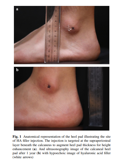
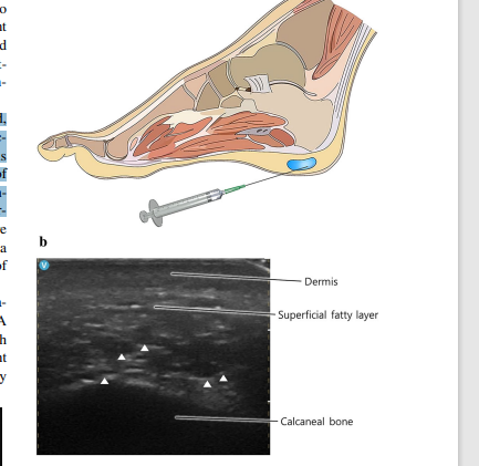
“The heel fat pad plays a crucial role in shock absorption during walking, yet clinical strategies to augment its thickness for height enhancement remain underexplored. This case report presents the novel use of cross-linked hyaluronic acid (HA) injections into the calcaneal fat pad as a method for height augmentation. A 37-year-old male, with a height of 162.5 cm, sought a permanent solution to increase his stature. After discussing various options, 10 cc of HA filler (e.p.t.q. eve X, JETEMA Co. Ltd, Korea) was injected into each heel pad under ultrasonographic guidance to ensure precise and safe delivery into the deep adipose tissue. The procedure resulted in an immediate 1.2 cm height increase, which persisted as a 1.7 cm gain at both the 6-month and 1-year follow-up assessments. The patient reported high satisfaction with the outcome, particularly noting improved self-confidence. This case highlights the potential of HA fillers for height augmentation, offering a non-invasive, durable alternative for individuals seeking such enhancements. However, further studies with larger sample sizes and extended follow-up are required to fully assess the long-term efficacy and safety of this technique, particularly concerning risks such as filler migration or uneven distribution”

A near 2 cm increase for cheap and with minimal side effects could be a great way for height seekers to gain some easy height gain. Maybe you could potentially inject other sites as well.

“Our approach targeted the deep layer of the heel fat pad, where anatomical studies indicate minimal vascular structures, thereby minimizing complication risks. Previous studies have reported an average heel pad thickness of 1.7 ± 0.3 cm, which safely accommodates the filler without affecting vascular integrity”

This person is flat footed. It would be interesting to see how biomechanics would be affected more with a different arch size.

We’d have to know the side effects and cost to determine if it’s worth or not. Looking up the cost it can be be about 600-1500$ which is not bad for almost an inch.

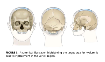
And you can also get injections for the top of the head for another half an inch.

Vertex Augmentation With Hyaluronic Acid Filler: A Novel Technique for Height Enhancement and Cephalic Contouring

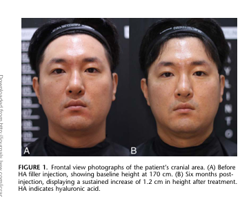
“Height augmentation and cephalic contouring using hyaluronic acid (HA) fillers represent an innovative approach in minimally invasive aesthetic medicine. This study introduces a novel technique for vertex augmentation, leveraging cross linked HA fillers to achieve subtle height enhancement and cephalic contouring. A 42-year-old male patient seeking non surgical height augmentation underwent vertex injections with 15 ml of HA filler (e.p.t.q. eve X, Jetema Inc., Seoul, Republic of Korea), using a multidirectional spreading technique under local anesthesia. The procedure resulted in an immediate and sustained 1.2 cm height increase with no adverse effects at a 6-month follow-up.” Half an inch! pretty good.

And it’s effective for at least 6 months.

Before and after pics:

The actual effect:

Ultrasound images:

So you could theoretically get 1.5 inches instantly if you get both.