Monthly Archives: April 2024

This paper shows why lateral and torsional loading are so effective in causing bone adaptation

A Wolff in sheep’s clothing: Trabecular bone adaptation in response to changes in joint loading orientation

Most loading to bones is axial but there is always some non-axial loads as the bones have shape thus there is a mix of tensile, compressive, and torsional loading. But loading bones in one axis will tend to produce the same stimuli, whereas doing things like lateral and torsional will induce unique stimuli. This will produce more effective adaptations including possibly growing taller if the loading is sufficient.

“This study tests Wolff’s law of trabecular bone adaptation by examining if induced changes in joint loading orientation cause corresponding adjustments in trabecular orientation. Two groups of sheep were exercised at a trot, 15 min/day for 34 days on an inclined (7°) or level (0°) treadmills. Incline trotting caused the sheep to extend their tarsal joints by 3–4.5° during peak loading (P b 0.01) but has no effect on carpal joint angle (P= 0.984). Additionally, tarsal joint angle in the incline group sheep were maintained more extended throughout the day using elevated platform shoes on their forelimbs. A third “sedentary group” group did not run but wore platform shoes throughout the day. As predicted by Wolff’s law, trabecular orientation in the distal tibia (tarsal joint) were more
obtuse by 2.7 to 4.3° in the incline group compared to the level group;{Can this change in trabecular orientation effect bone length?}
trabecular orientation was not significantly different in the sedentary and level groups. In addition, trabecular orientations in the distal radius (carpal joint) of the sedentary, level and incline groups did not differ between groups, and were aligned almost parallel to the radius long axis, corresponding to the almost straight carpal joint angle at peak loading. Measurements of other trabecular bone parameters revealed additional responses to loading, including significantly higher bone volume fraction (BV/TV), Trabecular num-ber (Tb.N) and trabecular thickness (Tb.Th), lower trabecular spacing (Tb.Sp), and less rod-shaped trabeculae (higher structure model index, SMI) in the exercised than sedentary sheep.
Overall, these results demonstrate that trabecular bone dynamically adjusts and realigns itself in very precise relation to changes in peak loading direction{it’s also possible that mixing up axial, lateral, and torsional loading could cause the trabecular to have to constantly dynamically adjust}, indicating that Wolff’s law is not only accurate but also highly sensitive”

“changes in posture will alter stress distribution within the bone and eventually, if they persist, the trabecular structure will readjust to the new stress trajectories.”

The paper mentions that different locomotive methods induce changes in trabecular orientation.

“As intended, the angle of the tarsal joint was more extended by 3.6° at
the time of peak GRF (midstance) in the incline group”

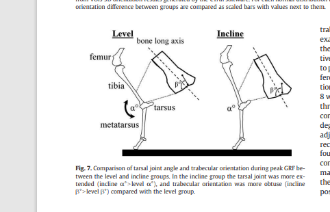
From the looks of this image the incline group looks like it stands taller even though bone length may be the same.

“a minimum level of loading is necessary to affect trabecular growth is evident from the almost total lack of trabeculae documented in the sagittal
ridges of the distal tibia and radius”<-So there must be sufficient weight used in lateral and torsional loading regimes.

“An additional limitation is that we studied only very young animals whose joints were still growing and remodeling.”

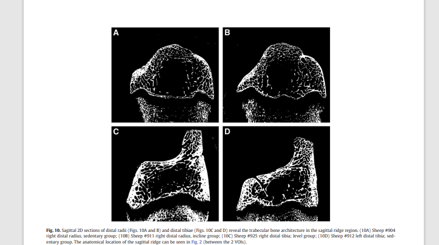
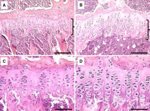
Note how much bigger and longer the epipihysis is in image B versus A but again these are growing animals. They said that trabecular did not really change much in the radius(groups A and B) but the two radius look much bigger.

In contrast the Group D looks smaller than group C which is the tibiae which is what changed.

Studies like Trabecular bone in the calcaneus of runners, were conducted on runners above age 20 so it’s likely still that trabecular bone is responsive to the direction of loading.

The paper Physical activity engendering loads from diverse directions augments the growing skeleton, shows that again loads from diverse directions are important thus perhaps typical axial loading is enough.

“Growing mice housed for three months in cages designed to emphasize
non-linear locomotion (diverse-orientation loading) were found to have enhanced trabecular and cortical bone in the proximal humerus compared to animals housed in cages that accentuated linear locomotion (stereotypic-orientation loading).”

These papers show why we must try to use experiment with lateral and torsional loading methods in attempts to grow taller.

Study shows that mechanical loading can alter the trabecular bone via the growth plate

This study shows that mechanical loading can alter the growth plate and the trabecular bone. This shows it is possible to use mechanical loading to alter height during development and also perhaps spinal height as there is a great deal of cartilage via the discs there.

Unveiling the Trabecular Connection: Exploring Morphological Adaptations in the Growth Plate and Their Response to Mechanical Stimulus

“The growth plate, also known as the epiphyseal plate, is a critical element in the longitudinal growth of long bones during development. This study aims to explore the connection between trabecular patterns and morphological alterations within the growth plate, particularly concerning their impact on shape and mamillary processes or trabecular patterns as well as the formation of ossification bridges{ossification bridges has the potential to reduce growth so studying them has potential}. Our objective is to investigate how the adaptations in the growth plate and the trabecular bone in its vicinity respond to mechanical stimulus, this approach is considered a new methodology to study endochondral growth and bone remodelling”

“Our investigation revealed insights into the development of the growth plate and its capacity to adapt its shape in response to the local mechanical environment. Previously, this environment had been predominantly modeled as a continuum, but our model allowed us to assess the impact of localized load transmission via the trabecular groups. Furthermore, our research demonstrated that the morphological changes within the growth plate in addition to bone adaptation in its vicinity, serve as an adaptive mechanism to withstand shear stress{so we would want to optimally induce shear stress, I believe that torsional loading is the best way to induce fluid shear}, contributing to increased bone density in specific regions”

“at birth the growth plates have a flat and smooth topology in humans, as age increases a wavy pattern is seen, this change is due to mechanical stresses”

“The thickness in the growth plate remains constant during childhood thanks to an equilibrium between chondrocyte proliferation and apoptosis. In puberty, when this balance is perturbed, the process of growth plate fusion starts, forming ossification bridges in different locations until the entire plate has been ossified, leaving behind a bone remnant, formally known as epiphyseal scar”

“animals, such as mice and rats, which are widely used as in-vivo models for endochondral ossification studies, do not entirely close their growth plates during maturation”<-but their growth plates do seem to be become senescent which indicates that there may be epigenetic factors that limit growth plates from growing indefinitely.

this looks like an example of the histological changes due to lateral synovial joint loading so joint loading definitely alters the growth plate morphology:

“the change in shape in the growth may occur as a consequence of the bone remodeling process in the vicinity of the growth plate, and at the same time trabecular formations adapt”

“A future study case for this model will be a therapy of hemiephysiodesis or guided growth, where this model can be used to quantify the degree of growth modulation along the entire growth plate, or the
change in geometrical descriptors; simply by including a transphyseal screw in the domain.”<-so there is potential for this research to be used for people to grow taller.

Passive diffusion and it’s potential role in growin taller

If articular cartilage endochondral ossification(and) is to be a viable with which to grow taller then the existing articular cartilage must grow and replenish while the rest ossifies. Vertebral and joint cartilage contributes to height so if there was a way to get them to grow then we could become taller. Since cartilage has poor blood supply it heals and grows by passive diffusion. So our goal would be to maximize passive diffusion.

Passive diffusion is where a molecule from high concentration to low to concentration.

The factors affecting diffusion are :

Temperature: Higher temperatures increase molecular energy, leading to faster movement and enhanced diffusion.

Concentration Gradient: The steeper the gradient (difference in concentration), the faster the diffusion.

Molecular Mass: Smaller molecules diffuse more rapidly than larger ones.

Medium: Different substances diffuse at varying rates through different media.”

So what we can influence is temperature and concentration gradient. Temperature is obvious but for concentration gradient we can increase supplementation and increase fluid flow as the more fluid is flowing the higher concentration is going to be and therefore the more compounds are going to be diffusing into the articular cartilage.

This coincides with anecdotal evidence that movement is vital for healthy cartilage and with the observation that sufficient force in chiropractic treatment is required to induce joint cavitation.

The Effect of Antibody Size and Mechanical Loading on Solute Diffusion Through the Articular Surface of Cartilage

” Therefore, the goal of this study is to investigate how the size of antibody (Ab) variants, as well as application of cyclic mechanical loading{the cyclic nature of the loading as the changing in load rate is important as well as overall load}, affects solute transport within healthy cartilage tissue. Penetration of fluorescently tagged solutes was quantified using confocal microscopy. For all the solutes tested, fluorescence curves were obtained through the articular surface. On average, diffusivities for the solutes of sizes 200 kDa, 150 kDa, 50 kDa, and 25 kDa were 3.3, 3.4, 5.1, and 6.0 μm2/s from 0 to 100 μm from the articular surface. Diffusivities went up to a maximum of 16.5, 18.5, 20.5, and 23.4 μm2/s for the 200 kDa, 150 kDa, 50 kDa, and 25 kDa molecules, respectively, from 225 to 325 μm from the surface. Overall, the effect of loading was very significant, with maximal transport enhancement for each solute ranging from 2.2 to 3.4-fold near 275 μm{so mechanical loading has a lot of potential to benefit passive diffusion and therefore cartilage growth}. Ultimately, solutes of this size do not diffuse uniformly nor are convected uniformly, through the depth of the cartilage tissue. This research potentially holds great clinical significance to discover ways of further optimizing transport into cartilage and leads to effective antibody-based treatments for OA.”