Monthly Archives: August 2022

Is LPP(Link Protein N-terminal peptide) a potential height increasing supplement?

I went through the papers to see if there’s any potential. LPP is linked to HMGA2 which does have height increase potential applications.

Link Protein N-Terminal Peptide as a Potential Stimulating Factor for Stem Cell-Based Cartilage Regeneration

<-the title right off the bat suggests potential as anything that suggests cartilage regeneration may be able to increase height if even only in the joints or spinal height.

Link protein N-terminal peptide (LPP) in extracellular matrix (ECM) of cartilage could induce synthesis of proteoglycans and collagen type II in cartilaginous cells{if the extracellular matrix of the joints or intervertebral discs is thicker that would overall make you taller!}. Cartilage stem/progenitor cells (CSPCs), the endogenous stem cells in cartilage, are important in cartilage degeneration and regeneration. We hypothesized that LPP could be a stimulator for stem cell-based cartilage regeneration by affecting biological behaviors of CSPC.  CSPCs were isolated from rat knee cartilage. We evaluated the promoting effect of LPP on proliferation, migration, and chondrogenic differentiation of CSPCs. The chondrogenic differentiation-related genes and proteins were quantitated. Three-dimensional culture of CSPC was conducted in the presence of TGF-β3 or LPP, and the harvested pellets were analyzed to assess the function of LPP on cartilage regeneration. LPP stimulated the proliferation of CSPC and accelerated the site-directional migration. Higher expression of SOX9, collagen II, and aggrecan were demonstrated in CSPCs treated with LPP. The pellets treated with LPP showed more distinct characteristics of chondroid differentiation than those with TGF-β3. LPP showed application prospect in cartilage regeneration medicine by stimulating proliferation, migration, and chondrogenic differentiation of cartilage stem/progenitor cells.”

So there is a possibility of LPP injects in cartilage regions to make people slightly taller of course with caveats as not everything that has potential works. In the paper there’s a lot about degeneration of articular cartilage so they so the potential for application in that area which would in turn result in potential height increase.

“Link protein, a glycoprotein that exists in human intervertebral discs as well as in the articular cartilage, plays an important role in strengthening the binding between aggrecan and hyaluronan. Link protein N-terminal peptide (LPP) is the cleaved N-terminal 16 amino peptide (DHLSDNYTLDHDRAIH) of link protein. LPP was thought to be the functional fragment of link protein as the cross-linker”

If you look at the doses figure 5 there seems to be an equilibrium effect with around 50ng/mL having the equilibrium effect.

This suggests that if you are not deficient in LPP it may have no impact on height whatsoever(LPP already exists in cartilage regions).

So LPP may have no impact on articular cartilage regions(but that doesn’t mean that it wouldn’t) but there is still the possibility of using LPP to induce stem cell differentiation into chondrocytes. But I don’t think the differentiation of stem cells into chondrocytes is the problem. In distraction osteogenesis there is already chondrogenic differentiation. The problem is likely a lack of stem cells in general in the articular and that bone is not capable of interstitial growth. Perhaps LPP could be used as part of microfracture surgery whose goal is to create microfractures to get stem cells to the articular cartilage but the problem is that the cartilage formed is fibrocartilage. So perhaps LPP could be used to make the cartilage purer.

Bone usually heals by bone remodeling perhaps LPP injections could encourage it to heal via endochondral ossification resulting in taller height over time?

Simultaneous Recruitment of Stem Cells and Chondrocytes Induced by a Functionalized Self-Assembling Peptide Hydrogel Improves Endogenous Cartilage Regeneration

“The goal of treating articular cartilage (AC) injury is to regenerate cartilage tissue and to integrate the neo-cartilage with surrounding host cartilage. However, most current studies tend to focus on engineering cartilage; interface integration has been somewhat neglected. An endogenous regenerative strategy that simultaneously increases the recruitment of bone marrow mesenchymal stem cells (BMSCs) and chondrocytes may improve interface integration and cartilage regeneration. In this study, a novel functionalized self-assembling peptide hydrogel (KLD-12/KLD-12-LPP, KLPP) containing link protein N-peptide (LPP) was designed to optimize cartilage repair. KLPP hydrogel was characterized using transmission electron microscopy (TEM) and rheometry. KLPP hydrogel shared a similar microstructure to KLD-12 hydrogel which possesses a nanostructure with a fiber diameter of 25–35 nm. In vitro experiments showed that KLPP hydrogel had little cytotoxicity, and significantly induced chondrocyte migration and increased BMSC migration compared to KLD-12 hydrogel. In vivo results showed that defects treated with KLPP hydrogel had higher overall International Cartilage Repair Society (ICRS) scores, Safranin-O staining scores and cumulative histology scores than untreated defects or defects treated with KLD-12 hydrogel, although defects treated with KLD-12 and KLPP hydrogels received similar type II collagen immunostaining scores. All these findings indicated that the simple injectable functionalized self-assembling peptide hydrogel KLPP facilitated simultaneous recruitment of endogenous chondrocytes and BMSCs to promote interface integration and improve cartilage regeneration, holding great potential as a one-step surgery strategy for endogenous cartilage repair.”

“LPP can induce directional migration of nucleus pulposus cells (NPCs) and cartilage-derived stem cells (CSCs)”

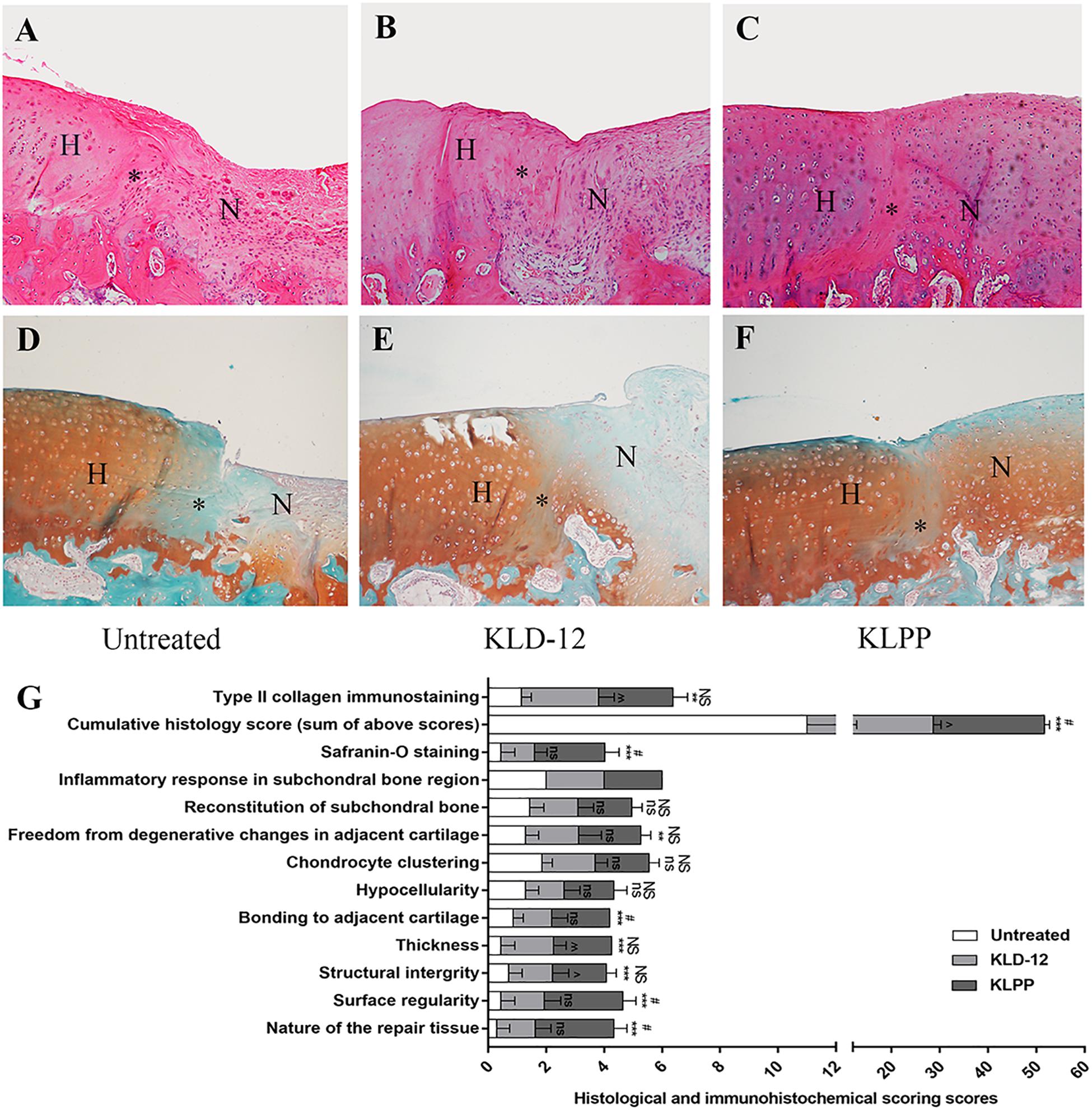
Now is this cartilage “taller” than before the injection? I can’t really tell from the images. IF you look at slide F it looks there might be some slight overgrowth.

Overall I’d say use LPP does have some height increase potential but probably very minor and it’s probably going to be a while before it can be used for height in practice.

Can Regeltec Hydrafil injections make you taller in the spine?

The short answer is yes of course. Loss of spinal height is loss of height. There was a limb lengthening surgery for the spine but it was only for immature pigs. And the growth achieved was very small. There is a growing rods technique for scoliosis patients but the lengthening achieved according to Spinal Lengthening With Magnetically Controlled Growing Rods is something on the order of millimeters. But that adds up.

The intervertebral discs are a component of height. Some people have claimed on forums to get taller in the spine via stretching and I would love to see those claims validated in some way.

Here’s the regeltec website.

Here’s a video with more about the technique:

There was a clinical trial but no results have been posted. I could find some data that the Hydrafil reduced backpain but no data on the impact on height. Of course how much height is added would vary on how much the disc is degenerated. And it would be interesting if you could somehow overfill the disc in order to add height above capacity.

There’s definitely a cost issue as there’s a lot of discs to inject hydrafil into. But there’s no surgery and you just have to inject it that should alleviate the cost. So it may not break the bank but it would probably be a significant sum like all surgical procedures.

But it may be a good non-invasive way to add a little bit of height?

LSJL studies 5: LSJL device design

This paper discusses a lateral bone loading device.  It mentions a capacity of 40N which I think won’t be enough for lengthening purposes as since lengthening post growth plate senesence is an abnormal task it probably requires very abnormal stimuli.  It’s interesting to look at the device though.

The study mainly mentioned the technical design of the device and no analysis of the applications.

A Mechatronic Loading Device to Stimulate Bone Growth via a Human Knee

“This paper presents the design of an innovative device that applies dynamic mechanical load to human knee joints. Dynamic loading is employed by applying cyclic and periodic force on a target area. The repeated force loading was considered to be an effective modality for repair and rehabilitation of long bones that are subject to ailments like fractures, osteoporosis, osteoarthritis, etc. The proposed device design builds on the knowledge gained in previous animal and mechanical studies. It employs a modified slider-crank linkage mechanism actuated by a brushless Direct Current (DC) motor and provides uniform and cyclic force.”

Here’s an example of what slider crank linkage looks like:

slider-crank-linkage

“The functionality of the device was simulated in a software environment and the structural integrity was analyzed using a finite element method for the prototype construction. The device is controlled by a microcontroller that is programmed to provide the desired loading force at a predetermined frequency and for a specific duration. The device was successfully tested in various experiments for its usability and full functionality.  The device works according to the requirements of force magnitude and operational frequency. This device is considered ready to be used for a clinical study to examine whether controlled knee-loading could be an effective regimen for treating the stated bone-related ailments{Hopefully bone length is one of those bone-related ailments unfortunately Ping Zhang’s name is not on this paper and he was always the one more interested in bone length}.”

“When a specific loading force is applied to the epiphyses of the femur and tibia, the trabecular bone tissue, which is characterized by axial stress resistance, resists this force from the opposite direction. This results in deformations in that area. These deformations create a variation of the fluid pressure in the intramedullary cavity. This pressure gradient allows the flow of fluids that carry essential nutrients to the bone cortex initiating osteoblast differentiation and osteogenesis, thus helping in repair and regeneration of the bone tissue. This unique reaction makes this procedure an effective treatment for bone rehabilitation. It helps in reduction of healing time of bone fractures and hastens recovery from bone-related injuries and diseases. The lateral stress application is also less strenuous to the knee bone and reduces the amount of force that needs to be applied to get this result.”

pressure-caused-by-fluid-flow

B is the force we’re looking for.  The pressure generated by fluid flow not just on the bone but on the stem cells to initiate chondrogenic differentiation.  The pressure on the intact bone may also allow the creation of cartilage canals to enable that requirement for a neo growth plate.

It’s also interesting to note that in the proposed knee loading device the load the entire lateral area of the epiphysis this may be a way to reduce slippage.

” it was decided that the proposed device should be robust enough to produce different magnitudes of linear force up to a maximum of 40 N”<-Since lengthening is not being considered in this study forces required for lengthening may be higher.

lsjl-dev-ice

The device doesn’t look wide enough for the knee really.  The dimensions of the device listed are:

Length: 0.3 m
Width: 0.1
Height: 0.2

There are about 39 inches in a meter so about 3.9 inches in width.  I don’t know if that’s enough.

Also the device looks more like this kind of clamp:

Then the other clamps we’ve been using.    Although you’d have to make new pads to actually adjust to knee.  Well actually more like:

But the pipe gets in the way of getting around the knee.  Although I’m not really sure that a pipe clamp is superior to the other clamps.  I’m just pointing out that it’s the clamp that looks most like the design mentioned in the study.

Here’s some more details on the device:

sensors-16-01615-g004

Here’s an actual physical prototype:

more-advanced-protoype

Here’s an update on that device:

“Dynamic loading to a knee joint is considered to be an effective modality for enhancing the healing of long bones and cartilage that are subject to ailments like fractures, osteoarthritis, etc. We developed a knee loading device and tested it for force application. The device applies forces on the skin, whereas force transmitted to the knee joint elements is directly responsible for promoting the healing of bone and cartilage. However, it is not well understood how loads on the skin are transmitted to the cartilage, ligaments, and bone. Based on a CAD model of a human knee joint, we conducted a finite element analysis (FEA) for force transmission from the skin and soft tissue to a knee joint. In this study, 3D models of human knee joint elements were assembled in an FEA software package (SIMSOLID). A wide range of forces was applied to the skin with different thickness in order to obtain approximate force values transmitted from the skin to the joint elements. The maximum Von Mises stress and displacement distributions were estimated for different components of the knee joint. The results demonstrate that the high load bearing areas were located on the posterior portion of the cartilage. This prediction can be used to improve the design of the knee loading device.”
Step 1 for improving effectiveness of the device is to prove that it loads where it’s expected to be loading.
“A lateral application of force on the knee joint was found effective in protecting bone tissue in the areas of distal femur and proximal tibia of the knee bone. The effects of the stimulation of bone regeneration are not limited to the applied areas but are seen along the length of the long bone. When a specific loading force is applied to the epiphyses of the femur and tibia, the trabecular bone tissue, which is characterized by axial stress resistance, resists this force from the opposite direction.”
“This results in reversable deformations in that area. These deformations create a variation of the fluid pressure in the intramedullary cavity. This pressure gradient allows the flow of fluids that carry essential nutrients to the bone cortex initiating osteoblast differentiation and osteogenesis thus helping in repair and regeneration of the bone tissue“<-this pressure gradient at minimum should help carry nutrients to the growth plate at a minimum.
There’s a lot more information within the paper itself about potential joint loading device design.

Lowering Fbn1 levels may increase bone length(Marfan’s Syndrome)

Even though it mainly seems like it’s an active growth plate thing, it’s possible that FBN1 deficiency could stimulate neo-growth plate activation as it does stimulate TGF Beta activation and there’s ectopic tendon calcification.  This Franscesco Ramirez scientist seems one to watch in understanding why Marfan’s Syndrome causes longitudinal bone overgrowth.   If we understand why Marfan’s Syndrome causes overgrowth then maybe we can use that to our advantage especially if it is connected to tendons which are connected to muscles and are therefore easier to manipulate.  Interesting that in the grant FBN1 inactivation is associated with greater TGF-Beta in the tendon/ligament but decreases TGF-Beta in the perichondrium.

TENDON-DEPENDENT CONTROL OF LONGITUDINAL BONE GROWTH

“Skeletal abnormalities caused by disproportioned bone overgrowth (LBO), are a common trait in Marfan syndrome (MFS), a connective tissue disease caused by mutations in the extracellular matrix (ECM) protein and TGFβ regulator fibrillin-1 (Fbn1). The cause of LBO in MFS is unknown and therapies are not available. Fibrillin-1 hypomorphic mouse model (Fbn1mgR/mgR) faithfully replicates MFS skeletal manifestations including elongated bones however, its early demise due aortic rupture limit the magnitude of LBO investigation.

To circumvent Fbn1mgR/mgR lethality and investigate the contribution of specific skeletal tissues to LBO, Fbn1 gene expression was targeted in developing limbs by crossing Fbn1Lox/Lox mice with Prx1-Cre, in or bone with Osx-Cre, in cartilage and perichondrium with Col2-Cre, in skeletal muscles with Mef2c-Cre, and ligaments and tendons with Scx-Cre. Bones length of Fbn1 conditional mice KO was measured and relevant histological, cellular and biomechanical parameters were assessed.

Fbn1Prx1−/+ and Fbn1Prx1−/− mice had longer limbs bones compared to WT mice and amount of fibrillin-1 in the limb matrix was inversely proportional to bone length. Interestingly, Fbn1 gene targeting in ligaments/tendons resulted in LBO, altered tissues’ mechanics and TGFβ-induced switch of tendon stem cells to chondrocytes{could we make tendon cells turn into chondrocytes as adults via TGF Beta?}. Gene targeting in other limb’s anatomical locations did not result in LBO thus ruling out the participation of surrounding tissues to this bone phenotype.

Fbn1 gene inactivation in ligament/tendon is associated with increased local TGFβ, altered biomechanical properties and LBO[longitudinal bone overgrowth]. As previously reported, ligaments/tendons respond to changes in mechanical load by increasing the levels and/or the activity of TGF-β while bones undergo morphological adaptation in response to muscle loads transmitted by tendons. We hypothesize that dysregulation of local TGFβ signaling and altered biomechanical properties of fibrillin-1 deficient ligaments/tendons affect endochondral ossification by improper load transmission to bone. By showing ligament/tendon-dependent regulation of postnatal longitudinal bone growth this study provides a paradigm-shift in tendon biology and it shades a new light on LBO pathophysiology in MFS, thus providing the bases for new pharmacological interventions for this and related skeletal conditions.”

So lower levels of Fbn1 means longer bone length and FBN1 deficient tendons and ligaments alter endochdondral ossification by altering load transmission to bone.  We can alter load transmission via mechanical stimulation without altering FBN1.

Here’s a grant(2016) related to the subject:

TENDON-DEPENDENT CONTROL OF LONGITUDINAL BONE GROWTH

“disproportionate increase of longitudinal bone growth that causes serious malformations of the limbs, anterior chest and Spine is the clinical hallmark of patients afflicted with Marfan syndrome (MFS), a connective tissue disease caused by mutations in the extracellular matrix (ECM) protein and TGFβ regulator fibrillin-1. Our preliminary studies of mice with tissue-specific ablated Fbn1 gene activity have revealed an unsuspected causal relationship between tendon/ligament (T/L) dysfunction and longitudinal bone overgrowth (LBO). Specifically, (1) Fbn1 inactivation in T/L cells was necessary and sufficient to promote linear bone overgrowth associated with dysregulated growth plate (GP) gene expression; (2) fibrillin-1-deficient tendons displayed abnormal tissue architecture and impaired mechanical properties, particularly at bone- insertion sites; (3) the relative amount of fibrillin-1 correlated with discrete changes in tendon mechanics; (4) tendon-derived stem/progenitor cell (TSPC) cultures deficient for fibrillin-1 differentiated improperly as result of increased latent TGFβ activation; and (5) ectopic tendon calcification of fibrillin-1-deficient tendons was commonly observed. fibrillin-1 assemblies normally restrict GP-driven linear growth of neighboring bones by specifying the mechanical properties of tendons through the control of ECM organization and TGFB-regulated TSPC differentiation. Accordingly, the scope of our proposal is two-fold; first, to characterize how fibrillin-1 deficiency translates into tendon dysfunction and tendon-associated LBO, and second, to establish how local TGFB hyperactivity in tendons promote tissue degeneration thereby leading to excessive linear growth of the adjacent, structurally normal bones. To this end, we will characterize the expression of molecular and cellular determinants of tendon development and maturation in mice deficient for fibrillin-1 in T/L matrices, in addition to employing computational approaches to identify probable disease-causing molecular abnormalities in the GP of these tendon-defective animals (Aim 1); apply data-driven statistical models to determine how graded fibrillin-1 deficiencies correlate with tendon mechanics and associated LBO (Aim 2); and assess whether systemic TGFβ neutralization modifies tendon pathology and LBO severity in fibrillin-1-deficient mice (Aim 3). The results of these investigations are expected to substantially advance our limited understanding of tendon function in health and disease and implicitly, of the cellular, molecular and tissue factors that coordinate the postnatal growth of musculoskeletal tissues. ”

Here’s the updated 2020 grant.

It seems exactly the same as the 2016 grant.

Fibrillin-1 deficiency in the outer perichondrium causes longitudinal bone overgrowth in mice with Marfan syndrome

“A disproportionate tall stature is the most evident manifestation in Marfan syndrome (MFS), a multisystem condition caused by mutations in the extracellular protein and TGFβ modulator, fibrillin-1. Unlike cardiovascular manifestations, there has been little effort devoted to unravel the molecular mechanism responsible for long bone overgrowth in MFS. By combining the Cre-LoxP recombination system with metatarsal bone cultures, here we identify the outer layer of the perichondrium as the tissue responsible for long bone overgrowth in MFS mice{the perichondrium is less mature than the periosteum so it is unclear whether manipulating the periosteum would have any impact on longitudinal bone overgrowth}. Analyses of differentially expressed genes in the fibrillin-1-deficient perichondrium predicted that loss of TGFβ signaling may influence chondrogenesis in the neighboring epiphyseal growth plate (GP). Immunohistochemistry revealed that fibrillin-1 deficiency in the outer perichondrium is associated with decreased accumulation of latent TGFβ-binding proteins (LTBPs)-3 and -4, and reduced levels of phosphorylated (activated) Smad2. Consistent with these findings, mutant metatarsal bones grown in vitro were longer and released less TGFβ than the wild-type counterparts. Moreover, addition of recombinant TGFβ1 normalized linear growth of mutant metatarsal bones. We conclude that longitudinal bone overgrowth in MFS is accounted for by diminished sequestration of LTBP-3 and LTBP-4 into the fibrillin-1-deficient matrix of the outer perichondrium, which results in less TGFβ signaling locally and improper GP differentiation distally.”

<-I could not get this full paper.  But also note that it’s the long bones that grow more with marfan’s syndrome so it’s possible that in different bones FBN1 has a different effect on TGFBeta

Interesting that less TGF Beta resulted in more longitudinal bone growth.  I’ve always thought that TGF Beta is good for height.  It could be that Smad 1/5/8 phosphorylation results in terminal differentiation.  According to Inhibition of TGF-β Increases Bone Volume and Strength in a Mouse Model of Osteogenesis Imperfectainhibition of TGF Beta increases bone strength(no mention of bone length however).

According to this study by Ramirez:
“limbs deficient for fibrillin-1 (Fbn1Prx1–/– mice) is accounted for by premature depletion of MSCs and osteoprogenitor cells combined with constitutively enhanced bone resorption. “

Tiffanie Didonato grew 14 inches with distraction osteogenesis

Tiffanie Didonato grew 14 inches with distraction osteogenesis but the limit for a normal person is about six inches. Why?

Looking at her instagram she seems to be doing fine.

According to Paley’s FAQ on limb lengthening surgery, “The total height gain with two lengthenings is up to 13cm (8cm in the femurs and 5 cm in the tibias. (8cm is not well tolerated in the lower leg (tibia) and
exceeding 5cm can lead to more serious complications such as equinus contracture [ballerina foot]). Most
patients will not tolerate more than 5cm in the tibias. Of course the cost of two lengthenings is nearly twice that of one lengthening. Although the Precice can lengthen up to 8cm, not every patient can safely achieve this much even in the femurs. We will only allow lengthening to the tolerance of the patient’s bone and soft tissues. SAFETY first. We will not risk a loss of function to gain one more cm. To get the full 8cm from both femurs and both tibias requires three lengthening surgeries (see option 5 below).”

Option five as mentioned is “Combined tibia (up to 4cm) and femur (up to 4cm) lengthening three
weeks apart: total 8cm followed by re-breaking femur and tibia with same nail in place and repeating up to 4cm femur and up to 4cm tibia lengthening one year or more later (up to total 16cm; 6.3 in.)”

So normal person can gain 6.3 inches but someone with achondroplasia can gain 14 inches. The answer may involve the ligaments.

According to the chandler project, “When we stretch the bones were also stretching the muscles, and the ligaments and the nerves in the blood vessels around it and some of those get tight, and when they get tight there can be other problems that come up” But for dwarfism, “[dwarves have] all the skin in the ligaments and everything like that to be average height, but her bones are shorter, and they just don’t get the signals to grow,” Lisa explained.”

So the reason that people with achondroplasia can grow taller with Limb Lengthening Surgery than people without it is because they have the ligaments etc of an average height person thus their muscles etc don’t stretch.

But why is the soft tissue such as problem in the first place with limb lengthening surgery?

Well the ligaments normally attach at the enthesis, and the enthesis is attached near the growth plate. It’s very likely that ligament and bone growth are connected in this way so that the ligaments can grow as needed to support longitudinal bone growth. ““Entheses are fibrocartilaginous organs that bridge ligament with bone at their interfaceMore info about the enthesis here.

So it’s likely that a superior option to limb lengthening surgery will develop that involves stimulating enthesis development as well.

A website has an interesting interpretation of what Lateral Synovial Joint Loading in

I have not been experimenting with Lateral Synovial Joint Loading as I’m focusing more on lateral impact because slippage was too much of an issue. I think joint loading may work more easily on wingspan then on height just because the joints are less cumbersome and the wrist is more moveable. The ankle is just a pain. In 2015 I report getting an increase of 1/2″ inch of wingspan from 74.5″ to 75″. My wingspan is still 75″ but it may be a more solid 75″. It is not measurement error for wingspan as easy to self measure lying down.

Anyways, here’s the site:

What is lateral synovial joint loading?

Question:

What is lateral synovial joint loading?

Synovial Joints:

Synovial joints are the types of joints in the body that allow movement. They are places where two or more bones meet yet are able to move at their connection point. Lateral is a term that means from or related to the side rather than above or below.

Answer and Explanation:

Lateral synovial joint loading is a process where lateral pressure applied to a joint increases the fluid pressure in the joint cavity. The pressure on the synovial fluid in the joint cavity causes it to become more viscous as a means of resisting the pressure. However, the increased hydrostatic pressure can cause stretching of the membranes containing the fluid and cause inflammation of the joint.”

Here’s more on hydrostatic pressure. More on the power of hydrostatic pressure. Hydrostatic pressure may be be linked to bone via the periosteum. So load on the joints can have an effect on bone whether that can increase height is TBD.