Category Archives: Uncategorized

China Banned Leg Limb Lengthening Surgery In 2006

Me: I remember reading from the Make Me Taller forum that the practice of leg and limb lengthening surgery was banned in China. However, that has not stopped some medical professionals from still setting up clinics in China to perform the limb lengthening surgery still. If I remember correctly the Chinese doctor Bai Helong who came up with what he said was a minimally invasive and NON-PAINFUL procedure had some trouble with the Chinese government and relocated his practice around to where I heard was in Shanghai last time. If I was to guess, the lure of the money will cause some doctors or people with medical degrees to be willing to go against the law and perform the surgery still for the many millions of people who would break the law for extra height.

From an internet source HERE

China Bans Illegal Leg-lengthening Surgery
  2006-11-04 22:34:30      AFP
China has banned leg-lengthening surgery that a largely unregulated beauty industry has been offering to customers who want to be taller.”Leg-stretching surgery for the image conscious has been banned by China’s Health Ministry after a spate of botched operations,” the Xinhua news agency reported Saturday.The operation involves breaking the patients’ legs and stretching them on a rack, and has led to several cases of disfigurement, Xinhua said, citing the health ministry.

Hospitals that conduct at least 400 orthopedic operations a year and offer post-surgery care and rehabilitation will be allowed to carry out such operations in future, but only on strictly medical grounds, according to a ministry circular.

The surgical procedure, popular among young Chinese professionals who believe height will help them climb the career ladder, was developed in Russia to help patients with birth defects such as dwarfism.

Botched operations carried out by unauthorized beauty clinics have left many patients physically and psychologically scarred, Xinhua said, without detailing the injuries.

Ten people, enticed by a promise of “height surgery without pain,” were reported to have been disfigured after they underwent the operation last year at a Beijing hospital.

The health ministry said hospitals must in future tell patients about the risks involved, while only qualified personnel would be allowed to carry out the procedure.

Low Magnitude, High Frequency Mechanical Stimuli Are Anabolic And Osteogenic To Trabecular Bone In Children

Me: This study helps in showing that even at low magnitudes for mechanical loads, as long as it is of high frequency the trabecular bones can have an anabolic effect. This pushes the idea further to show that the high magnitudes for loading which I had proposed before may not be needed and may be easier to create such a bone loading device than previously believed.

Analysis & Interpretation:

It would seem that for just s short amount of time with even small magnitudes in bone loading we find that the bone mass density (BMD) can be increased in kids who are still developing. The only thing that seems to be very important is to make the loading have high frequency.  The researchers wanted to test the loading on both the proximal tibia and spinal vertebrate area for bone mass density changes. The results showed that it was effective.

Implications For Height Increase:

This shows that at least for developing children, changing their bone density from low , which indicates bone fragileness, to high which would indicate strength and like chance for fractures is rather easy and quick to do. So many physicians have worried about the fact that many of the height increasing methods we have looked at may be bad for bone density since they can decrease BMD leading to weaker bones. This shows that their overall worrying and concerns about the loss of bone density is not really valid. We saw from a few of the recent posts with looking at the effect of chronic starvation, illness, and inflammation that the band or area of band actually increases in bone density in a developing child’s bone when analyzed histologically, however the longitudinal gorwth rate becomes slightly stunted during that phase.This means that when we are looking at any height increase idea or method we would not need to worry too much about the loss or effect on bone density. Bone density can be rather easily increased with this type of low magnitude high frequency loading or from taking certain bone increasing pills like statin.

As always let’s remember that this type of loading is very similar to the LSJL idea with the effect being bone growth or bone increase. However we are not really looking for bone growth, but bone lengthening. That requires cartilage. This would slightly infer that the chance of using the LSJL in children with their growth plates intact might be effective in increasing the rate of their longitudinal growth of long bones, ie. make them taller.

From PubMed study link HERE


J Bone Miner Res. 2004 Mar;19(3):360-9. Epub 2004 Jan 27.

Low magnitude mechanical loading is osteogenic in children with disabling conditions.

Ward K, Alsop C, Caulton J, Rubin C, Adams J, Mughal Z.

Source

Clinical Radiology, Imaging Science & Biomedical Engineering, University of Manchester, Manchester, United Kingdom.

Abstract

The osteogenic potential of short durations of low-level mechanical stimuli was examined in children with disabling conditions. The mean change in tibia vTBMD was +6.3% in the intervention group compared with -11.9% in the control group. This pilot randomized controlled trial provides preliminary evidence that low-level mechanical stimuli represent a noninvasive, non-pharmacological treatment of low BMD in children with disabling conditions.

INTRODUCTION:

Recent animal studies have demonstrated the anabolic potential of low-magnitude, high-frequency mechanical stimuli to the trabecular bone of weight-bearing regions of the skeleton. The main aim of this prospective, double-blind, randomized placebo-controlled pilot trial (RCT) was to examine whether these signals could effectively increase tibial and spinal volumetric trabecular BMD (vTBMD; mg/ml) in children with disabling conditions.

MATERIALS AND METHODS:

Twenty pre-or postpubertal disabled, ambulant, children (14 males, 6 females; mean age, 9.1 +/- 4.3 years; range, 4-19 years) were randomized to standing on active (n = 10; 0.3g, 90 Hz) or placebo (n = 10) devices for 10 minutes/day, 5 days/week for 6 months. The primary outcomes of the trial were proximal tibial and spinal (L2) vTBMD (mg/ml), measured using 3-D QCT. Posthoc analyses were performed to determine whether the treatment had an effect on diaphyseal cortical bone and muscle parameters.

RESULTS AND CONCLUSIONS:

Compliance was 44% (4.4 minutes per day), as determined by mean time on treatment (567.9 minutes) compared with expected time on treatment over the 6 months (1300 minutes). After 6 months, the mean change in proximal tibial vTBMD in children who stood on active devices was 6.27 mg/ml (+6.3%); in children who stood on placebo devices, vTBMD decreased by -9.45 mg/ml (-11.9%). Thus, the net benefit of treatment was +15.72 mg/ml (17.7%; p = 0.0033). In the spine, the net benefit of treatment, compared with placebo, was +6.72 mg/ml, (p = 0.14). Diaphyseal bone and muscle parameters did not show a response to treatment. The results of this pilot RCT have shown for the first time that low-magnitude, high-frequency mechanical stimuli are anabolic to trabecular bone in children, possibly by providing a surrogate for suppressed muscular activity in the disabled. Over the course of a longer treatment period, harnessing bone’s sensitivity to these stimuli may provide a non-pharmacological treatment for bone fragility in children.

PMID: 15040823    [PubMed – indexed for MEDLINE]

A Non-Surgical Method for Repairing Damaged Cartilage Using Viral Delivery Of Genes Encoding Growth Factors (Breakthrough)

Me: This for me is a huge breakthrough in the application of being able to combine the fields of genetic engineering, tissue engineering, and gene therapy. What we are seeing is that a single injection of growth factors, which are just proteins, when slightly altered from genetic engineering can then be injected onto the surface of the articular cartilage of the knee and result in cartilage formation meaning that more invasive approaches like using arthroscopy and/or microfracture surgery may not be needed for healing.

This proves the idea which I had proposed earlier that we can possibly cause cartilage to form just from an injection of growth factors into bone. At this point, the information is little but I am still a little confused on whether it is growth factors or genes which are injected into the knee area.

Let’s remember from our studies on pathways like the Wnt/Beta-Catenin, PI3K/ATK/mTOR, and MAPK signaling pathways like growth factors themselves are proteins, not cells. They act as ligands or substrates for the communication between the outside and inside of a cell outer membrane. We find that a lot of the growth factors we are familiar with like the TGF-Beta, BMP, GDF, IGF-1 don’t reach into the cell but only bind to receptors on the cell wall on the outside, which cause a certain pathway in the protoplasm/cytoplasm to be initiated. The signaling causes the nucleus to receive different types of signals resulting in certain genes being turned on or off.

Update 2/14/2013

After looking back at the claims and information made on this patent I found I have reached the conclusion that the injection of delivery is only growth factors, not genes. It would seem that the growth factors, in whatever combination or mixture they are in, gets injected to the surface or slightly underneath the surface of the articular cartilage which is starting to degenerate/deteriorate. The growth factors touch the cartilage, resulting in the diffusion effects which means the growth factors manage to get past the cartilage’s protective layers like the 2 layered perichondrium. The cartilage itself is not cell packed like so many other tissue but has the chondrocytes rather scattered around. It would require that the growth factors diffuse and seep around the extra-cellular matrix until they reach the surface of the chondrocytes. The chondrocytes themselves have certain receptors on the cell membrane which are willing substrates to the substrates/ growth factors. The growth factors will link to the receptor forming a complex, and causing the chondrogenetic signal pathways to begin. The signalling reaches to the nucleus, causing the genes that make the proteins for growth factors and mitosis possible. The cells also multiple resulting in more waste from the chondrocytes released, being collagen and proteoglycans. The net result and effect is that over time the cartilage manages to reform back. The thing I am wondering at this point is whether the cartilage that is formed is maybe the less fibrous arranged fibrocartilage or is it the hyaline cartilage that the researchers and us have been hoping for.

Implications For Height Increase:

This suggest that one of the critical steps in one my old proposed ideas on height increase is possible. However the existence of some cartilage already there for cartilage seed building makes it show that the 2nd step is possible, but the 1st step needs to have more research. WE can inject growth factors into the epiphysis but without the cartilage already there we may not form the type of cartilage culture or colony we want for possible a pseudo growth plate regeneration.

This new patent or invention I managed to find from this link HERE


A Non-Surgical Method for Repairing Damaged Cartilage In the Knee: Viral Delivery of Genes Encoding Growth Factors

A Non-Surgical Method for Repairing Damaged Cartilage In the Knee: Viral Delivery of Genes Encoding Growth Factors

Full description

Introduction/Background

Annually, more than 2 million Americans injure cartilage in their knees. There is a range of existing treatment options from arthroscopy and microfracture to osteochondral implant and autologous cell implant available to patients to repair damaged cartilage. In each of these cases, treatment requires invasive surgery and a substantial amount of rehabilitation.

Aims/Hypothesis

From a commercial perspective, a non-invasive treatment option would provide patients with a significant improvement over existing therapies and would likely become a front line therapy for repairing damaged cartilage.

Research 

Dr. Ernest Terwilliger and Dr. Magali Cucchiarini have developed a new method of repairing damaged cartilage tissue without the use of invasive surgery. Their novel method utilizes viral delivery of genes encoding growth factors that enable regeneration and repair of damaged cartilage tissue in the knee. In a rabbit animal model of cartilage damage Drs. Terwilliger and Cucchiarini have shown that delivery of a particular class of viral vectors encoding specific growth factors to the injured knee greatly promotes the cartilage regeneration and repair process.

Conclusion

Arthroscopy, microfracture, osteochondral implant, autologous cell implant and total knee replacement to repair damaged cartilage require invasive surgical procedures. This novel method developed by Drs. Terwilliger and Cucchiarini requires a simple injection at the site of injury, which would be considerably more desirable for the patient than invasive surgery.

Relevance/Opportunity

A Provisional US Patent application is pending. BIDMC is seeking a corporate partner to develop and commercialize the technology, which is available for licensing on an exclusive basis. In particular, Dr. Terwilliger is seeking support for additional preclinical experiments, potentially including large animal mammalian models, and eventually human clinical trials. Please enquire quoting reference no. 838.

Increase Height And Grow Taller Through Non Surgical Spinal Decompression Using Spinal Decompression Therapy

We have already discussed quite extensively the idea of vertebrate and spinal decompression as a way to increase height, even if it is just temporary. Apparently one of the easiest ways to get our vertebrate/spine decompressed is through Spinal Decompression Therapy. From my own rather superficial research (at this point) of this other way of therapy, the name really says it all. The therapy is to decompress the spine.

Analysis & Interpretation:

I would use three resources to look at the viability and practical usefulness of the idea of using spinal decompression therapy to possibly increase height. The websites from Wikipedia, WebMD, and the website for the American Spinal Decompression Association.

In terms of its medical benefits the spinal decompression therapy is a type of motorized traction that is used to help relieve back pain. The spine is slowly stretched. From the stretching, the amount of force being exerted on the area of the disks is decreased. This means that it might be possible that through the slow decompressed traction any type of bulging or herniated disk will be removed and the disk can go back to it’s less loaded form and heal. The spinal nerves that the herniated or bulging disk that it was touching will no longer be touching anything else which means that the pain trigger is not set off.

The 4 main types of issues that this non-surgical spinal decompression therapy is used to treat include.

  • sciatica
  • bulging/herniated disks
  • spinal joints that have degenerated
  • exposed or degenerated spinal nerve roots

Keep in mind that this type of physical therapy involves having a mechanical device that pulls different parts of the body away from each other. There are many other types of physical therapy which can probably also help with decreasing or alleviating back pain. Besides just the actual device being used to expand the body, the application of other familiar technologies like (electricity) PEMF , (ultrasound) LIPUS, and heat or cold is applied to relieve pain of assist the traction machine.

Overall, the technology is used to treat issues from the neck or lower back. The pulling causes the vertebrate to separate from each other causing a little bit of space between the stretching vertebral areas. The space that is induced will through successive treatments over a 2 month time period is theorized to cause any bulging or herniated disk spinal nerves to be retracted back into the vertebral spine cavity and result in no more nerve touching nerve. From the American Spinal Decompression Association they suggest that the repeated process of intervertebral cavity induction will result in water and nutrients moving from outside the vertebrate inside the spinal column to heal any fractures or holes made from the original decompression.

As for its medical application, the researchers and studies seem to suggest that it is probably more effective in relieving back pain than doing nothing at all. When it is compared to other vertebral/spinal decompression types of therapy, its effectiveness in being better than the others is hard to validate.

Implications For Height Increase:

What we find form the 3 main sources used is that apparently inversion therapy is a type of non-invasive spinal decompression therapy. Inversion therapy is basically using either the gravity boots and hanging upside down on a horizontal bar/pole or using an inversion table. With the inversion table one uses the feet to latch on a segment of the bottom of the table. The table is then swung around 180 degrees so the person is hanging upside down. I have already looked at the idea of the types of devices we can employ in inversion therapy. The posts were “Increase Height Using Gravity Boots” and “Grow Taller Using Inversion Table“. My conclusion is obvious for any of the regular readers of this website but for the first time visitor I will repeat this claim. “The inversion technique or therapy did allow for some height increase. Most people would easily guess that the height increase will be only temporary but what is not well known is that the height increase will actually only be for the time of day when one can reach one’s highest measurement. For most people who sleep the regular hours at night, this means that the effects of the inversion therapy will be noticeable after they wake up, when the are looking at the tallest height measurement of the day. The measured height at the end of the day will still be around the same amount.

As for the more general non-invasive spinal decompression therapy, let’s remember that inversion therapy is a type of non-invasive spinal decompression therapy. The therapy itself get the person to lie down horizontally and has a machine to pull the body. With inversion therapy there is no machine but uses the power of gravity to push down on the body. This could suggest that the therapy might more effectively for heavier set people. This means that the type of one’s body may determine whether the inversion or non-invasive spinal decompresssion therapy might be effective.

As for the overall effectiveness for some height increase, I would say that it is something real to consider.


From the WebMD website

Spinal Decompression Therapy

If you have lasting back pain and other related symptoms, you know how disruptive to your life it can be. You may be unable to think of little else except finding relief. Some people turn to spinal decompression therapy — either surgical or nonsurgical. Here’s what you need to know to help decide whether it might be right for you.

What Is Nonsurgical Spinal Decompression?

Nonsurgical spinal decompression is a type of motorized traction that may help relieve back pain. Spinal decompression works by gently stretching the spine. That changes the force and position of the spine. This will take pressure off the spinal disks, which are gel-like cushions between the bones in your spine.

Proponents of this treatment say that over time, negative pressure from this therapy may cause bulging or herniated disks to retract. That can take pressure off the nerves and other structures in your spine. This in turn, helps promote movement of water, oxygen, and nutrient-rich fluids into the disks so they can heal.

Doctors have used nonsurgical spinal decompression in an attempt to treat:

  • Back or neck pain or sciatica, which is pain, weakness, or tingling that extends down the leg
  • Bulging or herniated disks or degenerative disk disease
  • Worn spinal joints (called posterior facet syndrome)
  • Injured or diseased spinal nerve roots (called radiculopathy)

More research is needed to establish the safety and effectiveness of nonsurgical spinal decompression. To know how effective it really is, researchers need to compare spinal decompression with other less expensive alternatives to surgery. These include:

  • Nonsteroidal anti-inflammatory drugs (NSAIDs)
  • Physical therapy
  • Exercise
  • Limited rest
  • Steroid injections
  • Bracing
  • Chiropractic
  • Acupuncture

How Is Nonsurgical Spinal Decompression Done?

You are fully clothed during spinal decompression therapy. The doctor fits you with a harness around your pelvis and another around your trunk. You either lie face down or face up on a computer-controlled table. A doctor operates the computer, customizing treatment to your specific needs.

Treatment may last 30 to 45 minutes and you may require 20 to 28 treatments over five to seven weeks. Before or after therapy, you may have other types of treatment, such as:

  • Electrical stimulation (electric current that causes certain muscles to contract)
  • Ultrasound (the use of sound waves to generate heat and promote healing)
  • Heat or cold therapy

Who Should not Have Nonsurgical Spinal Decompression?

Ask your doctor whether or not you are a good candidate for nonsurgical spinal decompression. It is best not to try it if you are pregnant. People with any of these conditions should also not have nonsurgical spinal decompression:

  • Fracture
  • Tumor
  • Abdominal aortic aneurysm
  • Advanced osteoporosis
  • Metal implants in the spine

From the website for the American Spinal Decompression Association

Non Surgical Spinal Decompression

Non-Surgical Spinal Decompression is a revolutionary new technology used primarily to treat disc injuries in the neck and in the low back. This treatment option is very safe and utilizes FDA cleared equipment to apply distraction forces to spinal structures in a precise and graduated manner. Distraction is offset by cycles of partial relaxation. This technique of spinal decompression therapy, that is, unloading due to distraction and positioning, has shown the ability to gently separate the vertebrae from each other, creating a vacuum inside the discs that we are targeting. This “vacuum effect” is also known as negative intra-discal pressure.

The negative pressure may induce the retraction of the herniated or bulging disc into the inside of the disc, and off the nerve root, thecal sac, or both. It happens only microscopically each time, but cumulatively, over four to six weeks, the results are quite dramatic.

The cycles of decompression and partial relaxation, over a series of visits, promote the diffusion of water, oxygen, and nutrient-rich fluids from the outside of the discs to the inside. These nutrients enable the torn and degenerated disc fibers to begin to heal.

For the low back, the patient lies comfortably on his/her back or stomach on the decompression table, with a set of nicely padded straps snug around the waist and another set around the lower chest. For the neck, the patient lies comfortably on his/her back with a pair of soft rubber pads behind the neck. Many patients enjoy the treatment, as it is usually quite comfortable and well tolerated.

Non-Surgical Spinal Decompression is very effective at treating bulging discs, herniated discs, pinched nerves, sciatica, radiating arm pain, degenerative disc disease, leg pain, and facet syndromes. Proper patient screening is imperative and only the best candidates are accepted for care. Please go to the “Find a Physician” link to find a doctor in your area. You may also want to fill out the “Web Physician Consult” form to determine whether you are a candidate for this safe and effective treatment option.


From the Wikipedia article on Spinal Decompression

Spinal decompression is a term that describes the relief of pressure on one or many pinched nerves (neural impingement) of the spinal column.[1]

Spinal decompression can be achieved both surgically and non-surgically and is used to treat conditions that result in chronic back pain such as disc bulge, disc herniation, sciatica, spinal stenosis, and isthmic and degenerative spondylolisthesis.

Surgical spinal decompression may be performed using one of these common procedures:

Surgical spinal decompression

Microdiscectomy (or microdecompression) is a minimally invasive surgical procedure in which a portion of a herniated nucleus pulposus is removed by way of a surgical instrument or laser while using an operating microscope or loupe for magnification.[2]

Laminectomy (or open decompression) is an invasive surgical procedure in which a small portion of the arch of the vertebrae (bone) is removed from the spine to alleviate the pressure on the pinched nerve. This is an elective procedure for patients who have not had relief of back pain through more conservative treatment options.[3]

Non-surgical spinal decompression

In nonsurgical spinal decompression, a patient is strapped securely to a table. Mechanical traction slowly and temporarily alleviates spinal pressure.

Non-surgical spinal decompression is achieved through the use of a mechanical traction device applied through an on-board computer that controls the force and angle of disc distraction, which reduces the body’s natural propensity to resist external force and/or generate muscle spasm. This enhanced control allows non-surgical spinal decompression tables to apply a traction force to the discs of the spinal column reducing intradiscal pressure, unlike previous non-computer controlled traction tables.

Inversion therapy, which involves hanging upside down, is a form of mechanical traction used for spinal decompression.[4]

The practice is promoted as safe and effective without the normal risks associated with invasive procedures such as injections, anesthesia or surgery. Spinal decompression works through a series of 15 one minute alternating decompression (using a logarithmic decompression curve) and relaxation cycles with a total treatment time of 30 minutes. During the decompression [5] phase the pressure in the disc is reduced and a vacuum type of effect is produced on the nucleus pulposis. At the same time nutrition is diffused into the disc allowing the annulus fibrosis to heal. Very rarely is the nerve root compressed from the herniated disc and usually the back and leg pain associated with these conditions is a result of irritation to the nerve root sleeve by the inflammatory chemicals that are released as a result of inflammation in the disc.[6]

For the low back, the patient lies comfortably on his/her back or stomach on the decompression table, with a set of nicely padded straps snug around the waist and another set around the lower chest. For the neck, the patient lies comfortably on his/her back with a pair of soft rubber pads behind the neck. Many patients enjoy the treatment, as it is usually quite comfortable and well tolerated.[7]

The treatment has several varying versions, including articulating spinal decompression or range-of-motion (ROM) decompression, which enables the doctor or therapist to adjust the patient’s spinal posture during the decompression. Varying the spine’s posture enables the decompressive pulling forces to reach into spinal areas and tissues that basic linear decompression misses. The Antalgic-Trak is a brand name for an articulating decompression system.[8]

Theoretical foundations

The theory behind non-surgical spinal decompression is that significant distractive forces, when applied to the lumbar spine in variable directions, can create a negative pressure in the center of the intervertebral disc, thereby creating a suctioning effect or vacuum phenomenon in order to retract or reduce the size of the herniated or bulging disc’s gelatinous internal nucleus pulposus, thus diminishing or eliminating nerve compression, while at the same time creating an osmotic gradient which helps bring nutrients and water into the disc. Since intervertebral discs have poor circulation, they depend upon receiving their nutrition through diffusion across the end plates of the vertebrae above and below.

The appeal of non-surgical spinal decompression is that it is a non-invasive, non-surgical, drug-free alternative treatment for low back pain, sciatica, disc degeneration, disc bulges, disc herniations, and facet syndrome. There is copious anecdotal evidence of its effectiveness and more case studies are being published demonstrating very positive results in patients who have tried other conservative treatments that have failed.

History

Non-surgical spinal decompression was originally developed and pioneered by Dr. Allan Dyer, PhD, MD in 1985 and the first non-surgical spinal decompression table, the Vax-D was introduced by him in 1991.[9] This original device was controlled by a pneumatic system and gradually applied and released, with the traction force being applied to reduce muscle guarding and spasm. In 2004, Vax-D Medical Technologies introduced an enhanced version of this table called the G2 that replaced the pneumatic technology with more precise electrically driven components and also added an enhanced on board computer control system that instituted a logarithmic curve.[10]

Many other doctors, scientists, and corporations have developed other non-surgical spinal decompression tables, each with features believed to mimic or enhance the effectiveness of the original concept. Intervertebral differential dynamics (IDD) therapy[11] is a similar technique.

Effectiveness

In a small randomized study of 44 subjects, in which one author disclosed a proprietary interest in Vax-D, it was shown to have a clinical success rate of 68.4%.[12]

A 2004 report by the State of Washington Department of Labor and Industries concluded “Published literature has not substantially shown whether powered traction devices are more effective than other forms of traction, other conservative treatments, or surgery.”[13] A 2005 review of VAX-D (including the Sherry study above) by the Workers’ Compensation Board of British Columbia concluded “To date there is no evidence that the VAX-D system is effective in treating chronic LBP associated with herniated disc, degenerative disc, posterior facet syndrome, sciatica or radiculopathy.”[14]

A 2006 systematic review of studies of spinal decompression using motorized traction devices conducted between 1975 and October 2005 (including the two mentioned above) concluded that “…the efficacy of spinal decompression achieved with motorized traction for chronic discogenic low back pain [remained] unproved”, and called for “Scientifically more rigorous studies with better randomization, control groups, and standardized outcome measures … to overcome the limitations of past studies.”[15] A technology assessment conducted in 2007 by the Agency for Healthcare Research and Quality (for which the two studies cited above were included for analysis) said “Currently available evidence is too limited in quality and quantity to allow for the formulation of evidence-based conclusions regarding the efficacy of decompression therapy as a therapy for chronic back pain when compared with other non-surgical treatment options.”[16]

A 2007 critique of research studies, including the two cited above, said:

There is very limited evidence in the scientific literature to support the effectiveness of non-surgical spinal decompression therapy. This intervention has never been compared to exercise, spinal manipulation, standard medical care or other less expensive conservative treatment options which have an ample body of research demonstrating efficacy. Considering the cost-benefit relationship, many better researched and less expensive treatment options are available to the clinician.[17]

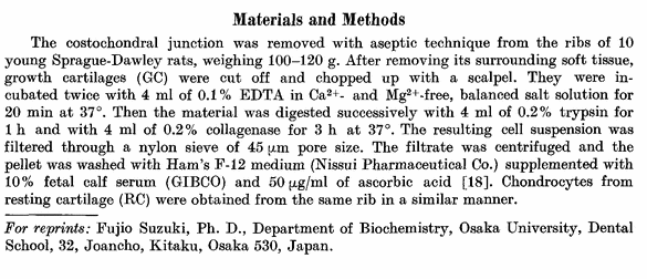
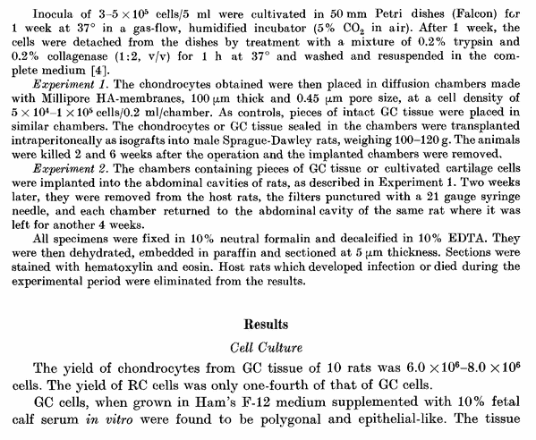
Studying Osteogenesis By Chondrocytes From Growth Cartilage

Update 2/12/2013: It would seem that I no longer can access through the link below to the full text/article without paying for it. This means that my ability to do research and learn more has been severely limited. Only a quick study from the abstract and introduction seems to be possible at this point.

From a paper/article of a study from SpringerLink “Osteogenesis by chondrocytes from growth cartilage of rat rib“.For the Full Article click HERE

Y. Shimomura, T. Yoneda,  F. Suzuki Ph.D. – Calcified Tissue Research – 1975/76, Volume 19, Issue 1, pp 179-187
Abstract

Chondrocytes were isolated from growth cartilage and resting cartilage of rat rib and cultivated in vitro. The cultivated chondrocytes were placed in Millipore diffusion chambers, which were then implanted into the abdominal cavities of rats for several weeks and prepared for histological analysis. The results indicate that growth cartilage cells have a remarkable osteogenic potential, even after cultivation in vitro, whereas resting cartilage cells show no osteogenic activity. However, growth cartilage cells alone do not form new bone but require the participation of certain host cells to initiate osteogenic differentiation.

Introduction

The process of replacing cartilage by bone is known as endochondral ossification. During the first stages of conversion into bone, epiphyseal cartilage cells
increase their rate of proliferation, enlarge, and become hypertrophic. It has been assumed in the past that the hypertrophic cells undergo degenerative changes and death [1, 2, 3, 8, 17, 22]. However, Holtrop, on the basis of experimental transplantation of growth cartilage labeled with 3H-thymidine [10, 11, 12], suggested that some of the cells survive and redifferentiate in the metaphysis to contribute to ossification. Shimomura and Ray have confirmed her findings that hypertrophic chondrocytes can survive and transform into bone-forming cells [19 ]. Recent investigations using electron microscopy have led to opposing interpre-
tations [3, 13, 14, 20], and controversy persists concerning the mechanism of endochondral ossification.

In the present study, hondrocytes isolated from growth cartilage of rat rib were cultivated in vitro and transplanted to investigate the mechanism of endo-
chondral ossification.

Screen Shot 2013-02-12 at 2.49.26 AM

Screen Shot 2013-02-12 at 2.49.40 AMAnalysis & Interpretation:

The reason why this article/study/paper is important is to show why it is more important to focus on the cartilage of the plate, then the bone area. We have sen in the past ideas like using Low Intensity Pulses Ultrasound (LIPUS) to possibly increase height, but from my own limited research so far it seems that any method or technique to increase the rate of bone growth or bone density will not do anything for our intention to grow taller ie. increase the length of the long bone in our body. Remember that commercial pills and supplements that are purported to be able to increase bone growth are true, but they don’t increase bone in the way we want. The bone growth happens inside the bone, often making the medullary cavity thinner and smaller, making the cortical bone layer thicker. Bone growth pills are sold as a way to treat or prevent bone degeneration in older people who might get osteoporosis.

LIPUS has been shown through maybe 1-2 PubMed studies to increase the rate of bone healing in distraction osteogenesis surgeries, but without the initial distraction, bone growth will not lengthen bone, at least within physically mature humans with no epiphyseal cartilage to widen and lengthen.

From the abstract, we learn that overall, the epiphyseal cartilage derived chondrocytes are special, diffierent from what we would find in other cartilage explants, like from the resting cartilage. When grown in the lab (in vitro) we find that the cartilage cells themselves already have bone creating properties.

It seems epiphyseal cartilage will always ultimately turn into bone in some way, whether through calcification, vascularization, etc. This suggest that when we are looking for a method to legnthen bone, we don’t really have to worry about the process of osteogenesis, but only have to worry about chondrogenesis. If we get the chondrocyte and cartilage formation correct from using growth plate derived cells, the osteogenesis and bone formation seems to automatically occur given time, and the right nutrients getting in, when the cultured cartilage or chondrocytes are finally implanted back into the subject.

Another thing that might be important to take away from the introduction is that maybe not all the hypertrophic chondrocytes in the hypertrophic zone of the growth plate completely die out through degeneration. Some researchers have suggested that the hypertrophic chondorcyte can actually differentiate even further going down deep into the calcification/ossification layer and turn into osteocytes/osteoblasts which start forming bone cells and bone elements.

So the big take-way from this article is to focus less on bone creation, osteogenesis, but focus more attention on cartilage formation, chondrogenesis.


Studying Changes In The Growth Plate By Restricting The Blood Vessel Supply, Ischaemia

Ever since I came across the study “SURGICAL STIMULATION OF BONE GROWTH BY A NEW PROCEDURE – PRELIMINARY REPORT” by ALBERT B. FERGUSON, M.D where it showed that it might be possible to induce increased longitudinal growth by severing the blood supply to the metaphysis, epiphysis, or the growth plate in growing children I have been curious over what other experiments have been done with a similar procedure and what were the results and effects seen in the growth plate. This study does that.

However I would guess that extensive study and reading of the study will only help contribute in adding to my personal understanding on how the blood vessels contribute to the growth plate in general. I have always believed that the more I know about the overall system, no matter how obscure or unrelated, it will only help contribute to the overall understanding of the system we are studying.

Note: The Actual PDF had many pictures which I have removed. For a more comprehensive view of the study, click the title link below.

Analysis & Interpretation:

The first thing to notice about this study is that unlike the other study being cited, the blood vessel being actually disrupted is going to the growth plate, not to the metaphysis. The old idea proposed fy Furgeson was that you can possibly stimulate higher than average levels of longitudinal growth by disrupting the inter-medullary blood vessels that contribute nutrients to the metaphysis of the tubular/long bone. This time the experimenters are disrupting the very blood vessels to the epiphysis, which would indicated that only decreased/stunted longitudinal growth is the likely outcome.

For the actual experimental part, the researchers took rabbits and drilled one small hole in the right medial side of the tibial epiphysis, then put a small spatula inside. The spatula is spinned around to cut & disrupt the blood vessles inside the epiphysis. The hole is drilled deep into the middle of the epiphysis. Then the hole is filled with some type of plastic to prevent re-vascularization. The lateral side of the right tibia was left as a control. The left tibia was treated similarly except for putting something on the other side for support.

Here is what the discussion has said about the blood vessels from the side, or the epiphyseal vessels. They are damn important. The proliferative chondrocytes need them to undergo further division. If any disruption of the vessels are done, lesions are developed which can cause breaks in the growth plate causing blood vessels to breakthrough causing vascularization, resulting in bone bridges, which would effectively stop all further longitudinal growth.

It would seem that there is at least two group sof blood vessels carrying nutrients to the growth plates, one coming from the side or epiphysis and the other coming from the middle, from the metaphysis. As for the source coming from the metaphysis the main roles and functions of it include carrying Vitamin D and Calcium as well as phosphates using the corpuscles which would cause calcification of the matrix, the removal of the cells that have disintegrated, and the use of laying down layers of bone material on the walls of the empty cavities in the matrix. The blood vessels from the metaphysis seem to hold no nutritional value to the hypertrophic chondrocytes but is what actually causes them to die. The level of alkaline phosphatase was also decreased in the experimental part where the blood vessels from the metaphysis was selectively removed. It would then suggest that only when you notice the hypertrophic chondrocytes releasing the compound alkaline phosphatase would the chondrocytes actually start to go through the process of dying through disintegration.

The researchers concluded that what will actually keep the hypertrophic chondrocyte alive and prevent them from dying is to prevent the process of calcification, not the presence of metaphyseal vascularization. When we compare the calcification rate of the matrix after vascularization in the normal way compared to say Vitamin D or A defficiency leading to Rickets, we can see that the whole process is very fast. The way that we should be remembering the growth plate ossification process is…

Calcification starts everything. The calcification leads to the blood vessels breaking in and getting the The hypertrophic chondrocytes starts releasing aklaline phosphatase signaling that they ready to die. They disintegrate, leaving the empty cavities to get coated with a layer of lamellae which will get filled then by osteoblasts which convert the pockets into real bone material.

The researchers end the study with this easy to take away lesson for the readers who wish to just take the basic idea away….

“The nutritional dependence of the proliferative cells on the epiphysial vessels has been established whereas the metaphysial vessels were seen to take part in calcification and ossification at the metaphysis.”

Interpretation & Implications:

This post has only helped reaffirm the idea that it may be possible to increase the lengthening of the bone in people with open growth plates through the disruption of the blood vessels that are reaching the growth plate from the metaphysis. Also, we definitely don’t want o damage the blood vessels that are supplying the blood and nutrients needed by the cells at the top of the growth plate process, around the reserve zone area.


From paper study “THE VASCULAR CONTRIBUTION TO OSTEOGENESIS, Changes in the Growth Cartilage Caused by Experimentally Induced Ischaemia

J. TRUETA, OXFORD, ENGLAND, and V. P. AMATO, SLIEMA, MALTA

From the Nuffield Orthopaedic Centre, Oxford.

In the two preceding papers of this series the vascular pattern close to the growth cartilage, and the relationship between vessels, cartilage cells and calcification, were studied under normal, or experimentally unaltered, conditions (Trueta and Morgan I 960, Trueta and Little 1960) and the findings were used as a control for the experimental work to be described in this and succeeding papers.

This study reports the changes in the epiphysial cartilage caused by the suppression of its blood supply. The changes caused by the interruption of the epiphysial blood flow will be described first and a description of the changes produced by interrupting the metaphysial circulation will follow.

METHOD

Forty six-weeks-old chinchilla rabbits, averaging 1,100 grammes in weight, were used. Operations were done on both tibiae under general anaesthesia (Nembutal and ether). In the right tibia a small hole was drilled on the medial side of the tibial epiphysis, avoiding so far as possible any direct damage to the growth plate. A small flat spatula was introduced through the drill hole until it reached approximately to the centre of the epiphysis when it was moved in a circular sweep in a plane parallel to the epiphysial cartilage. The cavity thus produced was packed with a strip of polythene film to prevent revascularisation. The lateral side of the epiphysis was left undisturbed to serve as a control. In the left tibia a similar technique was used but the block was placed on the metaphysial side of the growth cartilage and at a sufficient distance to prevent direct damage to it. All the forty rabbits were subjected to this procedure and out of the eighty tibiae operated upon seventy-eight were available for analysis : one tibia became infected and another fractured. The investigation proceeded in the following way: Group A-Twelve rabbits were killed at intervals of 1 , 2, 3, 4, 5, 6, 9, 12, 1 5, 18, 2 1 and 24 days after operation in order to study the changes in the epiphysial cartilage caused by ischaemia. Group B-A second operation was performed on all the remaining rabbits to remove the polythene sheath at intervals of 2, 4 and 8 days after the first operation. The rabbits were killed and injected at intervals of 2, 4, 6, 12, 16, 18, 21 and 24 days after the second operation in order to study the revascularisation that occurred.

RESULTS

INTERRUPTION OF THE EPIPHYSIAL BLOOD SUPPLY

The first changes occurred in the epiphysis itself. As early as the second day after operation many trabeculae were seen to be dead, with empty lacunae. By superimposing the stained histological preparations on the unstained sections it was seen that the dead trabeculae corresponded exactly to areas deprived of vascularity. These two changes-that is, the signs of bone death and the presence or absence of blood vessels-when seen in the terminal bone plate covering the growth cartilage, proved that the blood supply of the epiphysial plate had been cut off. When revascularisation was allowed to occur the evidence was still available, for the dead trabeculae could be seen enveloped by new bone (Fig. 1). The thick sections showed that the new blood supply in most instances was entering the epiphysis from the site of the operation. The newly established circulation was seen to correspond exactly with the areas where new bone was being laid down on the scaffolding of dead trabeculae in the terminal bone plate.

When the operation scar was placed far from the plate and the penetration was not deep enough into the central portion of the epiphysis there were only moderate changes attributable to interference with the circulation. The terminal bone plate appeared normal and all the vessels filled with dye. A transient widening of the growth plate was seen sometimes when it was compared with the undisturbed side of the plate. This widening was due both to a moderate increase in the number of cells of the hypertrophic layer and also to the increase in the size of the individual cells; apparently the proliferative layer remained unaltered although it is possible that accelerated division had taken place but, because of the great rapidity of this division, could not be detected, but could have produced the moderate increase in the number of hypertrophic cells. The widening of the growth cartilage was accompanied by a denser network of blood vessels in the region of the terminal bone plate and it was always more noticeable towards the periphery. The intercellular matrix took on a darker stain.

These effects were transient and could not be seen after a few days. Occasionally patchy damage was detectable in the plate. The thick sections showed this to correspond exactly to the area of ischaemia. The upper cells of the growth plate, referred to in a preceding paper of this series (Trueta and Little 1960) as the germinal cells, showed metachromasia; also a varying number of cells appeared dead, leaving empty V-shaped spaces consisting of intercellular matrix or matrix interspersed with dead or dying cells (Fig. 2). This minor degree of damage appeared capable of repair by a bulging inwards of the surrounding columns, so that the gap became closed off. The columns in such plates appeared fewer and farther apart, and the cytoplasm of many cells was granular (Fig. 3). The matrix was less well stained, appearing pale in colour.

In areas where the damage involved a larger number of columns the gap was too wide to become sealed off. Growth continued normally in the surrounding columns and resulted in a pulling out of the infarcted portion of the plate (Fig. 4). Long, empty, or partly empty tubes of cartilaginous matrix stretched from the epiphysis to the metaphysis, and as no further calcification took place this area was left behind in the metaphysis. If this stretching out process went too far, eventually a break occurred, usually at the metaphysial end, and blood vessels invaded the area (Fig. 5). New bone was laid down along these capillaries (Figs. 6 and 7) so that the end-result was usually a bone bridge between metaphysis and epiphysis. The shortest interval for the whole sequence of events to develop after this type of injury was found to be eight days from the operation. In some of the experiments in which the blood flow was allowed to return, the invasion of the blood vessels took place from the epiphysial to the metaphysial side, but the ultimate result-a bone bridge between the epiphysis and metaphysis-was the same.

In massive lesions, where the main source of supply to the whole epiphysis was successfully cut off, rapid death of the whole of the central part of the plate occurred. The cells became metachromatic, losing their polarisation and their morphological characteristics, and the matrix became very pale throughout (Figs. 8 and 9). The area that suffered least was a limited rim of the peripheral part of the cartilage which receives its blood supply from vascular anastomosis around the plate. The capillaries from the metaphysial side invaded the devitalised area on a wide front and new bone was very soon laid down in their wake. Epiphysiodesis occurred within a short while, usually ten days from the vascular interruption, and growth ceased altogether.

Direct injury to the terminal bone plate-In certain instances the epiphysial bone plate and the upper part of the growth plate, which is so intimately connected to it, were injured at the time of operation. Irreparable damage always resulted and an invasion of the blood vessels took place early, followed by partial epiphysiodesis (Figs. 10 and 1 1). As the medial side of the tibia was always used and as this type of injury was most likely to affect the peripheral end of the plate, a varus deformity developed.

Lesions of this area were often complicated by damage to the perichondral ring as will be mentioned later.

INTERRUPTION OF THE METAPHYSIAL BLOOD FLOW

After division of the main vessels at the metaphysial side of the growth plate this increases appreciably in width within twenty-four hours, entirely as a result of the accumulation of the cells of the hypertrophic zone. As stated by Trueta and Morgan (1960) the number of cells in the columns of the upper tibial epiphysis of the rabbit at six weeks varies from thirty-five to fifty, according to the mitotic activity of the proliferative part of the columns at the time the count is made. An average increase of approximately ten to sixteen cells occurs every twenty-four hours in the area of total suppression of the metaphysial blood flow. After six days the average width of the growth cartilage is increased by sixty to ninety hypertrophic cells (Figs. 12 and 13) and after eight days it may be as much as 100 cells or more above the normal average. After this extraordinary increase in width of the growth plate, the rate of expansion becomes limited and the metaphysial end of the columns of cartilage cells, now placed deep into the metaphysis, press against each other often at a depth of six or eight cells before the end. This permanency of the hypertrophic cells is accompanied by a total lack of calcification of the intercolumnar matrix. A close investigation of the morphology of these hypertrophic cells shows that along the whole length of the columns they do not exhibit any appreciable modification, so that individually the hypertrophic cells at the top of this section resemble those placed at its end, often fifty or more cells apart (Fig. 14). No degenerating cells are seen under these experimental conditions at the ends of the columns. The significance of this finding will be shown later. It is of particular interest to make a comparative study of the part of the growth cartilage placed over a metaphysial area of ischaemia and that which corresponds to a metaphysial area of normal blood flow. It is as ifall the activities so energetically engaged in the normal had been suspended, including calcification, preliminary ossification, bone reabsorption and final bone formation. Large zones of calcified cartilage and preliminary bone (Fig. 15) remain at a level in the metaphysis where, normally, remodelling is taking place (Fig. 16).

A somewhat surprising feature is the apparent survival of osteocytes in the metaphysial zone over the area of vascular suppression, suggesting that either the osteocytes, if they were dead. had no time to disintegrate or even change their stalnlng characterlstlcs or else that sufficlent blood flow still remained in the neighbourhood to provide the required transudates for their survival. Of these possibilities the latter seems the most likely, because in the injected specimens some of the per-fusing mass was scattered along vessels in the supposedly avascular area. The reason for this .l was the integrity of a number of perforating meta-physial vessels which, as explained elsewhere (Trueta and Morgan 1960) provide some of the blood, while the main blood flow from the nutrient artery is interrupted by the polythene film. In general, osteoclasts were absent and were seen only in reduced numbers in the proximity of the new, or the few spared, blood vessels. Another constant finding was the lack of alkaline phosFIG. 14 phatase concentration, characteristic of the

Detail of a “ tongue “ of epiphysial cartilage hypertrophic cartilage cells near the zone of showing the healthy hypertrophic cells with no empty columns at their sides, and the degenerating caIcli Icatlon. cells at the distal end of the columns. ( x 45.) Changes of the growth cartilage accompanying metaphysial vascular regeneration-Even without removal of the polythene film vascular proliferation from the perforating vessels invaded the ischaemic metaphysial area in less than twenty-four hours after the main blood flow had been blocked. Soon calcification and ossification proceeded along the now very elongated tubes surrounding the hypertrophic cells up to the normal level ofcalcification, with the usual eight or twelve hypertrophic cells left untouched, except that the last two or three were surrounded by calcified matrix, as in the normal (Fig. 17).

When the circulation was allowed to return freely by removal of the vascular block. the expanded growth plate calcified at a strikingly rapid rate from the periphery to the centre, each column starting at the farthest end in the metaphysis. The capillary loops entered at the bottom of the columns and in a few days travelled up-one loop to each column-to the level they would normally have reached had the vessels not been interrupted. New bone was laid down by the osteoblasts, appearing as a lining inside each tube. The trabeculae thus formed were thinner and more uniform than those of the control side and remodelling lagged behind. Alkaline phosphatase activity was increased and appeared to be related to the vascular progression. The last part of the growth cartilage to be calcified and ossified was always that in the centre, being the last to be reached by the incoming vessels. It often persisted as a long finger-like process of varying thickness and consisted of orderly columns of hypertrophied cells which on occasion numbered as many as 100 or more (Fig. 14). In thick sections a clear division between the old and the new blood vessels was seen, a division which exactly corresponded to the width of the finger-like process (Figs. 18 and 19). The vascular loops were always orientated in the direction of the columns, for, as has been stressed above, they only gained entrance to each column from its distal end. The widened growth cartilage did not tend, as a rule, to be broken up into islands of columns and cells but. on the contrary, calcification and ossification always proceeded very regularly. Occasionally. however, in the animals with the main metaphysial vessels obstructed for eight days or more an impenetrable barrier became established round the columns and their distal ends remained firmly closed. The vascular loops in these cases short-circuited the barrier by getting in between the long but normal-looking columns and those which appeared disorganised and unhealthy, and proceeded to lay down bone by the normal mechanism of vascular invasion of the hypertrophic cartilage.

In some instances if the central part of the epiphysial cartilage remained unossified for too long, permanent damage was caused by its tendency to get peeled off from the terminal bone plate, covering it at its epiphysial side. Thus a severe lesion of the germinal and proliferative cells was caused in breaking these away from their natural surroundings, particularly the epiphysial vessels ; they degenerated and soon were invaded by new epiphysial vessels which, by progressing towards the metaphysis, ended by establishing connections with the metaphysial vessels (Figs. 20 and 21). New bone was laid down and soon a central epiphysiodesis was established.

In the diaphysis the site of operation was always visible by its increased bone density; and its progress towards the shaft depended on whether or not a bony bridge had been established across the plate. Damage to the perichondral ring-The operation occasionally damaged the perichondral ring of the ossification groove (Lacroix 1951) and a large chondroma or osteochondroma resulted. It often spread down the metaphysis, and vessels from this region grew into it (Figs. 22 and 23). Normal moulding with organised growth did not take place and osteoclasts were absent from this region. There was a disorderly vascular invasion of the mass of cartilage cells, and bone was laid down as in the cartilage anlage before the growth cartilage is arranged in parallel columns ofcells (Fig. 24). The bone trabeculae were therefore irregular and the orderly laying down of bone characteristic of metaphysial bone formation was totally absent.

Bone bridging and partial epiphysiodesis-When a bone bridge became established between epiphysis and metaphysis it resulted in growth disturbances which followed a regular pattern according to the size and location of the bone bridge. Superimposition of thin and thick sections showed that a bone bridge was always immediately preceded by a vascular invasion across the area of damaged cartilage. If the two circulations could be kept apart no bridging occurred.

The effect upon growth after the establishment of a bone bridge was of interest and will be analysed elsewhere. It will suffice to mention that bone bridges of equivalent sizes caused greater deformities if placed at the periphery than at the centre of the growth plate, particularly if the perichondral ring was seriously affected, as in the experiments reported by Trueta and TrIas (1957).

DISCUSSION

The results of this investigation throw light on the respective role of each of the two systems of vessels (epiphysial and metaphysial) carrying the blood to the epiphysial cartilage. The technique used allowed the production of isolated disturbance to either of the two systems and offered the opportunity of studying the part each plays in the process of growth. It was established in a preceding work (Trueta and Morgan 1960) that the metabolic requirements of the reproductive cells of the epiphysial cartilage, and perhaps of all the cells in the columns, are supplied through the epiphysial vessels described as forming a ceiling under the roofofthe bone plate. There is very little doubt that these vessels are responsible for carrying the blood to the cells actively engaged in reproduction. The fact that any extensive damage to the epiphysial vessels causes irreparable lesions to the columns ofcartilage cells does not exclude the possibility that temporary ischaemia may be followed by an augmented vascularity responsible for causing an increase in the width of the growth cartilage. It is interesting that this is due to an increase in size of the hypertrophic cells and in their number, which in the absence of vascular impairment at the metaphysial side of the plate suggests an accelerated cell division at the proliferative segment of the column. The existence of the tubes empty of proliferative and hypertrophic cells in the middle of normal columns suggests that death of individual columns may be compatible with normal or nearly normal growth.

Once the responsibility of the epiphysial vessels for nourishing the epiphysial cartilage is established, it is interesting to consider the depth to which the transudates from these vessels may reach. In a normal epiphysial growth cartilage the proliferative cells begin to enlarge. and to become hypertrophic by the middle of its width. These new cells acquire the characteristics of the giant cells with vacuoles, oedema, etc. But no further changes occur to them as they become progressively separated from the epiphysial source of nutrition until they reach the area of matrix calcification. In the normal average cartilage this occurs from eight to twelve cells from the first showing hypertrophy. Only two or three cells farther down, degeneration sets in and, with the proximity of the metaphysial vascular loops, the final removal of the remains ofthe cartilage cells takes place. The nutritional dependence ofall the cells in the tubes on the epiphysial vessels is further shown by the disorganisation and death which occurs when the tops ofthe columns are pushed away from their normal position after the suppression of the main metaphysial blood flow, which is responsible for the growth cartilage enlargement.

The main role of the metaphysial vessels-carrying calcium and vitamin D in the serum and phosphates in the red corpuscles-was seen to be the calcification of the matrix, the removal of the degenerate cells and the laying down of lamellar bone along the inner side of the empty tubes. That the blood carried by these vessels is of no nutritional importance to the hypertrophic cells was shown by their healthy appearance once the majority of the metaphysial vessels were divided; with this no further calcification of the matrix occurred and the hypertrophic cells took a new lease of life, which suggests that the primary cause of their death is the presence of the metaphysial vessels-with normal blood-in their neighbourhood.

Alkaline phosphatase activity was also suspended by the arrest of the metaphysial blood flow, suggesting that with its production the hypertrophic cartilage cells must be on their way towards ‘ ‘ degeneration.” The long columns of hypertrophic cells resembled those which are characteristic of rickets (Fig. 25), thus pointing out that that which keeps the hypertrophic cells alive is not the absence of the metaphysial vessels but the absence of calcification. The vascular pattern in rickets will be studied elsewhere. Provided that-as in these experiments-there was no deprivation of vitamins A and D, calcification to the normal level occurred with extraordinary rapidity shortly after revascularisation was allowed. The columns of hypertrophic cells disintegrate and the tubes round them calcify up to the normal level, which suggests that vascular invasion depends on calcification-which will weaken the cell first-and that calcification can only occur close to a vessel which carries the appropriate blood, and when the matrix is ready to calcify. The method of metaphysial vascular suppression has allowed the study of the rate of new cellular additions in the growth cartilage in twenty-four hours. The evidence suggests that, in the rabbit’s tibia, from ten to sixteen new cells are added to each column in a day. That would mean about one-third to half of a millimetre in growth in length in twenty-four hours as an average, which is near the findings obtained in this centre by the measurement of growth on fine grain radiographs.

CONCLUSIONS

In this work the role of the blood vessels surrounding the epiphysial growth plate has been studied. The nutritional dependence of the proliferative cells on the epiphysial vessels has been established whereas the metaphysial vessels were seen to take part in calcification and ossification at the metaphysis. As it does not seem likely that the blood circulating in the two systems of vessels had a different constitution, particularly in hormones and vitamins, it seems permissible to assume that it is the characteristics, particularly in shape and number, of such vessels that make growth the orderly process it is, with the repeated birth of a cell at the top of a column and burial at the bottom end. But, despite this undeniable role of the vessels, growth depends on the ability of the cartilage cell to form a matrix which, in due course, will be avid for apatite crystals.