
Something that I finally have found evidence for and many height increase researchers have suspected have turned out to be true from recent research.
A finding in a Pubmed article “On vertebral body growth.” reveals a piece of information that most professional physicians would know but most amateur height increase researchers might not know is…
“…Unlike other long bones of the skeleton, vertebral body epiphyses never ossify, and after the end of the growth period of life they are reduced into thin plates of hyaline cartilage which are situated between vertebral body and intervertebral disc….”
I always thought that there was some layer of cartilage left from the ossification process of the normal bone development process and it seems that we might all be right about it. After typing the phrase “Intervertebral Disk Cartilage” into google, I found even more evident that there are cartilage in adult human vertebrate bones
From a webpage of a sort of advanced Biology or Anatomy Medical School course entitled “ANAT D502 – Basic Histology – Cartilage, Bone & Joints, Bone Formation Pre-Lab – revised 9.23.12″ which is from Indiana University – Purdue University Indianapolis.
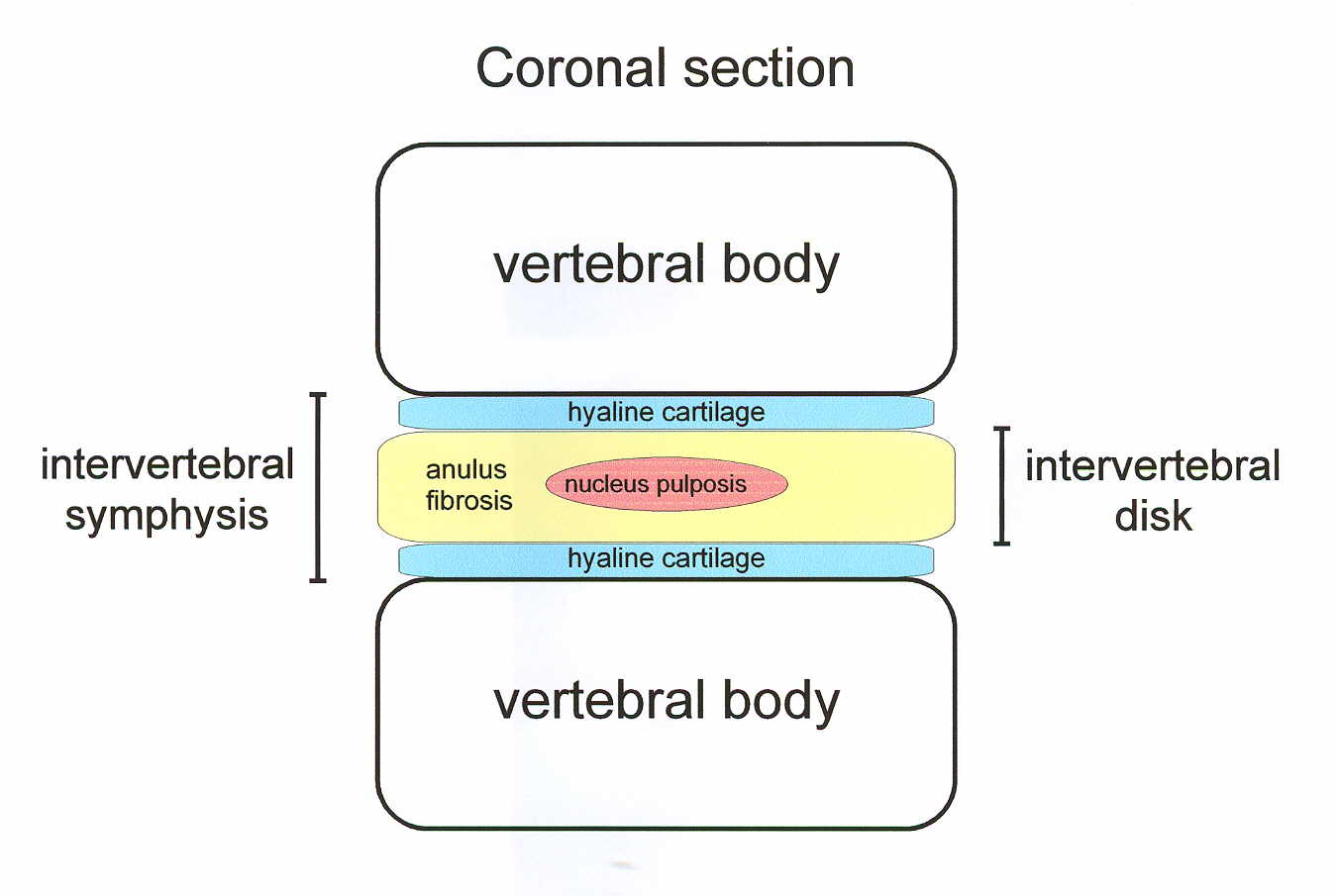
Something to notice is that there is supposed to be layers of hyaline cartilage surrounding the nucleus pulposis and anulus fibrosis.
While the intervertebral disk consists of only the…
- Anulus Fibrosis
- Nucleus pulposis
The two layers surrounding the intervertebral disks and are in the juntion between the disks and the vertebral bone are where the hyaline cartilage are supposed to be. The entire thing is called intervertebral symphysis. I would guess that the layer of hyaline cartilage is made much bigger to be used in the diagram but it should be there in adult humans.
Another PubMed article “Morphology of the cartilaginous endplates in human intervertebral disks with ultrashort echo time MR imaging.”
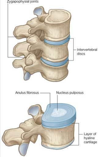
It definitely suggest that at the ends of the disks are these surfaces of the bone which are covered in cartilage, just like how the ends of a long bone, (ie femur) have a layer of articular covering them. More images show the same, thing although it is never explicitly stated that the diagrams are for adults. we know that in children who are still growing, there has to be some sort of endplate which acts like a growth plate. I am suggesting that the diagrams are for adults, which would give us hope.
 While this is sort of exciting, potentially good news, it is important to always be cautious in being too over-optimistic. I would refer to the study “Articular cartilage and intervertebral disc proteoglycans differ in structure: An electron microscopic study”
Abstract
Articular cartilage and the intervertebral disc tissues have different material and biological properties and different patterns of aging and degeneration. To determine if the proteoglycans of these tissues differ in structure, we used the electron microscopic monolayer technique to compare baboon articular cartilage proteoglycans with baboon annulus fibrosus, transition zone, and nucleus pulposus proteoglycans. Intervertebral disc and articular cartilage porteoglycans differed signficantly. Articular cartilage contained large proteoglycan aggregates formed from hyaluronic acid central filaments, multiple monomers, and large nonaggregated monomers. These molecules were identical to those of nasal cartilage, growth plate cartilage, chondrosarcomas, or menisci. In contrast, the intervertebral disc tissues contained only nonaggregated proteoglycan monomers and clusters of monomers without apparent central filaments. Intervertebral disc nonaggregated monomers were shorter and more variable in length than those from articular cartilage, and nucleus pulposus nonaggregated monomers were even shorter and more variable in length than transition zone and annulus fibrosus monomers. These observations suggest that significant differences in proteoglycan metabolism exist between articular cartilage and intervertebral disc.
Conclusion:
There is studies and anatomical diagrams showing evidence that there is a layer of very thin hyaline cartilage that is still there even in adult humans. If that is the case, we might have another source of mesenchyme we can try to work with to stimulate height increase. This new development will allow height increase researchers to push in an entirely new direction in research, through possible stimulation of the torso.
Cool info, this sounds promising.
And even if the hyaline cartilage is very thin, its still very useful, since we have so many movable vertabrae and spinal discs we conly need a small ammount of cartilage for every part, if each of those parts increase slightly in height then the overall height gain will still be very significant.
hopefully they will find a way to use this info for height gain as soon as possible
look this michael.. is about mgp – matrix gla protein (produced by vitamin k2)
and is the major inhibitor of human body tissue calcification
and seems to have much influence on chondrocytes:
http://jcb.rupress.org/content/154/3/659.full
bonus: Increases Testosterone:
http://www.ergo-log.com/vitk2testosterone.html
more about nitric oxide and chondrocytes apoptosis:
http://www.ncbi.nlm.nih.gov/pubmed/7856740
http://www.ncbi.nlm.nih.gov/pubmed/11840442
glutathione and NAC seem to help:
http://www.ncbi.nlm.nih.gov/pubmed/14673993
http://www.ncbi.nlm.nih.gov/pubmed/19725096
http://www.ncbi.nlm.nih.gov/pubmed/15501401
http://www.ncbi.nlm.nih.gov/pubmed/18311796
and the problem of NAC in relation to NADPH can be easily resolved with niacin, niacin is the precursor for NADPH