
Category Archives: Uncategorized

Update On Grow Taller Progress – April 1st, 2013

Note: I am not a big joke or prank guy so I don’t plan to ever joke or prank you guys with a silly April Fools joke like claiming that I grew 1-2 inches in the first month. That just doesn’t happen!

I had said in a post around the beginning of the last month in March entitled “It Is Official, I’m Going To Try To Grow Taller Myself, March 3rd” that I would try to take up exercising again to both try to gain any “hidden height” back which I might have lost in living such a sedentary life since I have been out of the country for the last 9 months.

I had made a vow with my gf that we would try to put ourselves through some type of exercise program to lose weight. We both noticed that we have been putting on the pounds for the last few months with our busy lives and hectic schedules. Or it could be that we are just too lazy to get fit.

So she is now walking much more to work these days (she takes the bus) and I have decided to try  getting back to swimming and stretching like I did years ago when I was trying to gain height. I guess I plan to try to loss weight, get healthy, and possibly also grow taller from my constant exercising and stretching.

She doesn’t realize that In addition to trying to lose weight and get fit, I am also slightly altering my exercise routine to focus more on stretching and decompressing my vertebrate for height increase reasons. She is around 5′ 2″ and has also expressed that she would love to be taller. At this point I haven’t told her that I write or operate this website yet. I am not sure how she would respond when she realizes that I run one of the largest websites on the internet which focus on this subject. Maybe she’ll think I am crazy.
tape-measureSo what is the update after almost 1 month?

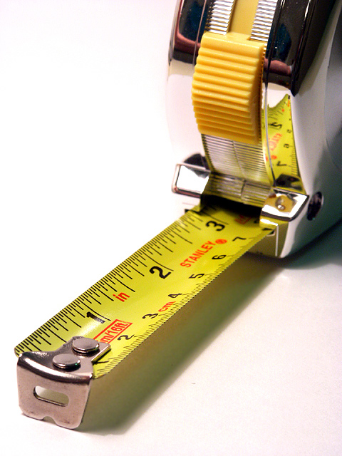
Absolutely very little. I have been reducing my food intake, eating one or two small portions of food each day and have been going swimming every other day. I found my old tape measure and looked at whether I had shrunk or grown. It seems that at last measurement my height is about the same as the last time I intentionally really made an effort in measuring myself, which was over 2 years ago. So at least I haven’t shrunk, which is something I do worry about. Whew!

My schedule in flying so much this recent month and running errands and driving to so many places forced me to not have enough time to really do the exercises I was hoping for. By the end of next month however I should have a lot more time and consistency in my exercises and stretching though.

Of course these days I have hair now and I would prefer to measure with my hair on instead of being shaved like before. The hair thing will lead to quite a bit of measurement error. I would like to keep my hair but still get a very accurate reading. Do you guys think I should get a stadiometer for my room?
Research At The University Of Washington Health Science Library

So I posted a video up of me talking at the Health Sciences Library at the University of Washington, which is the local university library since I am based from the beautiful city of Seattle.

Like I said before, I am going to take the research very seriously now and the only real way that I as a single researcher can make this research more legitimate is get off the scam and internet marketing websites from the internet and sit my ass down in a library and go do research the way my dad did it, by reading old stuffy medical journals and books.

As for my location, the only real good resource I can think of besides talking to a orthopaedic surgeon or endocrinologist is to go to a University with a Medical School which would also have a Medical School Library or Health & Medicine & Biology Library. Luckily, based in Seattle is the University of Washington and the University of Washington Medical School which has been ranked among the best National Hospitals and Medical Schools by US. Newsweek for at least the last few years. It may not be a John’s Hopkins or a Harvard or a Stanford in terms of prestige or name brand  but I believe it is still one of the best Medical Schools in the country

I am just showing you guys the types of books I am looking over these days. The thing I am looking for at this stage is to find any old, previously unknown articles which gives me more knowledge on how the growth plate in the postnatal human behaves.

At this point, I am doing more skimming at the sections and paragraphs because there is just so many books at the library I can go over. It will take me at least a couple of weeks to narrow down the types of subjects and books I want to really go deep on the details on.

The video is short and there is not a lot of content but you do see that I am doing some real research on the stuff and looking at the most relevant subjects.

NOTE: Turn up your volume to hear my voice! I am in a library so I had to lower my voice.

As always, you can check out the Natural Height Growth Youtube Channel or Subscribe by clicking HERE!


Estrogen’s effect on height may be mediated by chondrogenic effects

The effect of estrogen on the expression of cartilage-specific genes in the chondrogenesis process of adipose-derived stem cells.

“During adolescence, sex hormones play an important role in regulating proliferation, differentiation, maturation, and the scheduled death of chondrocytes.
In the present study, we used adipose-derived stem cells (ADSCs) to differentiate into cartilage. Differentiated cartilage cells were used in the control (without estrogen E2 in the culture medium) and experimental (with estrogen in the culture medium) groups to evaluate the expression of type II collagen and aggrecan as chondrogenic genes markers.
Estrogen leads to inhibition of type II collagen gene expression and reduction of aggrecan gene expression.
Estrogen probably has negative effects on chondrogenesis process of ADSCs”

“17-β estradiol (E2) increased the type X collagen, inhibited the type II collagen, and reduced the proteoglycan content of the cartilage”

Loading effects on bones and possible transdifferentiatiion

Since chondrocytes are what make up growth plates and growth plates are what make you taller.  This is huge.

Molecular events caused by mechanical stress in bone.

“The shape of bone changes as a result of bone remodeling corresponding to physical circumstances such as mechanical stress. The tissue which receives the loaded mechanical stress most efficiently is bone matrix. Recent studies revealed the function of osteocytes as mechanosensors in the early stage of bone remodeling. Loaded mechanical stress is converted to a series of biochemical reactions, and finally activates osteoclasts and osteoblasts to cause bone resorption and formation. Biochemical and molecular biological studies have recently resulted in the identification of the gene of which expression level is changed by mechanical stress. Nitric oxide (NO) and cAMP is secreted in response to mechanical stress in the immediate early stage. Genes encoding enzymes such as glutamate/aspartate transporter (GLAST), nitric oxide synthetase (NOS) and prostaglandin G/H synthetase (PGHS-2) are identified as mechanical stress-responsive. The expression level of IGF-I is enhanced under the control of PTH/PTHrP. The expression of c-fos is increased by loading of mechanical stress[LSJL upregulates C-Fos]. AP1, a heterodimer of c-FOS/c-JUN, functions as a transcription factor of downstream gene(s). Elements including AP1 sites, cyclic AMP response elements (CRE) and shear stress response elements (SSRE) are found in the promoter region of mechanical stress-response genes. The enhanced expression of osteopontin (OPN) in the osteocytes of bone resorption sites was demonstrated by in situ hybridization and immunohistochemistry and transdifferentiation of chondrocytes with the abundant expression of BMP-2 and -4 in the process of distraction osteogenesis was observed.”

“Mechanical stress loaded to bone causes the deformation of bone matrix and generates strain force, which is thought to initiate the mechanotransduction pathway. Since the bone contains a water solution, the strain causes fluid flow in the bone matrix. These represent secondary changes known as streaming potential and shear stress. The activities of ion channels, focal adhesion kinases, as well as the structure of the cytoskeleton of the bone cells, are also modulated by mechanical stress.”<-Wish LSJL we hope that this fluid flow induces differentiation of stem cells into chondrocytes.

“GLAST mRNA expression level in the osteocytes decreased in the loaded ulnae after 6 h of load on two occasions 24 h apart.”

“OPN can inhibit the formation of hydroxyapatite. Immunohistochemical studies demonstrated that OPN was localized only on the surface of matured bone. In contrast, much abundant and broad distribution of OPN was observed in embryonic bone, osteoid and fractured callus”<-OPN is upregulated in LSJL.

“OPN can promote the attachment of osteoclasts possibly via interaction of GRGDS amino acid sequence of OPN and the αvβ3 integrin of the osteoclasts ”

“OPN mRNA was detected predominantly in osteoclasts and osteocytes in the resorption site of the septum.”<-This could suggest that LSJL degrades existing bone to make room for growth plates.

“After 7 days of osteoectomy in the femur of an 11-week-old rat, 0.25 mm of distraction was carried out every 12 h for 28 days. Cartilaginous external callus with endochondral ossification, and endosteal callus formation with membranous ossification were observed at the osteoectomized site. In addition to these typical ossification patterns, we observed the direct bone formation by chondrocyte-like cells with the temporal appearance of fibrous tissue“<-So cells differentiated into chondrocytes with fibrous tissues as an intermediary giving evidence of transdifferentiation.

“The molecular/histological analysis in the process of transchondroid bone formation demonstrated the expression of type II collagen mRNA in the chondrocyte-like cells at the early stage of osteogenesis. With the decrease of type II collagen mRNA, the expression level of type I collagen became enhanced at later stages.”

“the cells present in the transitional region from cartilage to fibrous tissue express alkaline phosphatase mRNA, OPN mRNA and osteocalcin mRNA simultaneously. These results suggest that the differentiation of chondrocyte to hypertrophic chondrocyte is blocked by distraction force, and indicated that chondrocyte may transdifferentiate to the osteoblastic lineage.”

Some insights into the growth plate from fish growth plates

One of the key parts of LSJL is to cause new growth plate formation via inducing differentiation of epiphyseal bone marrow.  This study has insight that epiphyseal bone marrow may participate in the growth plate process and thus in turn may be capable of contributing to neo-growth plate formation.

The humerus of Eusthenopteron: a puzzling organization presaging the establishment of tetrapod limb bone marrow.

“Because of its close relationship to tetrapods{A tetrapod is a four limbed organism}, Eusthenopteron{a kind of fish} is an important taxon for understanding the establishment of the tetrapod body plan. Notably, it is one of the earliest sarcopterygians in which the humerus of the pectoral fin skeleton is preserved. The microanatomical and histological organization of this humerus provides important data for understanding the evolutionary steps that built up the distinctive architecture of tetrapod limb bones. Previous histological studies showed that Eusthenopteron’s long-bone organization was established through typical tetrapod ossification modalities. Based on a three-dimensional reconstruction of the inner microstructure of Eusthenopteron’s humerus, obtained from propagation phase-contrast X-ray synchrotron microtomography, we are now able to show that, despite ossification mechanisms and growth patterns similar to those of tetrapods, it also retains plesiomorphic{trait that only eusthenopteron’s have but not other tetrapods} characters such as a large medullary cavity, partly resulting from the perichondral ossification around a large cartilaginous bud as in actinopterygians. It also exhibits a distinctive tubular organization of bone-marrow processes. The connection between these processes and epiphyseal structures highlights their close functional relationship, suggesting that either bone marrow played a crucial role in the long-bone elongation processes or that trabecular bone resulting from the erosion of hypertrophied cartilage created a microenvironment for haematopoietic stem cell niches. ”

Now even though these fish and humans are different it suggests still that bone marrow has a capability to form new growth plates.

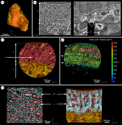
Here’s the anatomy of the mid-bone shaft of this fish(juvenile):

mid bone shaft

“(b) Virtual thin section (made along the longitudinal axis) showing the primary bone deposit of cortical bone and its connection to the spongiosa. Some remnants of calcified cartilage are still preserved at the location of Katschenko’s line (chondrocyte lacunae) and within the spongiosa (Liesegang rings).

So “micro-growth plates” within the bone marrow.

“At mid-shaft (figure 1a), the inner surface of the cortical bone is delimited by clusters of numerous large globular cell lacunae (cl, figure 1b,d) that can be identified as chondrocyte lacunae of cartilage. This suggests that remnants of Katschenko’s line are still present. Several stacks of Liesegang rings, typical of calcified cartilage, are also notable among the endochondral trabeculae”

“In extant tetrapods, the spongiosa forms when chondroclasts create erosion bays in the cartilage that are then lined with a thin peripheral bone layer, and it is common for small remnants of calcified cartilage to be left behind by the process; spongiosa formation in Eusthenopteron appears to have been similar.”

“The trabeculae in the spongiosa [of adult fish] are greatly remodelled and show no visible remnant of calcified cartilage”

“In extant actinopterygians, the fin endoskeleton develops from a blastema that differentiates into cartilages. Perichondral bone is deposited on the surface of the cartilages and continues to grow centrifugally as periosteal bone”

“a typical metapterygial bone is composed of a cartilaginous rod surrounded by a bony tube with cartilage projecting as condyles”

“In contrast to tetrapods, actinopterygian long bones have no haematopoietic bone marrow but only fatty tissues in the spaces created by the erosion of the cartilaginous rod. Neither do they show any evidence of a growth plate with longitudinally oriented columns of chondrocytes. The large longitudinal tubular mesh observed in Eusthenopteron humerus appears to constitute the earliest and phylogenetically deepest documented occurrence of a complex functional bone marrow in the tetrapod stem group.”

“As the tubular channels in Eusthenopteron are obviously connected to the epiphyses, the appearance of a complex bone marrow seems to be related to the appearance of tetrapod-like epiphyseal structures and elongation growth. Eusthenopteron lacks the comprehensive remodelling and trabecular resorption that creates an open medullary cavity in the majority of extant tetrapods, but the reduction of trabeculae between the longitudinal tubes in the adult compared with the juvenile may represent an evolutionary precursor of this process. ”

“Tetrapod bone marrow has been shown capable of degrading cartilage proteoglycans and inducing the initial stage of endochondral ossification”

X-Rays Of My Knees And Hands Showing Complete Epiphyseal Growth Plate Ossification

These x-rays shown in the video were taken maybe 3 years ago when I went to visit my parents and complained about pain in my lower back, knees, and left hand. They decided to send me to the hospital for a complete X-ray on the areas. You can see me explain the situation of what my growth plates are like in the video I uploaded to the Natural Height Growth YouTube channel.

Also: Subscribe to the Natural Height Growth channel by clicking HERE

So I actually got the X-rays, each costing around $250 each, and got myself checked out. I did see an orthopedic surgeon or doctor and we talked about what the pain could be coming from.

The conclusion was that I had some type of bone protrusion on the area on the top of my tibia or patella. This means that there was some type of bone longitudinal growth pushing up on the femur. This is what could be considered real longitudinal bone growth if it did happen. So the doctor said it was a type of bone spur which I had been suffering from. Apparently these “bone spurs” have a medical term for them, osteophyte.

After the talk with the “medical professional” I was not given any type of advice on how to treat the joint or bone pain. No treatment was advised except maybe to sometimes take ibuprofen or a type of NSAIDs (Non-Steroidal Anti-Inflammatory Drug).

After time passed the pain sort of disappeared and the bone issue was not brought up/ ONly recently did I find those old X-rays while I was packing and I thought it would be nice to show you guys that I do indeed have X-rays taken of parts of my limbs even from 3 years back which show that I don’t even have any type of epiphyseal growth plate ossification line any more. There is nothing there. Hope you guys stick around for tomorrow when I put up a video of the books and the intensive research I have been doing.