A radical switch in clonality reveals a stem cell niche in the epiphyseal growth plate.
“Longitudinal bone growth in children is sustained by growth plates, narrow discs of cartilage that provide a continuous supply of chondrocytes for endochondral ossification. However, it remains unknown how this supply is maintained throughout childhood growth. Chondroprogenitors in the resting zone are thought to be gradually consumed as they supply cells for longitudinal growth, but this model has never been proved. Here, using clonal genetic tracing with multicolour reporters and functional perturbations, we demonstrate that longitudinal growth during the fetal and neonatal periods involves depletion of chondroprogenitors, whereas later in life, coinciding with the formation of the secondary ossification centre, chondroprogenitors acquire the capacity for self-renewal, resulting in the formation of large, stable monoclonal columns of chondrocytes{if these chondroprogenitors never become senescent then we can grow forever}. Simultaneously, chondroprogenitors begin to express stem cell markers and undergo symmetric cell division. Regulation of the pool of self-renewing progenitors involves the hedgehog and mammalian target of rapamycin complex 1 (mTORC1) signalling pathways. Our findings indicate that a stem cell niche develops postnatally in the epiphyseal growth plate, which provides a continuous supply of chondrocytes over a prolonged period.”
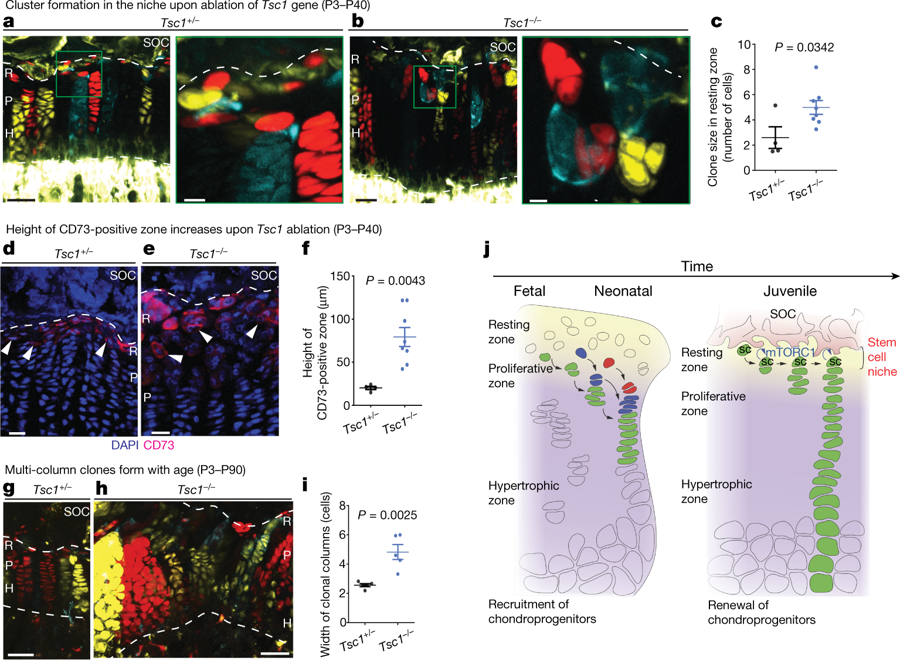
“CD73 (encoded by Nt5e) was among the most upregulated stem cell surface markers (4.9 ± 1.2-fold; P28 versus P2; P = 0.018) and immunohistochemical analysis confirmed de novo expression of CD73 at P28 (markers for cell proliferation (Ki67) and differentiation (MEF2C) were controls for developmental changes”
“Cells in the growth plate might also orient as a result of migration or stacking due to tissue polarization, which is reflected by the orientation of primary cilia. Cilia on flat chondrocytes, but not on chondroprogenitors, were polarized (with 61.3 ± 4.8% and 32.8 ± 2.2% of cilia, respectively, oriented longitudinally, P = 0.005, n = 3;”
3j is supposed to summarize it