Category Archives: Uncategorized

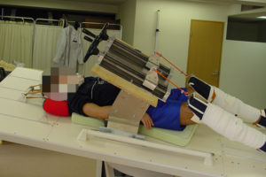
Ginza Kojima seems to have a new height increase device

Natural Height Growth has written about Ginza Kojima’s devices before.  The device appeared to be some kind of traction device with a rotational force applied.  The centripetal force could potentially stretch the body.  But all these forces ultimately have to lead to some kind of plastic deformation(permanent stretch of the bone or cartilage).  I speculated previously that the green light may be some kind of radiation that weakens the bone which allows it to be permanently stretched.

Here’s Ginza Kojima’s new website: http://www.shincho-nobashi.jp/

It looks like it applies a rotational/twisting force on the leg combined with some kind of pressure.

It looks like it’s mainly for small amounts of height as taken by this excerpt below.

In the case of the whole body course (guarantee course), it is 500,000 yen up to 1 cm, 1,300,000 yen up to 2 cm, and 2,300,000 yen up to 3 cm. (Excluding tax ※ Consumption tax will apply separately)

In the case of the knee and foot bone course (guarantee course), it is 1 million yen to 1 cm, 2 million yen to 2 cm and 3 million yen to 3 cm. (Excluding tax ※ Consumption tax will apply separately)

Please inquire for detailed charges for 3 cm and beyond.”

One limb length testimonial though advertised in the site is for 5 cm which you cannot realistically achieve without actual major bone growth.

The effects of the treatment are very individual differences and it is not known how long it will take without actually starting the treatment.

In our hospital, we guide you to receive the treatment once a week 24 times (about 6 months) as a rough guide. In addition, we offer a course that you can receive intensive treatment for a week as a short-term intensive course, so please consult with distant people.”

<-so looks like once a week for 6 months.

The current treatment schedule at our hospital is as follows.

  • 1st time 10:00 to 12:00 (30 minutes set time)
  • Second time 12:30-14:00
  • Third time 14:30-16:00

As mentioned above, an average of 3 sets is given daily. The second and subsequent treatments are basically the same, but we will make adjustments to suit your convenience as much as possible, so please contact us.”<-and 3 sessions of 30 minutes.

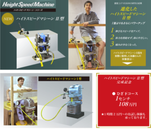
This machine advertised elsewhere on the site looks like a traction machine

Gina kojima explains the science behind his thinking here:

“First of all, I thought it would be good to pull. When I grabbed the bones of the foot from both sides and pulled it with a slight force according to the tug of war and the principle of childhood, I thought that it would be fine because the blood vessels became thinner and blood flow would improve. The heart is pumping 6 liters of blood a minute, so the blood vessels become thin if you pull a round blood vessel, and it is the theory that so much pressure will be applied to improve blood flow. It has been 40 years since I started making machines. To be honest, it will be dozens already. Anyway, it is said that if you look at it, why do you spend so much money to make stupid tools, and it keeps making it anyway.”

So this quote makes it seem like a traction device but a lot of traction devices don’t necessarily pull the bone to induce plastic deformation.

As far as traction devices go, you have to either induce plastic deformation in the bone or you have to stimulate the body to grow.

I think the whole body course is the traction machine and the knee course is the elevated height speed machine 2 device.

I think elevated height speed machine 2 device may just be another traction device but specifically for the knee.  Any japenese translators?

Articular cartilage versus growth plate cartilage

I am still working on finding a method to grow taller.  A lot of it is self testing as not a lot of the published papers published lately show promise of non surgical longitudinal bone growth that I could see.

Mechanisms of synovial joint and articular cartilage development

“Articular cartilage is formed at the end of epiphyses in the synovial joint cavity and permanently contributes to the smooth movement of synovial joints. Most skeletal elements develop from transient cartilage by a biological process known as endochondral ossification. Accumulating evidence indicates that articular and growth plate cartilage are derived from different cell sources and that different molecules and signaling pathways regulate these two kinds of cartilage{Could we inject these cell sources into the articular cartilage?  Could we mimic the molecules and signaling pathways with various stimuli?}. As the first sign of joint development, the interzone emerges at the presumptive joint site within a pre-cartilage tissue. After that, joint cavitation occurs in the center of the interzone, and the cells in the interzone and its surroundings gradually form articular cartilage and the synovial joint. During joint development, the interzone cells continuously migrate out to the epiphyseal cartilage and the surrounding cells influx into the joint region. These complicated phenomena are regulated by various molecules and signaling pathways, including GDF5, Wnt, IHH, PTHrP, BMP, TGF-β, and FGF. Here, we summarize current literature and discuss the molecular mechanisms underlying joint formation and articular development.”

“Matured articular cartilage is also referred to as hyaline cartilage because of its translucent appearance that reflects its unique constituents, such as type II collagen, glycosaminoglycans (GAGs), and low cellularity. In addition, articular cartilage does not have blood vessels, lymphatic vessels, or nerves. Articular chondrocytes produce extracellular matrices and maintain their environment with very little or no cell turnover{we want cell turnover, HGH increases cell turnover and HGH can make you very tall in certain circumstances}”

” most parts of articular cartilage derive from different lineages from the growth plate cartilage. The first signs of joint development are presented by the appearance of condensed flattened cells at the presumptive joint site within a pre-cartilage tissue known as the interzone, the origin of the joint. Removal of the interzone from a chick embryo leads to an uninterrupted long bone lacking joints, indicating that the interzone provides segmentation of skeletal elements in limbs. The interzone arises from mesenchymal/pre-cartilaginous tissue in which the cells initially express chondrocyte marker genes such as type II collagen, aggrecan, and matrillin-1 ”

“Instead of the decreased expression of these chondrogenic markers, the interzone cells acquire the expression of growth differentiation factor 5 (Gdf5), formerly known as bone morphogenetic protein 14 (BMP14), or cartilage derived morphogenetic protein 1 (CDMP1). Gdf5 is a representative marker for the interzone during early joint development. In addition to Gdf5, Wnt4, Wnt9a (formerly known as Wnt14), Wnt16, Erg, doublecortin, and Gli are also expressed in the interzone”

“Gdf5-expressing cell lineage gives rise to all mature joint structures including articular cartilage, meniscus, ligaments, and synovium ”

” joint components are formed by the integration of peripheral cells in joint development. Epiphyseal chondrocytes migrate into the interzone at early stages, and the external regions of joints such as the synovium/joint capsule and outer parts of the meniscus are mainly composed of lately integrated cells. Thus, the fate of embryonic interzone cells, the surrounding cells, and their progeny cells may be determined by their spatiotemporally environment. “<-so growth plate cells become integrated into the articular cartilage interzone cells but these cells are affected by the articular cartilage cells and microenvironment so that the articular cartilage overall environment is unaltered.  Maybe we can do the reverse and make the articular cartilage more growth plate like…

“IHH is produced from pre-hypertrophic chondrocytes and up-regulates PTHrP expression in peri-articular chondrocytes. PTHrP inhibits the differentiation of proliferating chondrocytes into pre-hypertrophic chondrocytes. This feedback loop determines the length of long bones. Before the feedback loop, pre-hypertrophic chondrocytes around the center of anlagen are associated with interzone generation through the secretion of IHH. The loss of IHH causes not only dwarfism but also joint fusion in distal limb joints ”

“excessive IHH signaling activity in the interzone progeny induces ectopic cartilage formation in the knee ”

“Unlike in endochondral ossification, IHH and PTHrP seem to be independent in interzone generation and joint development. The genetic alteration of PTHrP causes the impairment of endochondral ossification, but no severe changes in joints. Even after IHH signaling becomes silent, PTHrP-expressing cells exist in articular cartilage over a lifetime. Recombinant human PTH (1–34) suppresses osteoarthritis development, and PTH/PTHrP signaling induces lubricin”<-Can we use this fact to become taller?

Interesting study that explains why there are secondary ossification centers

Those who say that weightlifting stunts growth will have a field day with this one.  Small animals were used and if the loads mentioned in the study caused as much apoptosis then growth would be severly stunted by weight loading.  But if it indicated some cellular regeneration in regards to unplanned apoptosis due to mechanical that would be an amazing breakthrough.  We could trick the cells into thinking there was more apoptosis than there was and that would lead to overgrowth.

Secondary ossification centers evolved to make endochondral bone growth possible under the weight-bearing demands of a terrestrial environment

“The growth of long bones occurs in narrow discs of cartilage, called growth plates that provide a continuous supply of chondrocytes subsequently replaced by newly formed bone tissue. These growth plates are sandwiched between the bone shaft and a more distal bone structure called the secondary ossification center (SOC). We have recently shown that the SOC provides a stem cell niche that facilitates renewal of chondro-progenitrors and bone elongation. However, a number of vertebrate taxa, do not have SOCs, which poses intriguing questions about the evolution and primary function of this structure. Evolutionary analysis revealed that SOCs first appeared in amniotes[essentially egg laying reptiles, birds, and mammals] and we hypothesized that this might have been required to meet the novel mechanical demands placed on bones growing under weight-bearing conditions. Comparison of the limbs of mammals subjected to greater or lesser mechanical demands revealed that the presence of a SOC is associated with the extent of these demands. Mathematical modelling with experimental validation showed that the SOC reduces shear and normal stresses within the growth plate; while relevant biological tests revealed that the SOC allows growth plate chondrocytes to withstand a six-fold higher load before undergoing apoptosis{this provides evidence that too high a load could stunt growth}. Hypertrophic chondrocytes, the cells primarily responsible for bone elongation, were the most sensitive to loading, probably due to their low Young’s modulus (as determined by atomic force microscopy). Our present findings indicate that the primary function of the evolutionary delineation of epiphyseal cartilage into spatially separated growth plates was to protect hypertrophic chondrocytes from the pronounced mechanical stress associated with weight-bearing in a terrestrial environment.”

“hypertrophic chondrocytes undergo apoptosis or trans-differentiation, leaving their calcified extracellular matrix as a scaffold on which invading blood vessels and osteoblasts form new bone tissue.”<-interesting that the acknowledge the transdifferentiation theory.

“commonly to study bone growth (i.e., mice, rats and rabbits), the growth plate is separated from the articular cartilage by a bony fragment, the secondary ossification center (SOC). This skeletal element, formed during early postnatal development, splits the initially contiguous cartilaginous element into two independent structures, the growth plate and articular cartilage ”

“Growth plate chondrocytes appeared to be highly sensitive to load, with 40% dying upon application of a 1N load (as revealed by propidium iodide (PI) staining). At the same time, the SOC clearly protected these cells, allowing them to withstand a load an order of magnitude higher ”  An order of magnitude would be 10N

“directional compressive stress appears to be harmful to chondrocytes, especially hypertrophic chondrocytes”

Study finds chocolate increases longitudinal bone growth

Update:  I’m still working on a new non-LSJL method.

I don’t believe that chocolate can make you wildly taller but it tastes good.  I also think the optimal diet for longitudinal bone growth varies on developmental stage.

Chocolate and Chocolate Constituents Influence Bone Health and Osteoporosis Risk

“Bone loss resulting in increased risk of osteoporosis is a major health issue worldwide. Chocolate is a rich source of antioxidant/anti-inflammatory flavonoids as well as dietary minerals with the potential to benefit bone health. However, other chocolate constituents such as cocoa butter, sugar and methylxanthines may be detrimental to bone. Human studies investigating the role of chocolate consumption on serum bone markers and bone mineral density (BMD) have been inconsistent. A contributing factor is likely the different composition and thereby, nutrient and bioactive content amongst chocolate types. White, followed by milk chocolate, are high in sugar and low in flavonoids and most minerals. Dark chocolate (45-85% cocoa solids) is high in flavonoids, most minerals, and low in sugar with ≥70% cocoa solids resulting in higher fat and methylxanthine content. The aim of this review was to examine the relationship between consuming chocolate, its flavonoid content, and other chocolate constituents on bone health and osteoporosis risk. Studies showed postmenopausal women had no bone effects at moderate chocolate intakes; whereas, adolescents consuming chocolate had greater longitudinal bone growth{whether this fact can be used for any purpose though….}. Based on flavonoid and mineral content, unsweetened cocoa powder appeared to be the best option followed by dark chocolate with higher cocoa content in terms of supporting and preserving bone health. Determining dietary recommendations for chocolate consumption regarding bone health is important due to the growing popularity of chocolate, particularly dark chocolate, and an expected increase in consumption due to suggestions of health benefits against various degenerative diseases.”

“Diets that promote bone health have mainly focused on increasing Ca and
vitamin D consumption, but there is growing interest in phytochemicals[chemicals produced by plants]”

“Dietary polyphenols consist of a large group of plant-derived secondary metabolites
divided into four different classes, one of which is the flavonoids (diphenylpropanes C6-C3-C6).
Flavonoids contain several classes of bioactive compounds.  There is evidence
that intake of specific flavonoids may promote bone health including: soybean isoflavones (e.g.
genistein and daidzein), flavonols (e.g. aglycone quercetin) found in plums, and flavonones (e.g.
hesperidin) found in citrus pulp and juice.  Another food source noted for its flavonoid
content is chocolate.  Cocoa, a major constituent in chocolate, has the highest flavanol
content of all foods on a per-weight basis and contributes to greater total dietary intake of
flavonoids than tea, fruits, and vegetables ”

“Both animal models and human clinical trials have reported an inverse association
between reactive oxygen species (ROS) and bone health.  ROS can affect bone cells in
various ways including stimulation of osteoblast apoptosis and senescence and by upregulation
of receptor activator of nuclear factor kappa-B ligand (RANKL) to activate osteoclast
differentiation and bone resorption”<-I think though there is a role for ROS.  It’s just too many is a bad thing.  And bone can be inhibitory towards longitudinal bone growth but it’s degradation of bone(bone turnover) not degradation of bone building cells that would be beneficial.

“catechins preserved bone-forming osteoblasts by exerting anti-inflammatory actions”

” In a randomized, double-blinded placebo-controlled study, pre-pubertal (n=149, age 6.6-9.4 years) girls consumed two Ca-supplemented food products daily to achieve a Ca dose of 850 mg/d.  Results showed consuming Ca-supplemented chocolate bars, cakes or cocoa beverages for 48 weeks significantly increased height and bone mass acquisition in the radius and femur”

“Feeding murine dams chow diet supplemented with 400 mg unsweetened chocolate during pregnancy and lactation resulted in progeny with significantly shortened forefeet and hindlimbs.  Vascular endothelial growth factor (VEGF), which plays a role in ontogenesis and longitudinal bone growth related to angiogenesis in the epiphyseal growth plate, was significantly reduced in the femora of immature (age 4-weeks old) mice pups exposed perinatally to chocolate.  A follow-up study investigating the relationship of the chocolate constituent, catechins on angiogenesis and bone mineralization in the progeny of murine dams fed chocolate showed a negative correlation (r = -0.62, P<0.05) between embryo tissue epigallocatechin concentration and mean number of newly-formed blood
vessels.  Crystallinity of compact bone of diaphyses was 17% greater and femoral
epiphyseal cancellous bone was 30% greater in pups age 4-weeks old exposed perinatally to
chocolate compared to pups from control dams.  The authors suggested anti-angiogenic activity
of chocolate catechins disturbed the processes of bone elongation and mineralization “<-so chocolate may be better at different stages.

Here’s the study the chocolate study in girls:

Calciumenriched foods and bone mass growth in prepubertal girls: a randomized, double-blind, placebocontrolled trial.

“High calcium intake during childhood has been suggested to increase bone mass accrual, potentially resulting in a greater peak bone mass. Whether the effects of calcium supplementation on bone mass accrual vary from one skeletal region to another, and to what extent the level of spontaneous calcium intake may affect the magnitude of the response has, however, not yet been clearly established. In a double-blind, placebocontrolled study, 149 healthy prepubertal girls aged 7.9+/-0.1 yr (mean+/-SEM) were either allocated two food products containing 850 mg of calcium (Ca-suppl.) or not (placebo) on a daily basis for 1 yr. Areal bone mineral density (BMD), bone mineral content (BMC), and bone size were determined at six sites by dual-energy x-ray absorptiometry. The difference in BMD gain between calcium-supplemented (Ca-suppl.) and placebo was greater at radial (metaphysis and diaphysis) and femoral (neck, trochanter, and diaphyses) sites (7-12 mg/cm2 per yr) than in the lumbar spine (2 mg/cm2 per yr). The difference in BMD gains between Ca-suppl. and placebo was greatest in girls with a spontaneous calcium intake below the median of 880 mg/d. The increase in mean BMD of the 6 sites in the low-calcium consumers was accompanied by increased gains in mean BMC, bone size, and statural height. These results suggest a possible positive effect of calcium supplementation on skeletal growth at that age. In conclusion, calciumenriched foods significantly increased bone mass accrual in prepubertal girls, with a preferential effect in the appendicular skeleton, and greater benefit at lower spontaneous calcium intake.”

“Both statural height and body weight, however, were significantly greater among the spontaneously high- versus low-calcium consumers (129.160.7 vs. 126.560.7 cm, P , 0.001; 27.560.5 vs. 25.760.5 kg, P , 0.001, respectively). ”

There were several non calcium enriched foods in the study and not just chocolate so it’s likely the calcium and not the chocolate making the girls taller.
Old study on heat has minor breakthrough

I’m still working on height increase.  Just doing more independent research and learning more about anatomy and physiology and actions of cells as the research I’m looking at is a lot of the same old, same old.  Maybe the key is to look at older studies before things were set in their ways.  It seems in the old days they were willing to try new risks to get people to grow taller.

Essentially the study found that heat did not increase bone length however there was some promise in that decalcification could be caused by the heat and that could enable longitudinal bone growth.  And the study shows islands of cartilage which could be the creation of new growth plates which is quite promising.  But the heat itself actually didn’t stimulate the growth plate itself.  It was only the heat degeneration the calcified bone matrix and stimulating the creation of growth plate islands that incurred new growth.

The effect of heat upon the growth of bone

“GROWTH in length of long bones consists of two mutually independent processes, the division and palisade arrangement of cartilage cells, and the subsequent calcification of the matrix between these cells and its replacement by bone. It is generally agreed that growth is dominated by the activities of the cells of the reserve zone and those in the adjacent apex of the cartilage columns. The subsequent enmeshment, of hypertrophic cartilage cells within a calcified matrix must prevent elongation at other sites. Recent attempts to stimulate the growth of bone have been based upon the production of an irritative lesion within the metapliysis, stirnulating ossificaation rather than cartilage-cell division. ”

“Chapchal and Zeltienrust (1947-48) reported an inconstant increase in the rate OF growth in ths rabbit after the insertion of metal or ivory pins within the metaphysis. Wilson (1952) using copper and constantan wire in the dog produced similar results. The application of these methods to limb inequality in children has been reported by Pease (1962). Metal or ivory screws WPI’P inserted transversly into the metaphysis of the femur and tibia. Each of the seven children subjected to this procedure showed an increase in the rate of growth of the shorter limb, but equalisation of length was not attained. ”

“The overgrowth of bone which is constantly seen after fractures, in the presence of arteriovenus aneurysms, and in association with bone and joint infections”

arteriovenous aneurysm an abnormal communication between an artery and a vein in which the blood flows directly into a neighboring vein or is carried into the vein by a connecting sac.<-Lateral loading can increase blood flow.  Though this probably won’t work without existing growth plates.

“increase in the length of a limb following the production of an arteriovenous fistula in the dog”

“One of the clinical features of most cases of limb elongation with an increase in the temperature of the skin, and in the arteriovenous aneurysm this is associated with an increase in the temperature of bone”

“The rate of growth of the ulna was in some animals slightly depressed, in rabbit 480 markedly SO. In this animal there was considerable formation of new bone around tJie resistor, although the epiphyseal cartilage appeared normal. The overgrowth of the ulna noted in rabbit 465 was not associated with any abnormal histological appearance of the epiphyseal cartilage and the radiographs suggest that the discrepancy may have been due to a disturbance of growth in the control limb. The radio- graphs also show a relative decalcification of the the whole of the treated forelimb and this may have been associated with a generalized hyperemia{excess of blood in the vessels} of this limb.”

“Where the heating level was high, the epiphyseal cartilage is affected. The earliest changes in this region are an irregularity of the cartilage columns and a granularity of the matrix. When cellular destruction occurs it is at first confined to the region of the apex of the cartilage columns and is associated with fibrillation of the cartilage matrix. ”

“In only one animal (465) did an increase in bone length occur. This animal was heated for two days and, the wires having broken, the resistor was left unheated for a further 36 days. Although the increase in length of the limb was considerable, aid was associated with a generalized decalcification of the forelimb, suggesting an increase in the vascularity of this member, the difference in length of the limbs may have been due to some abnormality of growth on the control side. “<-So decalcification is key to growing taller.  High levels of heat can was found to cause necrosis which reduced growth but if decalcification can enhance growth.  Then maybe heat could enhance growth by decalcifying bones.

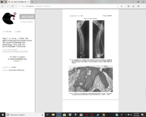
“heat has been shown to stimulate the production of cartilage around the resistor and to produce islands of endochondral ossification within the bony epiphysis and along the shaft of the radius.”<-islands of endochondral ossification is promising because it shows new induction of bone growth.

Islands of cartilage were produced within the bony epiphysis close to the resistor, and along the ulnar border of the radial shaft. Some of them islands showed endochondral ossification, but there was no increase of cellular activity in the epiphyseal cartilage. A large cartilaginous mass developed in the region where the resistor was buried, and, in many animals, the transverse diameter of the metaphysis was greatly increased. ”

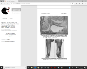
Figure 8 is an image near the resistor so there should be islands of cartilage visible.  I can’t tell If there are cartilage islands or not and if there are cartilage islands we can’t say for sure whether they are not just broken off cartilage from the growth plate.  Cartilage islands within the diaphysis would be key to seeing if heat can induce new growth plates.

Here’s mentioned overgrowth.

New updates on a semi-LSJL loading device

I’m still working on my own device.   I’m not sure if the device listed here is stimulatory enough to induce longitudinal bone growth.  The device may be useful if you have existing growth plates.  But to grow taller you’d need bone breakdown to occur faster than bone buildup{so that cartilage has room to grow} and the device is optimized for bone buildup.

Development of an Artificial Finger-Like Knee Loading Device to Promote Bone Health.

“This study presents the development of an innovative artificial finger-like device that provides position specific mechanical loads at the end of the long bone and induces mechanotransduction in bone{So you could theoretically use your own fingers to see what such an a device would do; use your own fingers to press on the epiphysis of bone}. Bone cells such as osteoblasts are the mechanosensitive cells that regulate bone remodeling{in order to induce height growth you’d need a lot more than bone remodeling, you need to degrade cortical bone and induce MSC differentiation into chondrogenic cells}. When they receive gentle, periodic mechanical loads, new bone formation is promoted{how this bone formation is promoted is of importance of whether such a method can induce height growth}. The proposed device is an under-actuated multi-fingered artificial hand with 4 fingers, each having two phalanges. These fingers are connected by mechanical linkages and operated by a worm gearing mechanism. With the help of 3D printing technology, a prototype device was built mostly using plastic materials. The experimental validation results show that the device is capable of generating necessary forces at the desired frequencies, which are suitable for the stimulation of bone cells and the promotion of bone formation. It is recommended that the device be tested in a clinical study for confirming its safety and efficacy with patients.”

Cortical bone is highly inhibitory towards longitudinal bone growth.  You need to generate sufficient fluid flow to induce degradation of the cortical bone or you likely will not be able to grow taller.  I think it would take a lot of fluid flow for that to happen.  Maybe with very high frequency and duration with this device it would be possible.

” if a small magnitude of mechanical stimuli is applied at a high frequency, an osteogenic response can be stimulated via mechanotransduction in bone cells.”<-we don’t care about stimulating bone cells EXCEPT for osteoclasts.  We need to simulate stem cells to differentiate into chondrocytes.

“Osteocytes are the most abundant type of cells in bone tissue, and they constitute more than 90% of the cells in bone matrix. They are rooted in the calcified bone medium, and communicate with each other and with bone-forming osteoblasts through slender processes and gap junctions. Osteocytes are highly mechanosensitive. Haversian system or osteon, one of the key components of a porous bone matrix, encloses a blood vessel in its center and sets up the canals known as Haversian canals or Volkmann’s canals. Osteogenesis is induced by the process of osteoinduction in which premature cells are recruited, stimulated and developed into pre-osteoblasts{we just need to induce a microenvironment where premature cells are induced into pre-chondrocytes instead and one way to do that is via a high hydrostatic pressure environment which could be induced by manipulating fluid flow}. Osteogenesis can also result from osteoconduction which is the passive process of bone growth on surfaces such as bone-implant surfaces”

“When rapid mechanical loading is applied at the end of long bone (e.g., knee), it is proposed that the interstitial fluid present around the osteocytes in the lacuna-canalicular network induces a pressure gradient and elevates nutrient transport throughout the porous network. “<-interstitial fluid flow could do more than this.  If you have a lot of fluid flow it could induce shear that degrades the cortical bone that prevents longitudinal bone growth.