Monthly Archives: June 2014

Cysteine for Height Growth?

ijmm_36_2_571_PDFCysteine induces longitudinal bone growth in mice by upregulating IGF-I.

“Cysteine (Cys) is known to exert various effects, such as antioxidant, antipancreatitic and antidiabetic effects. However, the effects of Cys on longitudinal bone growth have not been elucidate to date. Thus, the aim of the present study was to evaluate the effects of Cys on bone growth. Growth‑plate thickness and bone parameters, such as bone volume/tissue volume (BV/TV), trabecular thickness (Tb.Th), trabecular number (Tb.N), connectivity density (Conn.D) and total porosity were analyzed by means of micro-computed tomography (µCT). The levels of serum insulin‑like growth factor‑I (IGF‑I) were measured by enzyme‑linked immunosorbent assay (ELISA). Hepatic IGF‑I mRNA expression was analyzed by quantitative polymerase chain reaction (qPCR). The phosphorylation of Janus kinase 2 (JAK2) and signal transducer and activator of transcription 5 (STAT5) was investigated by western blot analysis. Our results revealed that Cys increased IGF‑I mRNA expression in HepG2 cells. The thickness of the growth plates was increased following treatment with Cys. Moreover, BV/TV, Tb.Th, TbN, Conn.D and total porosity were improved following treatment with Cys. Hepatic IGF‑I mRNA expression and serum IGF‑I levels were increased by Cys. The levels of phosphorylated JAK2 and STAT5 were elevated by Cys. The findings of our study indicate that Cys increases the thickness of growth plates through the upregulation of IGF‑I, which results from the phosphorylation of JAK2-STAT5. Thus, our data suggest that Cys may have potential for use as a growth-promoting agent.”

Cysteine is found in dietary sources but it may be possible to be deficient.

“Treatment with 50 mg/ml of Cys had the most prominent effect, and thus we evaluated the effects of treatment with 50 mg/kg of Cys in the next set of experiments, i.e., in vivo mouse models.”

“The thickness of the growth plates in the proximal tibias in the CON and PEM{protein deficient group} groups were 124.0±2.9 and 90.1±3.1, respectively; the growth-plate thickness in the Cys group was 117.0±4.3″<-Cysteine did not increase growth plate thickness versus control group.

“the levels of p-JAK2 and p-STAT5 in the liver were decreased due to malnutrition. However, the decrease in the levels of p-JAK2 and p-STAT5 was reversed by treatment with Cys”

“In general, endochondral cell proliferation in the growth plate results in bone growth. Thus, growth-plate thickness is a direct indicator of linear bone growth”

LSJL Studies 4: Some LSJL effects summarized

Unfortunately, not a lot of insight for LSJL for height growth as most of the effects are those related to the ability of joint loading to inhibit catabolism.

Mechanical intervention for maintenance of cartilage and bone.

Moderate loads to the synovial joint suppress the expression levels of matrix metallproteinases (MMPs), while loads above a threshold tend to increase their destructive activities{although some catabolic effects of MMPs may be good for height growth, as MMPs may degrade bone allowing for cartilage growth}.

“Moderate shear stress(2–5 dyn/cm2) reduced MMP expression levels, while high shear stress (10–20 dyn/cm2) increased them. Moderate hydrostatic pressure (1–5 MPa) suppressed MMP-1 expression, while higher loads (10 MPa) elevated it.”<-Since I have gotten more results with higher clamping force it could indicate that increased MMP expression is crucial to induce new length growth.

“The required magnitude of loads for joint loading is in general smaller than that for axial loading (e.g. 0.5 N for elbow loading and 2–3 N for ulna axial loading in mice). Bone is less stiff in a lateral direction than an axial direction.”<-Note that more than 0.5N(100N is mentioned) is likely required for humans. 0.5N is what was used in the mouse arm lengthening study.

“Joint loading periodically alters the pressure in the medullary cavity and activates molecular transport in a lacunocanalicular network in cortical bone.”<-It is our hypothesis that this increase in pressure in the medullary cavity induces chondrogenic differentiation.  The medullary cavity is continuous into the spaces of the spongy bone of the epiphysis.  It is these spaces where we aim to induce chondrogenic differentiation and thus induce endochondral ossification to grow taller.

“A pressure gradient in the medullary cavity generates oscillatory fluid flow in the porous bone cortex. Induced fluid flow then enhances molecular transport in the lacunocanalicular network and applies shear stress to osteocytes residing in lacunae”

“A pressure gradient in the medullary cavity generates oscillatory fluid flow in the porous bone cortex.”<-and fluid flow into the spongy bone spaces of the epiphysis and the increased MMP expression could allow the neo-growth plates to spread to other parts of the bone but this is highly speculative.

“Modulation of the intramedullary pressure with knee loading is exerted throughout the length of the tibia and the femur.”<-the epiphysis is part of the entire length thus knee loading like by LSJL alters pressure in the epiphysis.

“Proinflammatory cytokines such as IL-1β upregulate the expression and activity of MMP-1 and MMP-13. [In] cultured chondrocytes mechanical stimulation, given in a form of fluid flow shear stress, can suppress the IL-1β-induced upregulation of MMP-1 and MMP-13. In accordance with those in vitro results, joint motion in vivo is able to reduce inflammatory responses in a murine collagen-induced arthritis model. Additionally, in an antigen-induced arthritis model in rabbits, continuous passive motion suppressed transcription of IL-1β and synthesis of inflammatory mediator COX-2 and MMP-1. These mechanical signals also induced IL-10 synthesis, suggesting that moderate joint loading can generate anti-inflammatory signals.”<-MMP’s have complicated effects on height growth.  MMP-1 and MMP-13 are vital for the endochondral ossificatiion process.

“When knee loading was applied to one leg, the loaded tibia and femur were reported to be longer than the non-loaded contralateral bones. In response to knee loading, the number of cells in the growth plate of the proximal tibia increased and their cellular shape was altered.“<-If LSJL increases the number of cells in the growth plate by differentiation of stem cells into chondrocytes than LSJL will work in adults as well.  It’s possible that during knee loading only chondrocyte proliferation was increased but chondrocytes have a finite proliferative capacity and an increase in chondrocyte proliferation without increasing stem cell differentiation into chondrocytes should accelerate the the transition of proliferating chondrocytes into hypertrophic chondrocytes and not the number of cells in the growth plate.

Homeostasis of the articular cartilage is affected through interactions with the subchondral bone underneath the cartilage.  Both MMPs and ADAMTS need to be post-translationally activated, and this activation process is regulated by many factors including MMPs themselves and many proteoglycans.”<-Thus loading of the articular cartilage may itself play a role in the height gain by triggering a response in the subchondral bone in response to the stimulation of the articular cartilage.  Thus underlying the importance of loading the synovial joint.  The activation of MMPS and ADAMTS in the subchondral bone may play a role in neo growth plate formation.

“flexion of the joint in the presence of axial loads (5 N) increased the level of MMP-13 mRNA and its activity.”<-LSJL height growth can not be due to higher levels of MMP13 alone or axial loading would increase height!

“Whether mechanical loading can suppress or induce the integrated stress response is largely dependent on the loading intensity. This stress response leads to translational de-activation by a mechanism involving phosphorylation of eIF2α, with preferential translational activation of a particular set of proteins linked to cellular survival or apoptosis. In cultured chondrocytes, administration of thapsigargin and tunicamycin induces stress to the endoplasmic reticulum, which triggers an integrated stress response. In this response, the level of phosphorylated eIF2α was elevated together with the expression of MMP-13. Joint loading reduced the level of phosphorylated eIF2α by suppressing activity of Perk, one of the four known eIF2α kinases”

Substance P impact on endochondral ossification

All this may have an impact on LSJL but I’m not sure what it is yet.  However, nerves are something that you can feel.  However, different nerves have different effects so it’d be hard to gauge what the effect is.  But as alluded to in the study reduced pain sensitivity is a sign of reduced substance P and SNF.  So if you have reduced pain sensitivity due to LSJL you may have less SP and SNF.  The reduced mechanical stability may be beneficial to neo-growth plates also.  However, mesenchymal stem cells and chondrocytes stained heavily for SP so enhanced pain sensation may be more indicative of the success of LSJL.

Absence of substance P and the sympathetic nervous system impact on bone structure and chondrocyte differentiation in an adult model of endochondral ossification.

substance P pdf<-Read the full study.

“Sensory and sympathetic nerve fibers innervate bone and epiphyseal growth plate. The role of neuronal signals for proper endochondral ossification during skeletal growth is mostly unknown. Here, we investigated the impact of absence of sensory neurotransmitter substance P (SP) and removal of sympathetic nerve fibers (SNF) on callus differentiation, a model for endochondral ossification in adult animals, and on bone formation.
In order to generate callus, tibia fractures were set in the left hind leg of wild type (WT), tachykinin 1-deficient (Tac1-/-) mice (no SP) and animals without sympathetic nerve fibers. Locomotion was tested in healthy animals and touch sensibility was determined early after fracture. Callus tissue was prepared for immunofluorescence staining for SP, neurokinin1-receptor (NK1R), tyrosine-hydroxylase (TH) and adrenergic receptors α1, α2 and β2. At the fracture site, osteoclasts were stained for TRAP, osteoblasts were stained for RUNX2 and histomorphometric analysis of callus tissue composition was performed. Primary murine bone marrow derived macrophages (BMM), osteoclasts and osteoblasts were tested for differentiation, activity, proliferation and apoptosis in vitro. Femoral fractures were set in the left hind leg of all three groups for mechanical testing and μCT-analysis.
Callus cells stained positive for SP, NK1R, α1d- and α2b adrenoceptors and remained β2- adrenoceptor and TH-negative. Absence of SP and SNF did not change general locomotion but reduces touch sensitivity after fracture. In mice without SNF, we detected more mesenchymal callus tissue and less cartilaginous tissue 5days after fracture{so more sympathetic nerve fibers are pro-differentiation?}. At day 13 past fracture, we observed a decrease of the area covered by hypertrophic chondrocytes in Tac1-/- mice and mice without SNF, a lower number of osteoblasts in Tac1-/- mice and an increase of osteoclasts in mineralized callus tissue in mice without SNF. Apoptosis rate and activity of BMM, osteoclasts and osteoblasts isolated from Tac1-/- and sympathectomized mice were partly altered in vitro. Mechanical testing of fractured- and contralateral legs 21days after fracture, revealed an overall reduced mechanical bone quality in Tac1-/- mice and mice without SNF. μCT-analysis revealed clear structural alteration in contralateral and fractured legs proximal of the fracture site with respect to trabecular parameters, bone mass and connectivity density. Notably, structural parameters are altered in fractured legs when related to unfractured legs in WT but not in mice without SP and SNF.
The absence of SP and SNF reduces pain sensitivity and mechanical stability of bone in general. The micro-architecture of bone is profoundly impaired in the absence of intact SNF with a less drastic effect in SP-deficient mice. Both sympathetic and sensory neurotransmitters are indispensable for proper callus differentiation. Importantly, absence of SP reduces bone formation rate whereas absence of SNF induces bone resorption rate{maybe this could be beneficial as bone resorption would leave room for neo-growth plates}. Notably, fracture chondrocytes produce SP and its receptor NK1 and are positive for α-adrenoceptors indicating an endogenous callus signaling loop. We propose that sensory and sympathetic neurotransmitters have crucial trophic effects which are essential for proper bone formation in addition to their classical neurological actions.”

“Under rigid, stable fixation regimen, bone regenerates with no or only minor callus formation”

“When applying more flexible fixation regimens, bone healing occurs in consecutive stages which involve intense callus formation. Firstly, an acute inflammatory response and recruitment of mesenchymal stem cells (mesenchymal callus) occur in order to subsequently generate a primary cartilaginous callus populated mostly with chondrocytes (soft callus). Later, this cartilaginous callus undergoes revascularization and calcification (calcified hard callus) and is finally remodeled to fully restore a normal bony structure and architecture”<-The idea with LSJL is that perhaps the accute inflammatory respose and recruitment of mesenchymal stem cells can occur without a fracture.

“SP plays a role in pain transmission; tibial fractures cause an early and strong induction of sensory nerve regeneration and growth into the site of injury (sensory sprouting). The presence of NK1 receptors was demonstrated on bone cells”  SP can also affect proliferation in mesenchymal stem cells.

“skeletal growth or activity of bone tissue might be regulated by SNF”

“At day 5 after fracture, when chondrogenic differentiation starts, a substantial number of mesenchymal and chondrocyte-like cells stained positive for NK1R and some cells double-stained for SP. At day 9 after fracture, when most of the callus matrix has adopted a cartilaginous phenotype (soft callus), nearly all of the callus chondrocytes were SP- and NK1R-positive. At 13 days after fracture, when remodeling of the callus progressed toward tissue mineralization and the bony, hard callus was about to be formed, number of SP-positive callus cells appeared to be reduced compared to day 9 but NK1R staining seems to be unaltered in hypertrophic chondrocytes. SP- and NK1R staining pattern in sympathectomized mice was similar to WT”

“mesenchymal callus cells and periosteum stained positive for α1d adrenergic receptor 5 days after fracture whereas only few chondrocyte-like cells were α1d-positive”

Insight into cellular senescence(Why you stop growing)

Growth plate cellular senescence preceeds epiphyseal fusion.  It is the reason why people stop growing.  If we stop cellular senescence we can keep growing.  And this study states that stem cell growth is not based on demand.  Demand for stem cells too high to be kept up with may lead to cancer growth.  However, supplements, reduction of environmental stressors, and mechanical loading may be some ways to stimulate stem cell health and renewal.  The key to LSJL success on restoring growth renewal may be based on it’s ability to stimulate stem cell proliferation and impair cellular senescence.  As this study suggests that microcracks that occur during old age increase the demand for stem cells but the body doesn’t generate new stem cells or increase proliferation to compensate.  However, unlike microcracks due to degradation LSJL places forces directly on the cells themselves.  I will be posting some updates on the LSJL method as soon as the comments section is up(after Michael has resolved some legal issues).

Age-specific bone tumour incidence rates are governed by stem cell exhaustion influencing the supply and demand of progenitor cells

“Knudson’s carcinogenic model, which simulates incidence rates for retinoblastoma, provides compelling evidence for a two-stage mutational process. However, for more complex cancers, existing multistage models are less convincing. To fill this gap, I hypothesize that neoplasms preferentially arise when stem cell exhaustion creates a short supply of progenitor cells at ages of high proliferative demand. To test this hypothesis, published datasets were employed to model the age distribution of osteochondroma, a benign lesion, and osteosarcoma, a malignant one. The supply of chondrogenic stem-like cells in femur growth plates of children and adolescents was evaluated and compared with the progenitor cell demand of longitudinal bone growth. Similarly, the supply of osteoprogenitor cells from birth to old age was compared with the demands of bone formation. Progenitor cell demand-to-supply ratios are a good risk indicator, exhibiting similar trends to the unimodal and bimodal age distributions of osteochondroma and osteosarcoma, respectively. The hypothesis also helps explain Peto’s paradox and the finding that taller individuals are more prone to cancers and have shorter lifespans. The hypothesis was tested, in the manner of Knudson, by its ability to convincingly explain and demonstrate, for the first time, a bone tumour’s bimodal age-incidence curve.”

“Osteochondroma is the most common benign bone tumour, occurring as an abnormal osteocartilaginous outgrowth of the epiphyseal growth plate with a low rate (≤2%) of malignant transformation to secondary chondrosarcoma”

“In adolescents, growth plate senescence is followed by epiphyseal fusion when osteochondroma stop growing.”

“Osteosarcoma is the most common primary bone malignancy (excluding multiple myeloma), originating from the transformation of aberrant bone-forming mesenchymal stem cells (MSC), also known as marrow stromal cells”

“Differentiation is impaired by replicative senescence”

“Post-natal stem cell replication leads to telomere shortening in somatic tissues, as telomerase activity in humans is not at sustaining levels. At the Hayflick limit of cell division, telomeres reach a critical length, triggering cells to become senescent or apoptotic. A recent publication provides quantitative evidence that soft tissue organ mass loss in humans aged 25–70 is significantly associated with the log of shorter cell turnover times, implicating stem cell exhaustion and replicative senescence in normal ageing”

“MSC, like other stem cells, have been observed to have a self-renewal and proliferative capacity that diminishes with age, and that is regulated by different pathways, such as antioxidant defence, DNA repair, and protein turnover, before entering a senescence-associated proliferation arrest”

“There is a normal trade-off for a stem cell to self-renew or produce differentiated and differentiating progeny. High stem cell demand relative to supply appears to produce stem cell progeny with stopped differentiation but with self-renewal properties, perhaps triggered by senescent cell secretions”

Here’s a graph describing the stem cell pool:

stem cell pool

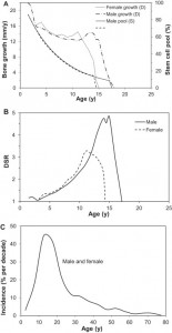
“A good marker of the skeletal demand for osteoprogenitors involved in bone formation (as opposed to osteoclast bone resorption activity) is serum BALP activity. This marker is particularly sensitive to physiological changes such as adolescent growth spurt and menopause. The demand for progenitor cells participating in bone formation, as measured by BALP activity, is highest in newborns, slowly declines until early adolescence in girls and late adolescence in boys, then decreases rapidly to its lowest level in young adults, before increasing again in the latter half of adulthood to cope with the decline in osteocyte density and repair of accumulated damage related to ageing, e.g. bone microcracks”

“senescent cells can arise due to “epigenetic senescence” (initiated by histones altering activity of genes), stress-induced senescence or replicative senescence”<-Stress-induced sensescence is the easiest to avoid and epigenetic senescence can be affected by supplements.  Replicative senescence may be influenced by mechanical loading.

“cell proliferation is not in itself a risk factor for tumogenesis; instead, neoplastic promotion or progression occurs preferentially when, for example, stem cell exhaustion creates a short supply of progenitor cells at ages of high mitogenic demand.”

” Ionizing radiation exposures produce free radicals and reactive oxygen species that excite a high proliferative demand on MSC involved in inflammation and subsequent fibrosis. When bone marrow is subjected to a high dose, there is an increase in expression of the ageing and senescent cell biomarker p16INK4a reducing the capacity for stem cell renewal and, consequently, causing a decline in cellularity commensurate with premature ageing”

Alexander Teplyashin Explains How To Regrow Severed Fingers

Alexander Teplyashin Explains How To Regrow Severed Fingers

Regrow Severed FingersI have talked before how there are already at least two research professors and teams in universities in the United States who have developed ways to regrow severed fingers. One of them developed this unique blend of “stem cell pixie dust” which is sprinkled like grounded pepper over the wound area of a severed finger. As much as the entire distal area of a finger can be cut off but the stem cell pixie dust would still work. Refer to the much older post “The Fingertips Of Young Human Children Can Regenerate If The Wound Is Not Closed And A Blastema Can Form” for more information. Others have figured out which genes are turned on when say a finger or arm of a salamander is cut off and the amphibian starts to regenerate their limbs back.

TeplyashinIt seems that our old friend the plastic surgeon Alexander Teplyashin has also been getting in on the field. He know how to regrow finger bones and make them longer.

(BTW: I have seen this type of thing being advertised in the Subway Stations in Gangnam Station in Seoul, South Korea when I was living there too. There would be these posters showing how the plastic surgeon company managed to correct their patients bow leggedness and how they figured out how to lengthen a finger bone with almost no surgery.)

Not only that, Teplyashin seems to be using a minimally invasive type of ilizarov distraction device.Alexander It looks similar to design and idea as what the Chinese Limb Lengthening Orthopedic Surgeon Dr. Bai Helong talked about back in 2010 about his rather original redesign of the older circular type external fixator model perfected back in the 80s in the former Soviet Union. 

You can see what the design of the new limb lengthening device looked like on the website news aggregator website entitled “Chinese doctor pioneers height-increase surgery

Notice how the distractor used by Teplyashin for bone lengthening is quite minimally invasive. We will be posted up something relevant to it in the coming days.

Note: To figure out where I am getting this information from go to the Russian/Cyrillic Based websites like Yandex.com or Rambler.Ru and type in the term александр тепляшин кость

кость is the cyrillic term for “bone” and хрящ is the cyrillic term for “cartilage”. Play around with it on the russian based search enginer Yandex.com to see what you guys can find on this guy. I’ve already found 4-5 different Russian based patents on this guy which I have translated using Google translator to see what he has been up to. Also search for his research partners Korjikoff Svetlana and this other person who I forgot the name of currently . I am guessing that it is the female researcher that shows up right next to Teplyashin’s name who is the real brains of the work. I found her Ph. D Thesis and the subject is on stem cell work. In comparison, Teplyashin’s Ph.D Thesis was on the prevention and treatment of septic complications that results from sugery. (Refer to the link here

Nate Robinson’s New Book “Heart Over Height” Reveals How To Deal With Heightism and Discrimination

Nate Robinson’s New Book “Heart Over Height” Reveals How To Deal With Heightism and Discrimination

I found out that Nate Robinson, who is one of the shortest players in the NBA, released his own book about his trials and tribulations recently. It is called Heart Over Height. I might have found out about the book from some online news aggregator but it gives a lot insight about how he at just around 5′ 9″ has had to compete and deal with the amount of “hate” and discrimination he has met choosing the career he has and being the size he is at.

The classical American hero story has always been about how the smaller guy managed to triumph over the larger, more dominant opponent and Nate’s story is a good example of it.

This last few days I have been out of town on an outdoors retreat with no laptop so I just picked up the book off of Amazon using the itunes app on my iphone at just $3.50 for Kindle just to see what this average sized man can tell us about how to deal with the internal issues that one might have from always being around people much bigger than him.

The thing is that Nate is not really short. He might be slightly below average when compared to the Average Caucasian Male these days, which is probably around 5′ 10″, but he is not that small. It is his chosen work that puts him in the type of unique situation (which he is forced to deal with internal issues over an aspect on who he is which he can’t control) every day. It is mentioned that his father is 6′ 1″ so he expected to be at least as big as his dad but that didn’t happen.

I do feel bad for the guy, since he didn’t get that 2nd big growth spurt that he hoped for. He has been 5′ 9″ since the age of 15-16.

Then again, we can’t all be 6′ 8″ like Lebron James. For every person who is blessed with the genetic lottery, 9 other person will loss out. It just means that for the people who weren’t so lucky at birth, we are forced to work much harder to get to where we want in life, just to break even with the luckier ones.

I will just quote some study (which everyone uses but can’t find the original study from) which said that for men, for every inch that they increase in height, they will earn about $800 more per year. Of course that is only in the area of life known as professional life. When it comes to our personal lives, it is much, much worst.

Yes, it is NOT FAIR. Yes, there is of course some type of double standard. Yes, there is an unequal distribution of reward to effort ratio when we compare the top 1% to the rest on the bottom, but that there is little that most of us can do about it. We are all sort of waiting for a Superman, who can rise up and save us from ourselves. Most of us feels so powerless to make the sweeping, all pervasive decisions which can really make a huge change in the world. Sure, some people like Kim Jung Un being dictators can change the little nation that they completely rule over but most of us don’t have that type of power.

I do read Reddit and the subreddit groups (like r/short) so I have an idea on what they talk about. This book might really help some people who are on those groups and want some type of light reading material to help get more validation or feedback on the issues they might have on being below average in height. There is not going to be any solutions, but it might be a good motivation and gives some inspiration. (You can read the summary of it here)