Monthly Archives: February 2015

Evidence that loading Ligaments near the epiphysis can encourage growth

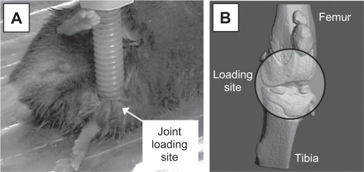
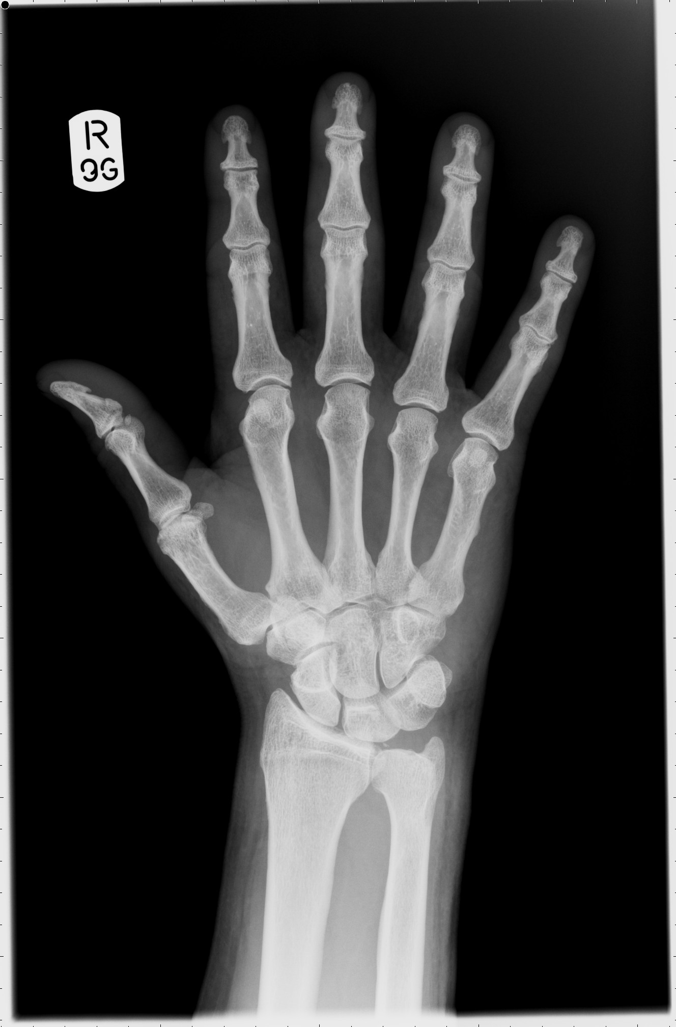
Loading bones against each other also loads the ligaments against the growth plate.  One method of LSJL involves pushing bones against each other at the points of the epiphysis.

Mesenchymal stem cell characteristics of human anterior cruciate ligament outgrowth cells.

“When ruptured, the anterior cruciate ligament (ACL) of the human knee has limited regenerative potential. Cells that migrate out of the human ACL constitute a rich population of progenitor cells and we hypothesize that they display mesenchymal stem cell (MSC) characteristics when compared with adherent cells derived from bone marrow or collagenase digests from ACL. ACL outgrowth cells are adherent, fibroblastic cells with a surface immunophenotype strongly positive for cluster of differentiation (CD)29, CD44, CD49c, CD73, CD90, CD97, CD105, CD146, and CD166, weakly positive for CD106 and CD14, but negative for CD11c, CD31, CD34, CD40, CD45, CD53, CD74, CD133, CD144, and CD163. Staining for STRO-1 was seen by immunohistochemistry but not flow cytometry. Under suitable culture conditions, the ACL outgrowth-derived MSCs differentiated into chondrocytes, osteoblasts, and adipocytes and showed capacity to self-renew in an in vitro assay of ligamentogenesis. MSCs derived from collagenase digests of ACL tissue and human bone marrow were analyzed in parallel and displayed similar, but not identical, properties. In situ staining of the ACL suggests that the MSCs reside both aligned with the collagenous matrix of the ligament and adjacent to small blood vessels.  The cells that emigrate from damaged ACLs are MSCs have the potential to provide the basis for a superior, biological repair of this ligament.”

“mobile population of MSCs within the ACL”

“cells derived from both ACL sources and bone marrow underwent chondrogenic differentiation in the presence, but not absence, of TGF-β1”

“MSCs migrate to sites of injury”

So ligaments near the epiphysis can differentiate into chondrocytes.

Chondrocyte phenotype and ectopic ossification in collagenase-induced tendon degeneration.

Chondrocyte phenotype and ectopic ossification in a collagenase-induced patellar tendon{in the new LSJL model the patella is a targetfor loading} injury model. Collagenase or saline was injected intratendinously in one limb. The patella tendon was harvested for assessment at different times. There was an increase in cellularity, vascularity, and loss of matrix organization with time after collagenase injection. The tendon did not heal histologically until week 32. Ectopic mineralization started from week 8. Tendon calcification was mediated by endochondral ossification, as shown by expression of type X collagen. viva CT imaging and polarization microscopy showed characteristic bony porous structures and collagen fiber arrangement, respectively, in the calcific regions. Marrow-like cells and blood vessels were observed inside calcific deposits. Chondrocyte-like cells as indicated by morphology, expression of type II collagen, and sox 9 were seen around and embedded inside the calcific deposits. Fibroblast-like cells expressed type II collagen and sox 9 at earlier times, suggesting that erroneous differentiation of healing tendon fibroblasts may account for failed healing and ossification in collagenase-induced tendon degeneration.”

“Chondrocyte markers were expressed in the clinical samples of calcific insertional Achilles tendinopathy”

“Chondrocyte-like cells as indicated by cellular morphology and expression of sox 9 and type II collagen were observed around the calcific deposits in collagenase-induced degenerative tendon injury”

“The differentiation of tendon progenitor cells into chondrocytes and bone cells was reported to be modulated by the expression of small leucine-rich repeat proteoglycans such as biglycan and fibromodulin, which control the differentiation process associated with BMP-2 activities ”

Tendons can differentiate into cells that undergo endochondral ossification.

knee tendons

The tendons are relatively close to the epiphysis as well.

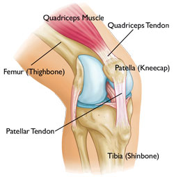
The origin points of the knee collateral ligaments: an MRI study on paediatric patients during growth

“Different femoral origins for both the medial collateral ligament (MCL) and the lateral collateral ligament (LCL) have been reported in the growing skeleton (epiphyseal and metaphyseal). This study assesses the femoral origins of the knee collateral ligaments in skeletally immature individuals.
MRIs of 336 knee joints (median age 15 years (range 2–18 years)) were retrospectively analysed to assess the distances between the femoral origins of the MCL and LCL to the distal femoral growth plate.
Both MCL and LCL ligament origins were invariably located on the epiphysis. Mean MCL origin–growth plate distance was 9.6 mm (SD 2.1 mm; range 2.2–13.6 mm) in boys and 8.6 mm (SD 1.5 mm; range 3.4–12.0 mm) in girls{The MCL ligament is very close to the growth plate}. Mean LCL origin–growth plate distance was 9.3 mm (SD 1.8 mm; range 4.3–13.0 mm) in boys and 8.2 mm (SD 1.5 mm; range 3.4–11.8 mm) in girls{The LCL ligament is very close to the growth plate}. The distance between the growth plate and both collateral ligaments as well as the length of the LCL correlated positively with patients’ age and body size.
During growth, the femoral origins of the MCL and the LCL are constantly located on the distal femoral epiphysis. There is a linear increase in the distances from the ligaments’ origins to the growth plate according to age and body size.”

“The LCL attaches slightly closer to the growth plate than the MCL. The distances between the origins of the ligaments and the distal femoral growth plate increase in a nearly linear pattern until closure of the growth plate.”

Where tendons and ligaments meet bone: attachment sites (‘entheses’) in relation to exercise and/or mechanical load.

“Entheses (insertion sites, osteotendinous junctions, osteoligamentous junctions) are sites of stress concentration at the region where tendons and ligaments attach to bone.”

“Tendons and ligaments can be regarded as machines with multiple moving parts (fibrils, fibres and fascicles) that perform the basic function of force transfer to and from the skeleton. They distribute the loads applied to them dynamically in order to execute movement patterns. Their complex response to loading allows for multi-axis bending, and this adds to the stress concentration in the region where they attach to bone. This attachment site will be referred to in this review as an ‘enthesis’, but it is also known as an ‘insertion site’, or an ‘osteotendinous’ or ‘osteoligamentous’ junction.”

The tissue at the enthesis insertion site is either fibrous or fibrocartilagenous.

“chondral–apophyseal entheses are found at the ends of the long bones and periosteal–diaphyseal attachments occur on the shafts.”

“At fibrous entheses, the tendon or ligament attaches either directly to the bone or indirectly to it via the periosteum. In both cases, dense fibrous connective tissue connects the tendon/ligament to the periosteum and there is no evidence of (fibro)cartilage differentiation”

“Fibrocartilaginous entheses are sites where chondrogenesis has occurred and thus four zones of tissue are commonly present: pure dense fibrous connective tissue, uncalcified fibrocartilage, calcified fibrocartilage and bone”

“The inclusion of a zone of ‘pure dense fibrous connective tissue’ and a zone of ‘bone’ at a fibrocartilaginous enthesis highlight the difficulty of defining with any degree of precision where such an enthesis begins and ends.”<-Thus the ligament that attatches to the epiphysis near the growth plate line may extend partially into the bone itself.

“the proportion of the enthesis subchondral bone plate which consists of calcified fibrocartilage increases with age, because of a thinning of the cortical bone”

“Although tendons and ligaments are often viewed as non-distensible, they do have the ability to stretch and recoil by approximately 6% of their original length without any obvious signs of damage.”

“entheses can act as growth plates for apophyses at tendon and ligament attachment sites.”

“cartilage at the enthesis is initially derived from that of the embryonic bone rudiment.”

“this hyaline cartilage is eroded during endochondral ossification and replaced by enthesis fibrocartilage that develops within the adjacent ligament by fibroblast metaplasia.”

“Bony spurs (enthesophytes) are well documented at numerous entheses as bony outgrowths that extend from the skeleton into the soft tissue of a tendon or ligament at its enthesis”<-our goal is a cartilage ingrowth into the bone from the tendon/ligament.

Magnetic resonance imaging of entheses. Part 1.
enthesis attachment

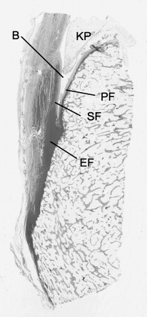
“Achilles tendon, sagittal, histological section stained with Toluidine blue. Enthesis (EF) sesamoid (SF) and periosteal (PF) fibrocartilages are seen. The retrocalcaneal bursa (B) lies between the tendon and the bone and contains the tip of Kager’s retromalleolar fat pad (KP).”

One of the interesting features of fibrocartilaginous entheses is the paucity[scarcity] of compact bone (often referred to as the cortical shell) immediately beneath the attachment site. The subchondral plate (i.e. the associated calcified fibrocartilage and cortical shell) is often very thin and in many fibrocartilaginous entheses, there are local areas where subchondral bone and calcified fibrocartilage are absent.“<-Thus, it would be easier for cells to migrate there.

“Fibrocartilage forms in tendons that are translocated around bony pulleys and regresses in tendons that are re-routed so that they are no longer subject to compression in the same region.”

Experimental Alternative-LSJL Routine Part 2


Here’s the new LSJL routine I’m trying.  It’s not quite the same as LSJL as it’s not based on loading the synovial joints really, it’s more about loading groups of bones that are connected close to another bone by it’s growth plates.  It’s based on the observation that my arms gained in length relatively consistently but not the rest of my body.  The manner in which I clamp my elbow is relatively unchanged versus how I clamped it into LSJL.

For example, in LSJL you clamp the synovial joint on it’s side.

If you clamp the knee on it’s side you are pressing ligaments against the growth plate.

However by pressing the patella against the growth plate line you are not just pressing the ligament near the growth plate line(growth plate remnant), you are pressing the bone against the ligament which would generate a new stimulus.

The philosophy behind LSJL was to induce lateral compression of the bones to induce fluid flow in the bone to generate hydrostatic pressure to induce mesenchymal condensation to form neo-growth plates to grow taller.

Ligaments and other connective tissue have stem cells and coincedentally some run directly into the growth plate region.  Osteoclasts could theoretically eat away at bone and ligament stem cells could migrate and form neo growth plates within this growth plate line.  I’ll be posting some things to explore this theory.

Here’s where and how I’m clamping.  I know the pictures are bad but even if the pictures were better you’d still have to feel and experiment with the optimal clamping position.  But the idea is to clamp one bone against another bone.  So you clamp the fibula against the tibia or the patella against the femur.  Since bones often are connected to each other near the growth plate region this allows for the possibility of stimulating growth plate regeneration.

Now I haven’t tested this routine that long and ligaments and soft tissue are more fragile then bone.  So do this routine at your own risk especially something like the patella clamp.  I also have pretty minimal evidence so far as the only clamp that’s proved to be decently effective is the elbow clamp.  Most I’ve done for these clamps is a count of 120 but I get results for elbow clamping with about that much.  I’m going for several sessions a day though.

I might have to do a video as well for each to explain how I find the right clamping spots.

Patella clamp:

20150218_153807


Fibula clamp:

20150218_153823


Ankle(Tibia and Fibula) Clamp:

20150218_153844

Cuneiforms and Metatarsals Clamp:

20150218_153857

Elbow(humerus clamp):

20150218_153926

Radius and Ulna(Wrist) Clamp:

20150218_153944

Metacarpal bones(clamp):

20150218_154001


Losartan

Losartan may or may not affect height growth as occassionally suppresion of osteoclasts decreases height growth.

Losartan increases bone mass and accelerates chondrocyte hypertrophy in developing skeleton.

“Angiotensin receptor blockers (ARBs) are a group of anti-hypertensive drugs that are widely used to treat pediatric hypertension. ARBs treat diseases such as Marfan syndrome or Alport syndrome has shown positive outcomes in animal and human studies, suggesting a broader therapeutic potential for this class of drugs.  ARBs [effect] adult bone homeostasis; however, its effect on the growing skeleton in children is unknown. We investigated the effect of Losartan, an ARB, in regulating bone mass and cartilage during development in mice. Wild type mice were treated with Losartan from birth until 6weeks of age, after which bones were collected for microCT and histomorphometric analyses. Losartan increased trabecular bone volume vs. tissue volume (a 98% increase) and cortical thickness (a 9% increase) in 6-weeks old wild type mice. The bone changes were attributed to decreased osteoclastogenesis as demonstrated by reduced osteoclast number per bone surface in vivo and suppressed osteoclast differentiation in vitro. At the molecular level, Angiotensin II-induced ERK1/2 phosphorylation in RAW cells was attenuated by Losartan. RANKL-induced ERK1/2 phosphorylation was suppressed by Losartan, suggesting a convergence of RANKL and angiotensin signaling at the level of ERK1/2 regulation. To assess the effect of Losartan on cartilage development, we examined the cartilage phenotype of wild type mice treated with Losartan in utero from conception to 1day of age. Growth plates of these mice showed an elongated hypertrophic chondrocyte zone and increased Col10a1 expression level, with minimal changes in chondrocyte proliferation. Inhibition of the angiotensin pathway by Losartan increases bone mass and accelerates chondrocyte hypertrophy in growth plate during skeletal development.”

The images suggest that Losartan makes bones less porous.

“The growth plate is divided into four discrete zones defined by morphology: 1) the small round chondrocytes at the end of long bone constitute resting zone (RZ); 2) flattened chondrocytes with a typical columnar organization constitute proliferative zone (PZ); 3) the enlarged post-mitotic chondrocytes form the hypertrophic zone (HPZ); 4) the transitioning cells between proliferative and hypertrophic chondrocytes are referred to as pre-hypertrophic chondrocytes. Prominent Agtr1 signal in the hypertrophic chondrocytes (HPZ) ”

“Proliferative chondrocytes express a lower level of Agtr1 than hypertrophic chondrocytes do, while resting chondrocytes exhibit nearly absence of Agtr1expression”

“To evaluate how AngII signaling inhibition affects cartilage development in the long bone, we analyzed the growth plate of mice with or without Losartan treatment in utero from conception until P1. The total length of the growth plate did not show a significant difference between treated and untreated mice”

New Experimental LSJL routine

Here’s the old routine and I am still doing that but I’m adding something else.

I was looking through some old LSJL studies and I noticed an interesting image.


knee loading image

This image is from the study Mechanical Intervention for the Maintenance of Cartilage Bone which is not a bone lengthening study but still pertinent.  You’ll note that the loading does not appear to be strictly on the lateral side of the knee but on the diagonal so the top and bottom of the knee is getting loaded a bit as well.

Here’s a mouse and rat knee:
mouse and rat knee

Here’s the explanatory text: “MRI images of a rat (Wistar) and mouse (C57BL/6) knee joint. (A) 3D spin echo MR image (117 × 114 × 144 μm) of a rat knee ex vivo displaying the anatomical landmarks of the articular joint: a = femur condyle, b = tibia, c = patella, d = patellar ligament, e = meniscus, f = articular cartilage and g = intrapatellar fat pad. (B) Histological image of the knee joint. The MR images provided an excellent visualisation of the rat knee anatomy, with detailed observations on the subchondral bone and in the articular synovial space. (a, b, c, d) Sequential fast spin echo multi-slices images (axial views from proximal to palmar) from the proximal region of the mouse knee (512 × 512 μm). The MR images displayed the bones of the area of knee joint (1 = patella; 2 = femur; 3, 3′ = femur condyles), providing good views of the subpatellar region and the synovial cavity (see arrows). Images acquired in a 9.4-T Varian scanner (Varian, Inc., Oxford, UK) with 100 G/cm gradient coils and a Rapid bird-cage RF coil. ”  You can also see that the patella has a position relatively close to the growth plate.

Mice and rats do have patellas.

Here’s the image of the loading under Lengthening of Mouse Hindlimbs with Joint Loading which is a lengthening study:
lengthening of mousehindlimbs
On the right diagram you can see that the patella is loaded as well.  What is on either side of the growth plate?  Bone and bone.  What is on the other side of the knee joint?  Bone and bone.  Loading bone directly against bone may have a unique response.

I gained in height initially but not so much lately but I have been gaining in wingspan.  On that post I reported a wingspan of 74.5″ but lately I’ve been getting closer to 75″.

Notice something about the elbow joint:
elbow xray

Lots of potential for bone on bone contact.

Now notice the knee joint from a lateral view:

lateral knee xray

Not much potential for bone on bone contact unless you load the fibula or patella.

I found evidence that my right metacarpal grew longer than my left but so much the other parts of the bone.  My left side was longer than my right before so growth may even be more than as noted.  Growth was about 0.8% of Right metacarpal versus left.  An 0.8% increase in height would be about half an inch.

What do you notice about the palm of hand bones(which includes the metacarpal) versus the finger bones?
hand xray

Much more bone on bone action.    I clamped the palm of my hand close to the index finger metacarpal.  I’ll have to post an image of how I did it next post.  But according to my theory the index finger grew from the thumb bone pressing down on it then my middle finger bone should’ve grown from the index finger pushing down.

Also, if you look at my first method for performing LSJL when I did initially gain length:


initial LSJL

I generated force like this and this would press the fibula against the tibia.  The reason gains begin to cease is that my ability to increase force was limited with this whereas clamping can generate much more force.

Interestingly I did not any results on ankle loading producing lengthening which gives me theory credence and Hiroki Yokota stated that he did not study it.

So expect some images of how I’m going to explore this theory and some studies on bone against bone(with tissue in between) especially loading against a growth plate region like patella against the femur.

Study with insight on why Marfan’s causes tall stature

If we know why people with Marfan’s stature grow taller we can incorporate that into our quest to grow taller.

It’s interesting to note that all the things that all the things that grow taller with Marfan’s have more joint mobility especially the fingers and toes.  What doesn’t grow with Marfan’s doesn’t have as much joint mobility such as the torso.  So Marfan’s syndrome creating additional joint spacing via looser ligaments could be key to growth.

The Fibrillin Microfibril Scaffold: A Niche for Growth Factors And Mechanosensation?

“The fibrillins, large extracellular matrix molecules, polymerize to form “microfibrils.”  Fibrillin microfibril scaffold is populated by microfibril-associated proteins and by growth factors, which are likely to be latent{So more microfibrils may catch more growth factors helping people grow taller?}. The scaffold, associated proteins, and bound growth factors, together with cellular receptors that can sense the microfibril matrix, constitute the fibrillin microenvironment. Activation of TGFβ signaling is associated with the Marfan syndrome, which is caused by mutations in fibrillin-1. Mutations in fibrillin-1 cause the Marfan syndrome as well as Weill-Marchesani syndrome (and other acromelic dysplasias) and result in opposite clinical phenotypes: tall or short stature; arachnodactyly or brachydactyly; joint hypermobility or stiff joints; hypomuscularity or hypermuscularity. These different syndromes are associated with different structural abnormalities in the fibrillin microfibril scaffold and perhaps with specific cellular receptors (mechanosensors). How does the microenvironment, framed by the microfibril scaffold and populated by latent growth factors, work?  Fibrillin microfibril niche [is] a contextual environment for growth factor signaling and potentially for mechanosensation.”

“fibrillins and LTBPs may bind and sequester different members of the TGFβ superfamily of growth factors.”

“Disruption of the fibrillin microfibril scaffold was implicated in the Marfan syndrome, and mutations in the gene for fibrillin-1 (FBN1) were shown to cause Marfan syndrome. Mutations in the gene for fibrillin-2 (FBN2) were found in congenital contractural arachnodactyly (now called Distal Arthrogryposis, Type 9 or DA9)”

“fibrillin-1 deficiency [may result] in abnormal activation of TGFβ signaling”

” fibrillins interact with the BMP-7 complex”

“TGFβ propeptides bind covalently to an 8-cysteine domain in LTBPs”

“TGFβ should be regarded as a “cellular switch”, and “its true function is to provide a mechanism for coupling a cell to its environment””

“Bound to the fibrillin microfibril scaffold, the large latent TGFβ complex, as well as BMP prodomain/growth factor complexes, require activation to initiate growth factor signaling. For activation of TGFβ, integrin binding to the propeptide might change the conformation of the propeptide such that the growth factor is allowed to interact with its receptors”

“If binding to fibrillin alters the conformation of the BMP prodomain such that its growth factor is not accessible to BMP receptors, mechanisms of activation will also be required for BMP signaling.”

“With the possibility of multiple interactions between growth factors and the fibrillin microfibril scaffold, tissue-specific microenvironments can be achieved through regulation of growth factor gene expression.”

New Home Page from LSJL Scientists

HIroki Yokota and Ping Zhang are two of the scientists responsible for studying mechanical load driven bone lengthening.

What can their latest website (you must visit this site to understand the rest of the post)tell us about their plans to study this further?

On their research page, both knee loading(LSJL) and spinal loading(perhaps developing a way to lengthening the spinal column) are mentioned but only as a means of strengthening bone.

They mention that they are studying the regeneration of cartilage tissue which means that it’s a possibility that they are studying growth plate regeneration but more likely they are focused on the regeneration of articular cartilage. Their method for cartilage regeneration mentioned does not involve stimulation of mesenchymal stem cells to differentiate into chondrocytes but rather stimulation of aggreccan and type II collagen expression.

Unfortunately it says that Ping Zhang is a collaborating past lab member and Ping Zhang was the driving force behind studying lengthening effects.  It mentions that PIng Zhang is studying mechanical loading, osteoperosis, and fracture healing but nothing on bone lengthening specifically.