Monthly Archives: October 2014

Aneurysmal Bone Cyst to increase height?

http://www.pedorthpath.com/about.html

Aneurysmal Bone Cyst<-may cause enlargement of affected bone.  Sometimes contain cartilage like-matrix.

Aneurysmal bone cyst of the mandible: A case report and review of literature

“Aneurysmal bone cyst (ABC) is rare benign lesions of bone which are infrequent in craniofacial skeleton. ABC’s are characterized by rapid growth pattern with resultant bony expansion and facial asymmetry. We describe a case of ABC in a 25 year old male patient affecting the body of the mandible with expansion and thinning of the buccal and lingual cortical plates. Treatment consisted of surgical curettage of the lesion. A one year follow- up showed restoration of facial symmetry and complete healing of the involved site.”

“ABC is a benign cystic lesion of bone, composed of blood-filled spaces separated by connective tissue septa containing fibroblasts, osteoclast-type giant cells and reactive woven bone”

 Spontaneous healing of aneurysmal bone cysts. A report of three cases.

” We report three cases of spontaneous healing of aneurysmal bone cysts (ABC). In one case histological material was obtained after resection of the already ossified expansile mass discovered as a lytic lesion seven months previously. In the two other patients, spontaneous ossification of a radiologically presumed ABC in the lytic and expansile phase was observed after nine and seven months respectively. The healed lesions have remained stable at 12, 32, and 36 months respectively. These findings suggest that when the diagnosis can be made with confidence, and the lesion is in a location and at a stage that does not entail any risk of fracture or compression, expectant management should be considered. Our three patients were aged 22, 19 and 18 years, older than usual for developing ABC. This is also true for many of the few other reported cases of spontaneous or almost spontaneous healing and suggests that ABC has a greater tendency to stabilise in older patients.”

If you look at the results the bone expansion seems to be permanent.


Cortical Bone erosion

Cortical bone blocks height growth so it’s erosion is beneficial but only if coupled with replacement tissue(ideally cartilage).

Bone erosion in rheumatoid arthritis: mechanisms, diagnosis and treatment

“Bone erosion is a central feature of rheumatoid arthritis and is associated with disease severity and poor functional outcome. Erosion of periarticular cortical bone, the typical feature observed on plain radiographs in patients with rheumatoid arthritis, results from excessive local bone resorption and inadequate bone formation[so bone erosion occurs in everyone the balance is just switched in RA]. The main triggers of articular bone erosion are synovitis, including the production of proinflammatory cytokines and receptor activator of nuclear factor κB ligand (RANKL), as well as antibodies directed against citrullinated proteins. Indeed, both cytokines and autoantibodies stimulate the differentiation of bone-resorbing osteoclasts, thereby stimulating local bone resorption. Although current antirheumatic therapy inhibits both bone erosion and inflammation, repair of existing bone lesions, albeit physiologically feasible, occurs rarely. Lack of repair is due, at least in part, to active suppression of bone formation by proinflammatory cytokines. This Review summarizes the substantial progress that has been made in understanding the pathophysiology of bone erosions and discusses the improvements in the diagnosis, monitoring and treatment of such lesions.”

“erosions can also be observed in forms of arthritis other than RA, such as gout, psoriatic arthritis, spondyloarthritis and even osteoarthritis,”

“bone erosions typically emerge at the site at which the synovium comes into direct contact with bone (known as bare areas), suggesting that anatomical factors render these areas of juxta-articular bone susceptible to erosion”

“Anatomical factors that predispose skeletal sites for erosions include: the presence of mineralized cartilage, a tissue particularly prone to destruction by bone-resorbing cells; the insertion sites of ligaments to the bone surface, which transduce mechanical forces to the bone and could induce microdamage; and inflamed tendon sheaths (termed tenosynovitis), which pass by the bone surface, and enable the spread of inflammation from the tendon to the articular synovium“<-maybe the transmission of mechanical forces via ligaments could be exploited to induce height gain.

“The small bone channels that penetrate cortical bone carry microvessels and bridge the outer synovial membrane and the inner bone marrow space; these channels are also prone to erosive change early in the course of RA. The microvessels located within these channels facilitate homing of osteoclast precursor cells to the bone, which, upon contact with bone and receipt of the appropriate molecular signals, differentiate into osteoclasts. Widening of cortical bone channels, as a result of osteoclast-mediated bone resorption, is a typical early change in animal models of arthritis”

“Osteoclasts, giant multinucleated cells derived from the monocyte lineage, are the only cells capable of resorbing bone in the body.2,3 Osteoclasts are designed to resorb bone by adhering tightly to the bone surface through interactions with both integrins and extracellular matrix proteins, as well as by assembling junctions, which seal the bone surface and the osteoclast and thereby separate bone from the surrounding extracellular space. Proton pumps along the osteoclasts’ ruffled border then create an acidic milieu, enabling solubilization of calcium from bone. Matrix enzymes synthesized by the osteoclast, including cathepsin K, matrix metalloproteinase 9 and tartrate-resistant acid phosphatase type 5 (TRAP), degrade the bone matrix”

“Development of osteoclasts occurs locally in the synovial tissue as a result of expression of the two essential osteoclastogenic mediators, macrophage colony-stimulating factor 1 (M-CSF)and receptor activator of nuclear factor κB ligand (RANKL; also known as TNF ligand superfamily member 11). This process involves the migration of monocyte-lineage cells from the bone marrow into the secondary lymphatic organs and finally into the joints. The differentiation step at which monocytes enter the joint is unclear. Indeed, monocytes could be already committed to a certain monocyte lineage, such as M1 or M2 macrophages, dendritic cells or osteoclast precursor cells when they enter the joint. Some data suggest that TNF, a key proinflammatory cytokine expressed in RA synovial tissue, stimulates the migration of osteoclast precursor cells from the bone marrow into the periphery. In addition, TNF stimulates expression of surface receptors such as osteoclast-associated immunoglobulin-like receptor before the precursor cells enter the joint, and these receptors facilitate differentiation.  Once within the microenvironment of the joint, these cells are exposed to M-CSF and RANKL, and differentiate toward osteoclasts. Final differentiation into bone-resorbing osteoclasts is then achieved following contact with the bone surface.”

Inflammation plays a role in osteoclast formation.

Another potential LSJL design mentioned in a scientist paper

Todd Dodge is a scientist who works at Hiroki Yokota’s lab which developed the beginnings of LSJL.  Most of the paper is not relevant to LSJL except for his data that compares the inside of the control bone and an axial loaded bone.  The difference in the cracks is not as dramatic as the control and LSJL loaded bone.    This indicates that LSJL may alter bone structure more dramatically than typical loading and hopefully in such a way that facilitates neo-growth plate formation.  The key takeaway is the sample LSJL device provided in the picture at the end of this article and in the appendix of Todd Dodge’s paper.

Experimental and Computational Analysis of Dynamic Loading for Bone Formation<-Link to thesis at end of article

This paper is written by Todd Dodge who works closely with Hiroki Yokota who was involved in the initial ideas behind LSJL.

“Curvature in the structure of bone was hypothesized to enhance its damping ability and lead to increased bone formation through bending. In addition, loading at frequencies near the resonant frequencies of bone was predicted to cause increased bone formation, specifically in areas that experienced high principal strains due to localized displacements during resonant vibration“<-Is there a specific frequency for cartilage formation?

“Many types of applied mechanical loading of the skeleton have been proposed as potential treatments for osteoporotic conditions, including whole body vibration, axial loading or bending of long bones, and lateral joint loading. Each mechanical loading modality is thought to strengthen bone by causing dynamic fluctuations in intramedullary fluid pressure in areas that experience enhanced stresses and strains due to the applied load. This dynamic pressure gradient{This dynamic pressure gradient may also lead to stem cells differentiating into chondrocytes to form neo-growth plates} may cause fluid flow through the lacunocanalicular network of pores in the bone, causing a shear stress to be applied to osteocytes that inhabit those pores and channels. This shear stress may excite the osteocytes, causing activation of osteoblast activity and initiation of the bone remodeling process. Mechanical loading may also lead to application of force directly to osteocytes as strain in the bone matrix changes the shape of the lacunae and canaliculae{plastic changes in the bone matrix could lead to permanent lengthening? But it is unlikely given that bone is known for not being capable of interestitial growth}, imposing deformations on the osteocyte and its processes”

Todd Dodge used lateral knee loading on OVX rats.  1N of load was used at 5Hz for 5 min for a group of rats that was tail suspended.  Loading was done once a day for five straight days.  For a group of rats that were OVX 15Hz for 3 min for 10 days(5 days on, then one off)  12 week-old female sprague dawley rats.  So sort of medium along in the growing process with it starting to taper off.

“The slight increase in BMD in hindlimb-suspended mice may be attributed to additional mechanical stress being applied to the lumbar section of the body, possibly increasing bone growth in that area.”

Unfortunately longitudinal growth was not measured.

Here’s the tibia response to axial loading:

tibia load

Compare this to the LSJL holes which are much bigger between control and loaded and are larger.

“Future studies may incorporate not only cortical bone but also trabecular bone and growth plates, as well as surrounding tissues such as muscle, skin, and joints.”<-We may need to wait for further studies to get the results we need for LSJL.

“the e ffects of loading may not only be localized to nearby bones, but may have remote impacts in the spine.”

Here’s the knee loading device that he recommends, note though it is designed for BMD not for lengthening:

Knee Load device

Grow taller with prostacyclin?

Prostacyclin is available by prescription in brands such as Flolan.

Prostacyclin regulates bone growth via the Epac/Rap1 pathway.

“Growth plate cartilage is distinct from articular cartilage with respect to COX-2 mRNA expression: whereas articular chondrocytes express very little COX-2, COX-2 expression is high in growth plate chondrocytes, and is increased by Insulin-like Growth Factor-I (IGF-I). In bovine primary growth plate chondrocytes, ATDC5 cells and human metatarsal explants, inhibition of COX activity with non-steroidal anti-inflammatory drugs (NSAIDs) inhibits chondrocyte proliferation and ERK activation by IGF-I. This inhibition is reversed by PGE2 and PGI2, but not by PGD2 or TXB2. Inhibition of COX activity in young mice by intra-peritoneal injections of NSAIDs causes dwarfism. In growth plate chondrocytes, inhibition of proliferation and ERK activation by NSAIDs is reversed by forskolin, 8-Br-cAMP and a prostacyclin analog, iloprost. The inhibition of proliferation and ERK activation by celecoxib is also reversed by 8CPT-2Me-cAMP, an activator of Epac, implicating the small G protein Rap1 in the pathway activated by iloprost. Prostacyclin is required for proper growth plate development and bone growth.”

“Within the growth plate, chondrocytes resting within the reserve zone are recruited to the proliferative zone, wherein IGF-I stimulates cell division, which occurs along the long axis of the bone.”<-Can this recruitment be mimiced in adults to cause new growth plate formation?

“Terminally differentiated cells are found in the hypertrophic zone, wherein glycogen accumulation leads to dramatic cell hypertrophy.”

“IGF-I changes the ratio of activities of the mitogen activated protein (MAP) kinases, ERK1/2 and
p38; relatively high ERK1/2 activity drives proliferation, whereas relatively high p38 activity favors differentiation. The kinase cascades in which ERK1/2 and p38 participate presumably have indirect transcriptional effects, but little is known about the downstream transcriptional
targets of IGF-I in these cells.”

“chondrocytes treated with IGF-I that the growth factor significantly increases the expression of COX-2 in primary bovine growth plate chondrocytes. COX-1 and COX-2 catalyze the same rate-limiting step in the synthesis of prostaglandins, the conversion of arachidonic acid to prostaglandin H2 (PGH2). PGH2 is further metabolized by specific isomerases to produce a
variety of prostanoids, such as prostaglandins, prostacyclins and thromboxanes. COX-1 is constitutively expressed in almost all tissues. COX-2 is normally undetectable in most tissues, but its expression is rapidly induced by injury and proinflammatory factors, and in some tissues mitogens increase COX-2 expression”

“Growth plate chondrocytes release PGE2, which enhances their proliferation”

“whereas COX-2 expression is low in articular chondrocytes, the growth plate expresses
high levels of COX-2 mRNA, and IGF-I increases COX-2 expression and activity in these cells; the ability of IGF-I to stimulate chondrocyte proliferation andERKactivation is dependent on the presence of PGE2 or PGI2; PGI2 is more potent than PGE2 in supporting proliferation and ERK
activation; and PGI2 signals via cAMP activation of the Epac/Rap1 pathway.”

“ibuprofen and celecoxib blocked IGF-I-stimulated proliferation in both primary chondrocytes and in ATDC5 cells at concentrations near or above the IC50 for COX-2 (eg, 50 M ibuprofen, or 2 M celecoxib for primary chondrocytes).”<-Does ibuprofen stunt growth?

“Both the ibuprofen and celecoxib-treated mice were significantly smaller than the mice that received vehicle only”<-and they specifically used doses comprable to those used in children.

“the effects of the protein kinase A inhibitor H89 in chondrocytes is dose-dependent;
low doses tended to increase IGF-I stimulated proliferation slightly, whereas higher doses (such as 10 M), blocked chondrocyte proliferation”

“COX-2 inhibition negatively affects growth plate development in mice by blocking the ability
of IGF-I to activate ERK1/2 and stimulate chondrocyte proliferation; that PGI2 (and to a lesser extent PGE2) and their analogs are able to reverse this blockade suggests that the ability of NSAIDs to suppress chondrocyte proliferation is due to direct inhibition of COX activity. At
least in vitro, we found that the ERK inhibiting effects of celecoxib on chondrocytes are rapidly reversed by a PGI2 analog, suggesting that the dwarfism in mice treated with NSAIDs is due to direct inhibition of COX activity.”

Creep strength and it’s applications to LSJL

If LSJL can physically deform the bone then perhaps it can deform the bone in such a way as to make it longer.

Creep of trabecular bone from the human proximal tibia

“Creep is the deformation that occurs under a prolonged, sustained load and can lead to permanent damage in bone{So to get creep with LSJL the duration of the loads should be longer}. Creep in bone is a complex phenomenon and varies with type of loading and local mechanical properties. Human trabecular bone samples from proximal tibia were harvested from a 71-year old female cadaver with osteoporosis. The samples were initially subjected to one cycle load up to 1% strain to determine the creep load. Samples were then loaded in compression under a constant stress for 2 h and immediately unloaded. All tests were conducted with the specimens soaked in phosphate buffered saline with proteinase inhibitors at 37 °C. Steady state creep rate and final creep strain were estimated from mechanical testing and compared with published data. The steady state creep rate correlated well with values obtained from bovine tibial and human vertebral trabecular bone, and was higher for lower density samples. Tissue architecture was analyzed by micro-computed tomography (μCT) both before and after creep testing to assess creep deformation and damage accumulated. Quantitative morphometric analysis indicated that creep induced changes in trabecular separation and the structural model index. A main mode of deformation was bending of trabeculae{stretchinig of the trabeculae would be what would make it longer although it’s possible that bending would make trabeculae longer if done in a certain way}.”

” One manifestation of creep damage is the decreased stature of elderly individuals due to the creep damage of vertebrae”

” trabeculae from vertebral areas are mostly rod-like, while in the metaphyses and epiphyses of long bones there is a more balanced mixture of plate-like and rod-like trabeculae. Furthermore, it was shown that these cellular structures degrade differently during the progression of osteoporosis, resulting in differences in the degree of anisotropy of mechanical properties”

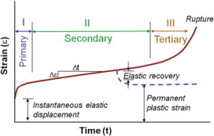
” Creep occurs in three stages: primary, secondary and tertiary. Primary creep is the brief initial period during which the creep rate decreases and is accompanied with some material relaxation. In secondary creep, which is the longest and most studied regime, a steady state creep rate (dε/dt) is observed. Tertiary creep, occurring at the end of the secondary regime, is associated with an accelerated creep rate and more damage accumulation that results in eventual failure if the load is sustained. If the creep test is stopped during the secondary stage and the load removed, there is some elastic and viscoelastic strain recovery.”

creep strain

“under a prolonged load, microcracking of the bone matrix (and not fracture of the whole trabeculae) was the primary mode of deformation during creep. On the other hand, it was shown that even a small damage, barely distinguished on radiographs, to older vertebrae accelerates creep, and leads to a visible contribution to a vertebra deformity”

Here’s a paper that shows possible positive benefits of creep deformation:

In vitro torsion-induced stress distribution changes in porcine intervertebral discs.

“A cadaveric porcine spine motion segment experiment was conducted.

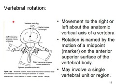
To test the hypothesis that small vertebral rotations cause increased stress in the anulus while decreasing stress in the nucleus through stiffening of the anulus.

Stress profiles of the intervertebral disc reportedly depend on degeneration grade and external loading. Increased stress in the anulus was found during asymmetric loading. In addition, depressurization of the nucleus combined with an instantaneous disc height increase was found when small (<2 degrees ) axial vertebral rotations were applied{So twisting could increasing height?}.

vertebral rotation

Seven lumbar porcine cadaveric motion segments consisting of two vertebrae and the intervening disc with ligaments were loaded in the neutral position with 340 N of compression. Stress profiles were obtained in the neutral position, then after 0.5 degrees and 1 degrees axial rotation of the bottom vertebral body. The distribution of compressive stress in the disc matrix was measured by pulling a miniature pressure transducer through the disc along a straight path in the midfrontal plane. Stress profiles were measured in vertical (0 degrees ) and horizontal (90 degrees ) orientation.

Deformation of the anulus by small axial rotations of the lower vertebra instantaneously decreased the horizontally and vertically measured stress in the nucleus while increasing stress in the anulus. A 1-hour period of creep loading decreased the stresses in the nucleus and the anulus 20% to 30%, depending on the orientation, but the effect of an increasing stress in the anular region after axial rotation persisted.

The compressive Young’s modulus of the composite anulus tissue increases instantaneously when small axial rotations are applied to porcine spine motion segments. This is accompanied by decreased stress in the nucleus pulposus, increased stress in the anulus fibrosus, changes in the stress profile superimposed on and independent of prolonged viscoelastic creep and dehydration, and changes in stress distribution independent of horizontal and vertical orientation.”

“the nucleus acts as a sealed hydraulic system, in which the fluid pressure rises substantially when volume is increased (by fluid injection) and falls when volume is decreased (by surgical excision or endplate fracture). “Stress” peaks in the anulus reportedly are increased by prior loading and degeneration.”

“[The] pressure reduction in the nucleus pulposus under torsion coincided with an increase of disc height ”

Magnitude of loads influences the site of failure of highly curved bones.

“The structure and material properties of bones along with applied boundary conditions determine the region of peak stresses, where fracture is expected to occur. As the site of peak stresses is not influenced by the magnitude of applied load, the fracture site is not expected to change during fatigue loading of whole bone at different loads. However, in a highly curved bone such as the rat ulna, the magnitude of applied loads was found to influence the fracture site. Fatigue loading was conducted under load control on intact rat forearms and on excised ulnae. The distance to the site of failure from the proximal olecranon process of ulnae was determined. In intact forearms, the site of failure demonstrated a linear progression distally, towards sites with lower moment of inertia (or sites exhibiting lower section modulus). Intact rat forearms and excised ulnae loaded to failure at low loads fractured 2-3mm distal to where they failed when applying high loads. This indicates a shift in the site of failure by approximately 10% of whole bone length just by varying the applied load magnitude. The site of failure in excised ulnae was similar when loading at 2Hz or at 4Hz, suggesting that this was frequency independent in this range and indicating that strain rate was not an important contributing factor. Creep loading of excised ulnae also demonstrated similar changes in the site of failure, indicating that magnitude of loads, and not type of loading were important in determining the site of failure.”

“the shape of whole bone can be such that failure occurs at sites other than the smallest cross-section and/or smallest moment of inertia. This is especially true for curved bones such as the ulnae and tibiae of commonly used rodent strains.”

“Loading was conducted by placing the excised ulnae between brass U-cups, with the load being applied in the direction parallel to the length of the ulna (left-to-right). During this loading, the natural curvature of the ulna causes bending induced tensile and compressive stresses on the lateral and medial surfaces, respectively”

“a gradual shift is observed as the site of failure of the ulna moves from 15.2 mm to 17.3 mm as the peak compressive load decreases from 26 N to 14 N”

“In excised ulnae, the site of failure shifted distally as the load was decreased, with failure occurring slightly distal to the mid-shaft at high loads and even more distally at lower loads, irrespective of the frequency of cyclic loading (2 Hz or 4 Hz) or the mode of loading (creep or cyclic).”

“Failure occurred at extremely short time scales at high loads, and at considerably longer time scales when loading at low loads. When loading at high loads, times to failure were not dependent on loading frequency or mode of loading (creep or cyclic). When loading at low loads, the time it took for the bones to fail decreased with increased frequency of cyclic loading  and was the shortest during creep loading. Dring cyclic loading, failure occurs in fewer cycles at greater loads, irrespective of the frequency of loading. At the same loads, loading at a higher frequency (2 Hz vs 4 Hz), there was no difference in the numbers of cycles to failure when loading was conducted at different frequencies (2 Hz or 4 Hz).”

“As expected, loading at high loads causes bones to fail in shorter times (within 30 s) as opposed to loading at low loads (periods of a week or two).”<-Loading for a week or two would be extreme.

Metacarpal growth to prove LSJL

Below I will submit evidence of LSJL.  It is not perfect proof but with the resources available it is not possible to generate perfect proof.

However, I will submit the following facts as evidence that there is a very strong possibility that LSJL works.

The basis on whether this is proof of LSJL depends on the answers to the following questions:

    • What is the probability that every bone shaft on my left side of the body is longer than the right except for the one metacarpal which I performed LSJL?
    • What is the probability that my LSJL loaded finger is longer likely due to lengthening of the metacarpal when everything on my left side is longer(can provide additional evidence of my left side being longer if needed)?

20140804_144910

Is this not reasonable proof that LSJL works?


So the current hypothesis is that LSJL lengthened my right index finger metacarpal.  The right index finger metacarpal is about 1% longer than the left and since my left bones are almost universally longer than my right bones(at least for all the bones that I have identified) that probably means about a 2% growth(with 1% being a correction of the discrepency).  The remaining bones of the index finger may have grown as well but not beyond the initial discrepency.

img006img007

Here are my two hand images.  If I could proof that the fourth and fifth metacarpals were longer on my left side on my right that would provide evidence that the fact the index finger metacarpal is longer on my right side is an outlier and is due to loading from LSJL.  The middle finger is not taking into account because it is close to where I loaded with LSJL so there might be some byproduct lengthening of the middle finger.

hand bones

Here’s a labeling of the bones of the hand.  It would be easy to measure if it was like but the bones are not that organized.

wrist-labeled-xray

Here’s a labeling of the bones on an actual skeleton.  You can see how the hamate mucks up with an accurate measurement.


4th metacarpal5th metacarpal

From these images you can see how hard it is to get an accurate measurement of the 4th and 5th metacarpal.  The shaft of the fourth metacarpal is definitely longer on the left side and eyeballing the shaft of the the 5th metacarpal the left bone looks like a little bit longer.  In terms on the exact beginning and end of the bone it is hard to tell because of the hamate.

Here’s the left and right index finger:


frontal index metacarpals

The shaft of the right bone looks longer in addition to the overall bone being longer.

I submit that the fact that the fourth and fifth metacarpal all have the left shaft as longer than the right like all the other bones I have compared with the exception of the right and left metacarpals of the index finger of LSJL induced lengthening.