Monthly Archives: October 2016

Alexander Teplyashin’s Research Increases Length Of Finger Bone

anna-tsitsyankouMost of the time we don’t get any useful messages or emails. Only very occasionally do we get something that is actually worth reporting. Here is something.

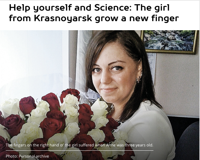
The original source was from – http://www.kp.ru/daily/26526.3/3544077/ – From google translator, converting from russian to english, a young russian female by the name of Anna Tsitsyankou of Krasnoyarsk (25 years old). Anna’s mother, Olga, says that her daughter’s middle finger is shorter than the others, relatively from some injury when she was just 3 years old which damaged the growth plate cartilage area in her right middle finger bone. 

Using the automatic translator, we get “...Teplyashin” preparing to conduct a unique experiment – to grow its own bone to lengthen the phalanx person who, for whatever reasons, this is not the phalanx.” Further “…scientists will grow on the basis of collected stem cells from her piece of the missing finger bone and then transplanted it to her piece”

So when I read this part, what I am assuming is that the Teplyashin’s team, or any tissue engineering research team, is trying to cut off a piece of anna’s bone, so that they can extract the stem cells in that bone, and use it to turn into a new bone piece. However, it is obvious that it would not be possible, without anna losing even more length of her finger bone for that. Here is how his research team has got around that.

The next paragraph – “A team of scientists of our Center of cellular technologies developed and patented in the US” and the European Union a unique method: 28 days mineral harvesting in vitro in a special solution is saturated with stem cells, and then the surgeon implanting it in the rest of the patient’s native finger. Grown phalanx survives, and no one even noticed after three months, that it in this place once was not!

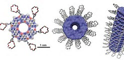
My interpretation of this following paragraph is that in the lab, a scaffold was created, and anna’s stem cells were extracted and placed into the scaffold where they multiplied for almost a month. After that time, the stem cells completely invaded the structure of the scaffold. Once that was done, somehow the surgeon attached that scaffold to her finger. The surgery was a success and after 3 months, she has a new right middle finger, and the casual person walking by would not notice at all that her right middle finger was unusual at all.

What I am not sure of is whether Teplyashin’s team choose to first cut Anna’s phalanx somewhere in the middle, and attach that scaffold where the cut was, or whether they just attached that scaffold to the finger tip her most distal phalanges bone. However, this story does show that Teplyashin’s team is still working with tissue engineering and regenerative medicine.

The way that this news article explains it, it suggests that the surgical idea was to implant the scaffold which is saturated with stem cells somewhere in the middle of Anna’s phalanges bones. If that is the case, since the article says that the experiment has already been tried, tested, and been successful on lab animals for years (I am assuming they are talking about the old sheep leg experiment which we reported on years ago), maybe we can assume that the Finger growth technique that was patented was not just to attach a lab grown organic piece to the end tip of a finger.

The good news is that this surgery will not cost the girl any money, and the surgery’s cost will be paid for by the researchers. However, she will be responsible for paying for the flight to get to their surgery clinic and her hotel. The article mentioned trying to get Anna 100,000 rubles, which converts to about $1,614 USD to fly her to the surgery clinic.

It would be the news source or website “Komsomolskaya Pravdawhich will be reporting on the updates of this story. If you guys want to donate some money for this finger bone lengthening surgery to take place for the russian young woman, then you can contact the following. aveligzhanina@yandex.ru

LSJL Update 10-12-2016

Here’s the link to the last LSJL update.

It seems like my feet have continued to grow from clamping.

20161012_180735

Actually there seems to be rather remarkable growth of the 2nd phalanx/phalange bone.  Right foot is loaded with LSJL and left foot is unloaded.  It’s hard to tell if my big toe has grown anymore.  I’ve changed my clamping strategy rather than trying to specifically clamp the epiphysis of the bone.  I’m clamping a part of the bone close to the epiphysis where I can avoid slippage.  This change in clamping strategy may be the cause of the second phalanx growth too. By focusing more on the force rather than location I notice a rush of blood/fluid flow to regions when I release the clamp this may be a beneficial stimulus towards longitudinal bone growth.

My hands look like they’re growing too but there’s no need for pics all I need to is get an xray of one hand and compare them to the existing ones I have.  I may be growing in height again too but until it’s more definitive it’s better to focus on things that are easier to measure.

The Bone Growth Effects Of Growth Hormone On Rich Piana

There is a rather famous YouTuber and bodybuilder Rich Piana who has very publically admitted to his long term consistent usage of steroids as well as growth hormone (Please note that technically steroids and growth hormones are 2 different types of chemicals but for the average person using common words, we can say that growth hormones are in the “steroids” category.). In a couple of his videos, he has talked about what consistent usage of growth hormone has done to his body over the years.

Here are the changes in his bones which he has claimed.

  • Rich has used growth hormones for 10 years straight
  • His shoe size has gone from a size 12 to a size 15.
  • His head size (skull size??), when measured through the wearing of hats, has gone from 7 3/8ths (fitted cap) to 7 3/4ths or even 7 5/8ths
  • His hands have grown
  • His fingers have grown
  • His wrists have grown – by his claim, his wrist has grown 3/4th of an inch (I am assuming in circumference here)
  • His fist have grown 1/2th of an inch in width

So basically he said that the GH injections have grown every single tissue in the body. Here are a few of his other claims, which he used the word “probably” on…

  • heart has grown (probably)
  • brain has grown (probably)
  • intestines have DEFINITELY grown – The evidence for this claim is that his stomach has grown thicker over the years.

It seems that the growth of his stomach was what caused Piana to stop doing GH.

Overall, there are 3 chemical compounds Piana talked about…

  1. Growth Hormone (somatotrophin)
  2. Insulin
  3. IGF-1 (Insulin like Growth Factor Type 1)

His usage of all 3 types of bodybuilding chemicals has caused his intestines to grow. His claim that these same chemicals causing his heart to grow as well will mostly cause him to die at a younger age, which he has sort of fully accepted.

Some Other Information That Piana Talked About

The Cost of Steroid Cycle

  • To compete on the national level of bodybuilding, you would need to be taking about 18 IU – 20 IU of growth hormone a day. The exact type of growth hormone Piana took was Serostim
  • You can buy this growth hormone from a pharmacy, and a kit of Serostim would cost about $2000/kit.
  • There is 7 bottles in 1 kit. He would use 4 bottles a month.
  • The Serostim is about $400-$600/kit
  • If you got it from a doctor, the serostim is about $8000/mth

The usual first cycle that most bodybuilders do, which is sort of like the gateway steroid to the harder stuff, is TEST and DECA, which is a mild cycle – Dosage: 1 cc a week

For Piana, he claimed that after he went through the first trial of steroids ever when he was younger, he put on 28 lbs in 8 weeks, and 22 of those lbs was muscle.

What We Can Take Away From Rich’s Claims

Most people would have never heard of this guy unless they are really deep into the bodybuilding community. This guy is very loud, and makes grand gestures and likes to show off. That is fine since everyone has their own way of life and they will live it in their own style.

We can however learn quite a bit about the long term effects that growth hormone usage will have. The biggest thing is that his shoe size went up 3 sizes. Did the growth hormone cause the irregular bones in his feet to grow bigger? Or maybe the growth hormone caused the ligaments and tendons that connect the bones to muscles and bone to bone to grow” thus expanding the area and distance between each of the bones in the feet, causing the feet to thus expand?

Let’s note that Rich never talked about him noticing any changes in his height after he started using GH.

When he says that his wrist have gotten wider too, I am not sure whether that is just normal appositional bone growth for males between the ages of 20-30 or the result of the GH.

It is obvious that the expansion of the soft tissues like the  ones found in the intestines that are the most notable, and that side effect of GH was what caused Piana to stop using GH, at least on a consistent regular basis.

MATN3 and Rosette Nanotube for growth plate regeneration

LSJL does upregulate MATN3.  A rosette is a hexameric disc shaped aggregate.
Here’s what a rosette Nanotube looks like.  The novel aspect of this is that it can be injected as a liquid.

GROWTH PLATE CARTILAGE REPAIR VIA NOVEL MATRILIN3/ROSETTE NANOTUBE HYBRID MATRIX

“Approximately 15% to 30% of all childhood fractures are growth plate fractures. Because the growth plate determines the length and shape of a mature bone, this type of fracture may result in severe growth abnormalities in children. Pathologically, the growth abnormality is caused by the formation of a bony bridge in the injured growth plate cartilage. Currently, the clinical treatment of growth plate fractures includes the surgical removal of the bony bridge and insertion of autologous fat or cartilage tissue into the empty space to discourage bony bridge reformation. Such surgical procedures are invasive and result in unsatisfactory outcomes. In addition, this treatment is only useful after the bony bridge has formed. Our long-term goal is to understand how to prevent bony bridge formation and improve growth plate cartilage regeneration at cellular and molecular levels and develop the first preventive and therapeutic approach for growth plate fracture. Specifically, the primary objective of this proposal is to evaluate the therapeutic effects of a nano-matrix assembled from matrilin-3 (MATN3) and rosette nanotube (RNT) in a preclinical growth plate fracture model. Our central hypothesis is that the MATN3/RNT nano-matrix specifically promotes chondrocyte growth and enhances chondrogenesis of mesenchymal stem cells (MSCs), while it also inhibits vascularization and osteogenesis at the fracture site{these two things may increase growth plate generation especially since this is supposed to be used for growth plate fracture}. This is the cellular basis for such nano-matrix to improve growth plate cartilage regeneration and prevent bony bridge formation. We will test our central hypothesis and achieve the objective of the proposal by pursuing two specific aims: 1) to determine the ability of MATN3/RNT to prevent bony bridge formation; and 2) to determine the ability of MATN3/RNT to deliver growth factors for further improvement of chondrogenesis and growth plate cartilage regeneration. To achieve the two aims, our overall research strategy includes: 1) optimization of the ratio and dose of MATN3/RNT and its ability and bioactivity for loading growth factors in vitro; and 2) determination of the therapeutic efficay of the nano-matrix in our established growth plate fracture model in rats in long term. The proposed research is innovative: 1) biologically, it simultaneously promotes cartilage regeneration and inhibits bony bridge formation; 2) therapeutically, MATN3 and RNT can be injected as a liquid in a minimally invasive manner, and form a nano- matrix at the fracture site; 3) structurally, the nano-matrix concentrates bioactive MATN3 locally at the fracture site as well as binds TGF-β1 and IGF-1 to achieve multi-functional delivery. With the results of the two specific aims, we expect to 1) realize a synergistic strategy to specifically promote chondrogenesis while inhibiting osteogenesis and vascularization; and 2) develop an injectable approach for the localized delivery of cartilage growth factors. These outcomes have an important positive impact in developing novel, perhaps the first, preventive and therapeutic approach for growth plate cartilage repair. ”

Here’s more info about nanotubes:

Helical rosette nanotubes: a more effective orthopaedic implant material

“Due to the nanometric properties of some physiological components of bone, nanomaterials have been proposed as the next generation of improved orthopaedic implant materials. Yet current efforts in the design of orthopaedic materials such as titanium (Ti) are not aimed at tailoring their nanoscale features, which is now believed to be one reason why Ti sometimes fails clinically as a bone implant material. Much effort is thus being dedicated to developing improved bioactive nanometric surfaces and nanomaterials for biospecificity. Helical rosette nanotubes (HRN) are a new class of self-assembled organic nanotubes possessing biologically-inspired nanoscale dimensions. Because of their chemical and structural similarity with naturally-occurring nanostructured constituent components in bone such as collagen and hydroxyapatite, we anticipated that an HRN-coated surface may simulate an environment that bone cells are accustomed to interacting with. The objective of the present in vitro study is therefore to determine the efficacy of HRN as a bone prosthetic material. Results of this study clearly show that both HRN-K1 and HRN-Arg coated Ti displayed enhanced cell adhesion when compared to uncoated Ti. Enhanced cell adhesion was observed even at concentrations as low as 0.005 mg ml−1. These results point towards new possibilities in bone tissue engineering as they serve as a starting point for further mechanistic studies as well as future manipulation of the outer chemistries of HRN to improve the results beyond those presented here. One such effort is the incorporation of peptide sequences on the outer surface of HRN and/or growth factors known to enhance bone functions. “

LSJL studies 5: LSJL device design

This paper discusses a lateral bone loading device.  It mentions a capacity of 40N which I think won’t be enough for lengthening purposes as since lengthening post growth plate senesence is an abnormal task it probably requires very abnormal stimuli.  It’s interesting to look at the device though.

The study mainly mentioned the technical design of the device and no analysis of the applications.

A Mechatronic Loading Device to Stimulate Bone Growth via a Human Knee

“This paper presents the design of an innovative device that applies dynamic mechanical load to human knee joints. Dynamic loading is employed by applying cyclic and periodic force on a target area. The repeated force loading was considered to be an effective modality for repair and rehabilitation of long bones that are subject to ailments like fractures, osteoporosis, osteoarthritis, etc. The proposed device design builds on the knowledge gained in previous animal and mechanical studies. It employs a modified slider-crank linkage mechanism actuated by a brushless Direct Current (DC) motor and provides uniform and cyclic force.”

Here’s an example of what slider crank linkage looks like:

slider-crank-linkage

“The functionality of the device was simulated in a software environment and the structural integrity was analyzed using a finite element method for the prototype construction. The device is controlled by a microcontroller that is programmed to provide the desired loading force at a predetermined frequency and for a specific duration. The device was successfully tested in various experiments for its usability and full functionality.  The device works according to the requirements of force magnitude and operational frequency. This device is considered ready to be used for a clinical study to examine whether controlled knee-loading could be an effective regimen for treating the stated bone-related ailments{Hopefully bone length is one of those bone-related ailments unfortunately Ping Zhang’s name is not on this paper and he was always the one more interested in bone length}.”

“When a specific loading force is applied to the epiphyses of the femur and tibia, the trabecular bone tissue, which is characterized by axial stress resistance, resists this force from the opposite direction. This results in deformations in that area. These deformations create a variation of the fluid pressure in the intramedullary cavity. This pressure gradient allows the flow of fluids that carry essential nutrients to the bone cortex initiating osteoblast differentiation and osteogenesis, thus helping in repair and regeneration of the bone tissue. This unique reaction makes this procedure an effective treatment for bone rehabilitation. It helps in reduction of healing time of bone fractures and hastens recovery from bone-related injuries and diseases. The lateral stress application is also less strenuous to the knee bone and reduces the amount of force that needs to be applied to get this result.”

pressure-caused-by-fluid-flow

B is the force we’re looking for.  The pressure generated by fluid flow not just on the bone but on the stem cells to initiate chondrogenic differentiation.  The pressure on the intact bone may also allow the creation of cartilage canals to enable that requirement for a neo growth plate.

It’s also interesting to note that in the proposed knee loading device the load the entire lateral area of the epiphysis this may be a way to reduce slippage.

” it was decided that the proposed device should be robust enough to produce different magnitudes of linear force up to a maximum of 40 N”<-Since lengthening is not being considered in this study forces required for lengthening may be higher.

lsjl-dev-ice

The device doesn’t look wide enough for the knee really.  The dimensions of the device listed are:

Length: 0.3 m
Width: 0.1
Height: 0.2

There are about 39 inches in a meter so about 3.9 inches in width.  I don’t know if that’s enough.

Also the device looks more like this kind of clamp:

Then the other clamps we’ve been using.    Although you’d have to make new pads to actually adjust to knee.  Well actually more like:

But the pipe gets in the way of getting around the knee.  Although I’m not really sure that a pipe clamp is superior to the other clamps.  I’m just pointing out that it’s the clamp that looks most like the design mentioned in the study.

Here’s some more details on the device:

sensors-16-01615-g004

Here’s an actual physical prototype:

more-advanced-protoype

Weightlifting and stunted growth

Light weight lifting during development may enhance growth.  Metatarsals are feet bones.

Influence of loading on bone growth at the growth plates in immature rat metatarsals

“Growth of different bones in children is facilitated by different mechanisms according to the anatomical site and function of the bone. Longitudinal bone formation in long and short bones occurs in the cartilaginous growth plates located at each end of the growing bone through a process known as endochondral ossification. This growth continues until a child becomes full-grown at which point the growth plate calcifies to solid bone. It is still unclear how mechanical and biological factors affect bone growth. For the purpose of this study, immature rat metatarsals have been subjected to varying number of cycles (1, 5, 10 and 50 cycles) in order to better understand the effect that mechanical loading has on bone growth. This has been done using two consecutive trials. The trends in these trials were analyzed and compared. Specimens subjected to 5 cycles exhibited the most prominent effect of loading over the course of 16 days. The results of the trials reveal that immature bones are sensitive to cyclic compressive loading. The results revealed a potential threshold below which the loading resulted in an increased growth. Furthermore, simulations of longitudinal bone growth using a thermal-structural coupled analysis, with the findings from the experiment, has been performed. The model results in a stress free structure that is comparable to the growth of the experiments to a certain extent. The model also allowed incorporation of the bent growth that is observed in the experiments.”

“The piston was displacement controlled at 0.01 mm/s up to a predefined maximum load of 0.05 N. After reaching the maximum load, the bones were immediately unloaded. The loading sequence was carried out with varied amount of cycles”

“Compressive loading (static and dynamic) initially reduced the growth rate and growth plate height significantly compared to nonloaded specimens. However, continuing the experiment over a longer time period the results between the groups started to level out. Additionally, growth resumption was observed after loading removal for both statically and dynamically loaded specimen “<-so loading reduced growth rate but not “final” bone length

“At the end of the trial, specimens subjected to 5 cycles exhibited an average percentage growth of 190.9% while the specimens subjected to 50 cycles had an average percentage growth of 166.6%. The control bones grew 166.3% on average. ”

“. Their results showed that both static and dynamic compressive loading initially reduced the growth rate significantly compared to nonloaded specimens. However, continuing the experiment over a longer time period the results between the groups started to level out “<-So you need to change the stimulus to keep getting benefits.

So according to this study, compressive loading exercise should at least alter growth rate.