Monthly Archives: August 2016

Myxedema

Myxedema increases hydrostatic pressure by resulting in increased deposition of connective tissue elements like hyaluronic acid and GAGS(chondroitin).  Maybe there’s a way to use the pathology of this disease to safely increase the deposition of connective tissue to possibly increase height.

Increases in deposition of this elements can result from scar tissue.  Perhaps the separation in limb lengthening surgery can be thought of as a form of scar.  Increases in Fibroblast levels also could increase accumulation of connective tissue.  It is worth it to note that FGFR3 decreases height.  Maybe an interesting possibility is that FGFR3 reduces circulation Fibroblast levels and decreases hydrostatic pressure and results in a height increase that way. Thyroid hormone is thought to increase these connective tissue elements.

Maybe in pregnancy the elevated thyroid causes the bump in the stomach during pregnancy and possibly causes the increase in shoe size and height.  Being pregnant can increase the size and production of the thyroid.

Actors like Feldman who had graves disease was not tall at 5’7″.

Other famous with Graves:

Missy Elliott-F 5’2″

Rodney Dangerfield-M 5’10”

1st President Bush-M 6’2″

Maggie Smith-F 5’5″

According to this paper Graves’ disease–acceleration of linear growth., Grave’s may cause an acceleration of linear growth but I could not find anymore beyond the title.

Body height and weight of patients with childhood onset and adult onset thyrotoxicosis.<-Thyrotoxisis is another name for hyperthyroidism

“The present study has compared body height and weight of thyrotoxic female patients of childhood onset and adult onset. The body height of 141 out of 143 (99%) adult-onset thyrotoxic patients was within the range of mean +/- 2SD for the age-matched general Japanese female population. On the other hand, in 42 patients with childhood-onset thyrotoxicosis, 6 (14%) had their height being greater than the mean + 2SD of general population, and 34 (81%) were taller than the mean value. In 86 patients with siblings, 42 (49%) were at least 2 cm taller than their sisters, and 26 (30%) were more than 2 cm shorter than their sisters. The body weight of 27 out of 42 (68%) patients younger than 20 years was not decreased but was even greater than the mean value for the age-matched general population. The results indicate that excessive thyroid hormone in vivo enhances body height in humans. The increased body weight in some young patients suggests that enhanced dietary intake due to increased appetite in hyperthyroidism has overcome the energy loss with increased metabolism.”

If you look at figure 1 you get quite a interesting figure that shows that of females with adult onset hyperthyroidism tend to be taller than the mean(this is not a longitudinal study so hyperthyroidism could not be a direct measurement of height increase).  I could not excise the figure out of the study you will have to look at it directly.

46, XY pure gonadal dysgenesis: a case with Graves’ disease and exceptionally tall stature.

“Growth was arrested with height remaining at 187 cm after normalization of the thyroid function by treatment with an antithyroid agent, although follow-up to monitor growth was limited to 3 months. In some cases of gonadal dysgenesis, then, Graves’ disease may contribute to an abnormally tall stature.”

So we see that Grave’s disease has an impact on height but that the affect on height is variable sometimes an increase and sometimes a decrease.  Maybe there’s some other variable like FGFR3 levels that influence the effects on height.

This provides more evidence that hydrostatic pressure influences height but perhaps some other stimuli is needed like CNP as increases Fibroblastic stimuli would result in more FGFR3 stimuli in some cases.  CNP would cancel that out.

Tubacin for height growth

Tubacin is not over the counter but you can buy it but it’s for lab research only so there’s no guarantee that it’ll work and we don’t know if there’s side effects.  “Tubacin is a highly potent and selective, reversible, cell-permeable HDAC6 inhibitor with an IC50 of 4 nM in a cell-free assay, approximately 350-fold selectivity over HDAC1.”

HDAC6 deficiency or inhibition blocks FGFR3 accumulation and improves bone growth in a model of achondroplasia.

“Mutations that cause increased and/or inappropriate activation of FGFR3 are responsible for a collection of short-limbed chondrodysplasias. These mutations can alter receptor trafficking and enhance receptor stability, leading to increased receptor accumulation and activity.  wildtype and mutant activated forms of FGFR3 increase expression of the cytoplasmic deacetylase HDAC6 (Histone Deacetylase 6) and that FGFR3 accumulation is compromised in cells lacking HDAC6 or following treatment of fibroblasts or chondrocytes with small molecule inhibitors of HDAC6. The reduced accumulation of FGFR3 was linked to increased FGFR3 degradation that occurred through a lysosome-dependent mechanism. Using a mouse model of Thanatophoric Dysplasia Type II (TDII) we show that both HDAC6 deletion and treatment with the small molecule HDAC6 inhibitor tubacin reduced FGFR3 accumulation in the growth plate and improved endochondral bone growth. Defective endochondral growth in TDII is associated with reduced proliferation and poor hypertrophic differentiation and the improved bone growth was associated with increased chondrocyte proliferation and expansion of the differentiation compartment within the growth plate.”

“The effects of activating FGFR3 mutations on growth plate chondrocytes are mediated by the activation or modulation of several interconnected downstream signaling programs, including the MAPK, BMP and Hedgehog pathways. These pathways converge on key downstream targets such as STAT, SMAD, Snail and Sox9 transcription factors that play important roles in chondrocyte proliferation, differentiation and survival”

“HDAC6 controls FGFR3 accumulation by regulating its protein stability and that tubacin-dependent destabilization of FGFR3 is more robust in chondrocytes than in MEFs.”

“the absence of HDAC6 resulted in modest increases in tibia and femur OB length, but not in ulna or humerus”

” since HDAC6 regulates FGFR3 accumulation, increased HDAC6 might trigger a positive feedback system whereby HDAC6 and FGFR3 promote and maintain each other’s expression. Elevated FGFR3 in this scenario is predicted to behave analogously to mutant versions of FGFR3 and have a negative effect on both chondrocyte proliferation and differentiation. In addition or alternatively, elevated HDAC6 in achondrodysplasia-related disorders might contribute to disease by deacetylation of proteins such as α−Tubulin and Cortactin which can alter cell mobility and adhesion ”

Requirement of HDAC6 for activation of Notch1 by TGF-β1.

“TGF-β1 is enriched in the tumor microenvironment and acts as a key inducer of epithelial to mesenchymal transition (EMT) {Mesenchymal to Epithelial translation may be a key growth plate stage but knowledge of the reverse may be important to our understanding} in lung cancer. The NOTCH signaling pathway is conserved across species and is an essential pathway for development, cell differentiation, and cancer biology. Dysregulation of Notch signaling is a common feature of non-small cell lung cancer (NSCLC) and is correlated with poor prognosis. Crosstalk exists between the NOTCH and TGF-β signaling pathways in EMT. Herein we report that histone deacetylase 6 (HDAC6) modulates TGF-β1-mediated activation of the Notch pathway. HDAC6, a primarily cytoplasmic deacetylase, mediates TGF-β1-induced EMT in human lung cancer cells. Inhibition of HDAC6 with a small molecule inhibitor, namely tubacin or with siRNA attenuated TGF-β1-induced Notch-1 signaling. We show that TGFβ-1-induced EMT is accompanied by rapid HDAC6-dependent deacetylation of heat shock protein 90 (HSP90). Consistently, inhibition of HSP90 with its small molecule inhibitor 17AAG attenuated expression of TGF-β1-induced Notch-1 target genes, HEY-1 and HES-1. These findings reveal a novel function of HDAC6 in EMT via mediating the TGF-β-Notch signaling cascade, and support HDAC6 as a key regulator of TGFβ-induced EMT in NSCLC.”

“Epithelial-mesenchymal transition (EMT) is a process in which epithelial cells lose their characteristic cell-cell junctions and polarization of cell-surface molecules while acquiring properties typical of mesenchymal cells”

Could an MMP supplement help you grow taller?

Some supplements affect MMP expression.  Zinc suppresses MMP2 and MMP9(Zinc Regulates Lipid Metabolism and MMPs Expression in Lipid Disturbance Rabbits.) and Selenium decreases MMP2 and TIMP1 levels(Selenium’s effects on MMP-2 and TIMP-1 secretion by human trabecular meshwork cells).

Effects of matrix metalloproteinases on the fate of mesenchymal stem cells.

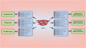
“Mesenchymal stem cells (MSCs) have great potential as a source of cells for cell-based therapy because of their ability for self-renewal and differentiation into functional cells. Moreover, matrix metalloproteinases (MMPs) have a critical role in the differentiation of MSCs into different lineages. MSCs also interact with exogenous MMPs at their surface, and regulate the pericellular localization of MMP activities{so you could supplement with exogenous(external from the body) MMPs to manipulate MSC and hopefully induce chondrogesis}. The fate of MSCs is regulated by specific MMPs associated with a key cell lineage. Recent reports suggest the integration of MMPs in the differentiation, angiogenesis, proliferation, and migration of MSCs{all these four things are likely factors related to chondroinduction}. These interactions are not fully understood and warrant further investigation, especially for their application as therapeutic tools to treat different diseases. Therefore, overexpression of a single MMP or tissue-specific inhibitor of metalloproteinase in MSCs may promote transdifferentiation into a specific cell lineage{this could help you grow taller if it’s transdifferentiation towards a chondrogenic lineage the official term for neo growth plate formation would be something like interosseous chondrofication}, which can be used for the treatment of some diseases. In this review, we critically discuss the identification of various MMPs and the signaling pathways that affect the differentiation, migration, angiogenesis, and proliferation of MSCs.”

“Collagenases (MMP-1, MMP-8, MMP-13, and MMP-18) cleave fibrillar collagen types I, II, and III, and they can cleave other ECM proteins. Gelatinases (MMP-2 and MMP-9) have high activity against gelatin, and degrade other ECM molecules including collagens, laminin, and aggrecan. Stromelysins (MMP-3, MMP-10, and MMP-11) digest a number of noncollagen ECM molecules, and their domain arrangement is similar to that of collagenases. The membrane-type MMPs (MT-MMPs) (MMP-14, MMP-15, MMP-16, MMP-17, MMP-24, and MMP-25) are intracellularly activated transmembrane molecules, and their active forms are expressed on the cell surface. There are other less characterized members including MMP-7, MMP-12, MMP-19, MMP-20, MMP-22, and MMP-23”

“MMPs have critical role in the differentiation of MSCs to adipocytes, osteocytes, and chondrocytes. MSCs interact with exogenous MMPs at their surface and activate proMMP-2 and proMMP-13, regulating the pericellular localization of MMP activities. They have the capability to regulate exogenous MMP-2 and MMP-9 by the expression of TIMP-2 and TIMP-1, protecting the perivascular niche from their high levels”

“silencing of MMP-2 by siRNA impaired chondrogenic differentiation, and increased the protein level of fibronectin and β1 integrin. Treatment with a MMP-2 activator resulted in the activation of chondrogenesis.

“MMP-2 is involved in the chondrogenic differentiation of MSCs via downregulation of focal adhesion kinase (FAK)–β1 integrin interaction, which leads to phosphorylation of FAK”

“the degradation of MMPs enhances the chondrogenic differentiation of MSCs by allowing the morphological changes and increasing the contents of glycosaminoglycans (GAG) and expression of chondrogenic markers. A chondrogenic cell line (ATDC5) was examined for chondrogenic differentiation, and the expression of MMP-2, MMP-9, and MT1-MMP was upregulated during the early stages of chondrogenic differentiation with a downregulation in the expression of reversion-inducing cysteine-rich protein with Kazal motifs (RECK)”

“t the increase in production of MMP-13 drives the MSC differentiation to chondrocytes by degrading the cleavable components of the ECM, and regulating integrin-binding peptides”

“Stimulation of MSCs in chondrogenic media with IL-β1 resulted in increased expression of MMP-2, MMP-3, and MMP-13 while IL-6 downregulated the expression of MMP-3 and MMP-13 compared with control without stimulation”

” Commitment of MSCs to differentiate into a specific lineage, proliferate, or migrate is regulated by many factors in the local tissue microenvironment.”<-So finding a way to stimulate MMP2 on it’s own may not be enough on it’s own to create chondroinduction.

mmp-differentiation-regulation

Hydrostatic skeleton: Key studying point for height growth

One way to develop new height growth techniques would be to study how the hydrostatic skeleton works and since hydrostatic pressure is a key way that hydrostatic skeletons stay structurally sound.  We can use the methods that hydrostatic organisms generate hydrostatic pressure and use those methods on our endoskeletons.

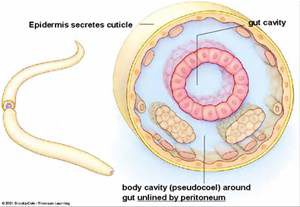
“A hydrostatic skeleton or hydroskeleton is a structure found in many soft-bodied animals consisting of a fluid-filled cavity, the coelom, surrounded by muscles.”

Here’s an image:

hydrostatic skeleton

Another description: ”

A hydrostatic skeleton is one formed by a fluid-filled compartment within the body: the coelom. The organs of the coelom are supported by the aqueous fluid, which also resists external compression. This compartment is under hydrostatic pressure because of the fluid and supports the other organs of the organism. This type of skeletal system is found in soft-bodied animals such as sea anemones, earthworms, Cnidaria, and other invertebrates .”

“earthworms move by waves of muscular contractions (peristalsis) of the skeletal muscle of the body wall hydrostatic skeleton, which alternately shorten and lengthen the body”<-earthworms grow shorter and taller almost at will.

Another description:

“A hydrostatic skeleton is a structure found in many cold-blooded and soft-bodied organisms. It consists of a fluid-filled cavity, which is surrounded by muscles. The cavity is called a coelom and in some animals this cavity is filled with a blood-like substance called haemocoel. The fluid presses against the muscles, which in turn contract against the pressure of the fluid. The fluid is incompressible and thus maintains a constant volume against which the muscles can contract.”

Maybe high hydrostatic pressure in bone serves as a temporary hydroskeleton which allows for temporary loss of osteo-structural components and allow for things such as the growth plate.  Maybe we need to stop think of it is an osteochodral skeleton but a hydroosteochondral skeleton where the hydroskeleton allows the skeleton to function without all the osteocomponents.

ONTOGENETIC SCALING OF HYDROSTATIC SKELETONS: GEOMETRIC, STATIC STRESS AND DYNAMIC STRESS SCALING OF THE EARTHWORM LUMBRICUS
TERRESTRIS

“Hydrostats are constructed of an extensible body wall in tension surrounding a fluid or
deformable tissue under compression. It is the pressurized internal fluid (rather than the rigid levers of vertebrates and arthropods) that enables the maintenance of posture,
antagonism of muscles and transfer of muscle forces to the environment”

“The major source of static load on the body wall of a hydrostatic skeleton is internal pressure (P). Pressure can be generated by the contraction of muscles in the body wall surrounding the incompressible fluid and/or by mechanisms such as ciliary pumps (e.g. in sea anemones), osmotic pressure (e.g. notochords) and gravitational pressure (the gradient of pressure
produced in a static fluid by its own weight”

“hydrostats tend to be highly deformable”

“The main source of loading on the skeleton of most terrestrial organisms with rigid skeletons is body weight. In earthworms, the main source of loading on the skeleton is internal pressure (generated by body wall muscles contracting against a constant volume of internal fluid)”

“The upper limit to the size of hydrostatic skeletons is unclear, but some of the possible limitations to giant earthworms include (1) a decreased respiratory surface area
due to the low surface-to-volume ratio compared with that of smaller earthworms, (2) an increased importance of gravitational pressure as a source of load on the body wall, (3)
an increased frictional resistance to burrowing, and (4) the exponential increase in the cost of tunnel construction with increasing body diameter”<-An upper limit to hydrostatic skeletons would not be good as it would imply limitations on generation hydrostatic pressure but none of these reasons would seem to impede a structural limitation on hydrostatic skeleton size.

Scaling of the hydrostatic skeleton in the earthworm Lumbricus terrestris.

“We used glycol methacrylate histology and microscopy to examine the scaling of mechanically important morphological features of the earthworm Lumbricus terrestris over an ontogenetic size range from 0.03 to 12.89 g. We found that L. terrestris becomes disproportionately longer and thinner as it grows. This increase in the length to diameter ratio with size means that, when normalized for mass, adult worms gain ~117% mechanical advantage during radial expansion, compared with hatchling worms. We also found that the cross-sectional area of the longitudinal musculature scales as body mass to the ~0.6 power across segments, which is significantly lower than the 0.66 power predicted by isometry. The cross-sectional area of the circular musculature, however, scales as body mass to the ~0.8 power across segments, which is significantly higher than predicted by isometry. By modeling the interaction of muscle cross-sectional area and mechanical advantage, we calculate that the force output generated during both circular and longitudinal muscle contraction scales near isometry.”

Negative Pressure Cupping any link to height increase?

Negative pressure is a potential novel technique that can be used to manipulate height growth but there is nothing in the evidence to suggest that it will increase height growth.  The main selling point behind it is that it’s novel and is inversely related to the concept of LSJL(which wants to increase pressure).  The inverse relation may actually eventually prove useful as you could do rapid sunctioning and unsunctioning to build pressure or you could cup every area other than the epiphyseal region of the bone(But the blood just seems to rise to the area of sunction not to other areas).

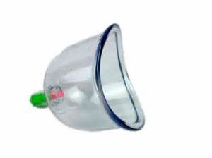
Here’s a cupping device:

cupping

Cupping essentially manipulates fluid and blood flow.  So is there a way to use such a cupping device on the bone or maybe.

The device is not expensive:

Rather than cupping the back, you would cup the synovial joints or epiphyseal region if this had any potential to work.  The bruising redness often seen with cupping is reported to be caused by a discharge of blood from the vessels but can this be used to induce height growth.

First, this would have to manipulate the blood within the bone itself and it’s possible that it does because blood vessels are interconnected but in addition it would have to manipulate blood flow to increase hydrostatic pressure in the epiphyseal region.  You would either have to put the cup on the target area or everywhere but the target area.  If cupping makes the blood flow it has to go somewhere and unfortunately it seems to head into the red spots seen post cupping.

One thing that could be done is to rapidly cup and uncup a region creating a pressure gradient.

I think cups like this with might work better for that:

According to this cupping website, cupping can activate the secretion of synovial fluids but I’m not sure that can cause height growth unless their are nutrients in the synovial fluid that can stimulate endochondral chondrogenesis.

Cupping is basically the inverse LSJL.  LSJL involves lateral tissue compression whereas cupping is negative pressure.

According to Effect of Negative Pressure on Human Bone Marrow Mesenchymal Stem Cells In Vitro:

“The aim of this study was to determine how low-intensity intermittent negative pressure affects the differentiation and proliferation of human mesenchymal stem cells (MSCs), as well of OPG and OPGL mRNA expression in MSCs. MSCs were isolated from adult marrow using the density gradient separation method, passaged for three generations, and divided into the vacuum group, which was administrated at pressure of −50 kPa{So LSJL involves positive pressure and this involves negative pressure}, for 30 min at a frequency of 2/d, and a control group. The differentiation of MSCs was examined through inverted phase contrast microscopy, measurement of alkaline phosphatase activity, alizarin-red staining, and immunohistochemistry for type I collagen, hypoxia-inducible factor-1α (HIF-1a), and vascular endothelial growth factor (VEGF). The MTT assay and flow cytometry were used to measure proliferation and apoptosis. Real-time PCR detected the expression of mRNA from OPG/OPGL. Compared to the control group, there was a decrease in the proliferation of cells in the vacuum group. The number of cells in S phase was reduced by 62.4%, while the rate of apoptosis, the activity of ALP, and calcium release all increased under vacuum conditions. Calcium nodes could be observed through alizarin-red staining, and the expression of collagen type I, VEGF, and HIF-1a were increased significantly. Expression of OPG mRNA was increased and the expression of OPGL mRNA decreased in the vacuum group relative to the control group. In conclusion, low-intensity intermittent negative pressure can inhibit the proliferation of human MSCs, induce differentiation to bone cells, promote the OPG mRNA expression, and reduce OPGL mRNA expression.

This sounds like something unhelpful for height growth unless you make MSCs undergo a chondrogenic versus an osteogenic lineage but you never know.


Finger progress/methodology update

I found an interesting observation while evaluating my finger progress.  I realize that a lot of this may be confusing to understand as I’m not explaining it very well.  But the core idea is that when clamping you tend to clamp the same way every time and that may be supoptimal to growth.  That may foster angular growth which may slow the overall longitudinal bone growth process so if I find a way to balance the clamping I can grow straighter/faster.  I’m also trying this on the bigger bones in the legs and arms too.  It’s just a lot easier to monitor progress in the fingers and I don’t have to worry about clamping force being a limiting factor.  The entirety of my body can generate enough force to clamp the finger.

If you don’t understand don’t worry about it.  I’m just letting you that I had a methodology epiphony and am still working on finding a way to grow taller.

The InfoVisual.info site uses images to explain objects.


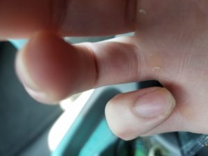
20160804_123712

You can see that the right pinky finger is slightly crooked.

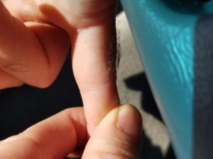
20160804_124101

You can see that the pinky is crooked in solitude as well.  This is likely due to the fact that the way I’ve been clamping has resulted in the bone tilting in a certain direction.  If only one side of the bone has been growing that won’t result in as much longitudinal growth as could be as it will be weighed down by the shorter side.  Naturally you tend to clamp the same way every time so I’m adjusting it to try to see if I can fix the angular growth.

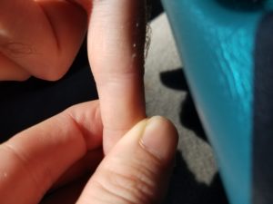
20160804_123815

So for instance rather than clamping like this.  I’m going to be feeling what parts of the bone are underclamped and try to focus on clamping at that angle.  Underclamped regions of bone don’t feel as thick and tend to feel to smoother than the other regions of bone.  For instance like:

20160804_123801

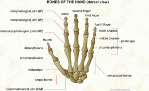
I’m clamping the right hand pinky phalanges on the epiphysis of the distal phalange on the inner side but the middle phalange on the outer side this could result in crooked growth.  For the lower areas it’s inner medial/outer proximal and inner proximal/outer metarcapal.  It wasn’t intentional but I tended to clamp the same style every time.  Actually when I was clamping my thumb I was clamping around the proximal phalanx on both sides and I do not see signs of curved growth but I will try clamping more on the distal phalanx to see if that can inspire more growth

I realize this may be hard to understand but I’d rather focus on doing it rather than trying to draw some diagrams.  It’s possible that I will go back in the future.  So now what I’m going to do is invert it.  Clamp the middle phalange on the inner side and the proximal phalange on the outer side.  It’ll be very difficult to do this on the proximal/metacarpal intersection this way due to the web intersection.

If inbalanced growth is an issue and this new hand clamping strategy corrects it then this will be the LSJL proof I’ve been looking for as angular growth can severely reduce overall growth.  If only one side of the bone is growing you’re going to get angular changes but not much longitudinal bone growth until both are growing so if I manage to correct this it will be a huge improvement.

Whether you be a skeptic or a believer regardless I’m going to try to change my clamping technique and I’ll either have strong evidence of LSJL or not but adjusting clamping this way already feels different as one part of the epiphysis feels a lot more underdeveloped than the other.