Monthly Archives: May 2016

Study that shows that Fluid flow stimulation like induced by LSJL stimulates MSCs

Dynamic Fluid Flow Mechanical Stimulation Modulates Bone Marrow Mesenchymal Stem Cells.

“Osteoblasts are derived from mesenchymal stem cells (MSCs){Also chondrocytes are derived from MSCs}, which initiate and regulate bone formation. New strategies for osteoporosis treatments have aimed to control the fate of MSCs. While functional disuse decreases MSC growth and osteogenic potentials, mechanical signals enhance MSC quantity and bias their differentiation toward osteoblastogenesis. Through a non-invasive dynamic hydraulic stimulation (DHS), we have found that DHS can mitigate trabecular bone loss in a functional disuse model via rat hindlimb suspension (HLS). To further elucidate the downstream cellular effect of DHS and its potential mechanism underlying the bone quality enhancement, a longitudinal in vivo study was designed to evaluate the MSC populations in response to DHS over 3, 7, 14, and 21 days. Five-month old female Sprague Dawley rats were divided into three groups for each time point: age-matched control, HLS, and HLS+DHS. DHS was delivered to the right mid-tibiae with a daily “10 min on-5 min off-10 min on” loading regime for five days/week. At each sacrifice time point, bone marrow MSCs of the stimulated and control tibiae were isolated through specific cell surface markers and quantified by flow cytometry analysis. A strong time-dependent manner of bone marrow MSC induction was observed in response to DHS, which peaked on day 14. After 21 days, this effect of DHS was diminished. the MSC pool is positively influenced by the mechanical signals driven by DHS. Coinciding with our previous findings of mitigation of disuse bone loss, DHS induced changes in MSC number may bias the differentiation of the MSC population towards osteoblastogenesis, thereby promoting bone formation under disuse conditions. the mechanism of MSC induction in response to mechanical loading [is time sensitive], and for the optimal design of osteoporosis treatments. ”

Whether MSCs are driven towards osteo or chondrogenesis could be related to the microenvironment.  So how strong the existing bone is could be a key as to whether the MSCs are driven torwards chondro or osteogenesis.

“DHS was delivered through a custom-made inflatable cuff placed around the right hind limb above the tibia. An oscillatory actuator-driven syringe, a force-controlled syringe and a pressure sensor, were connected to the stimulation cuff. The actuator-driven syringe was controlled by a programmable 100 MHz waveform/signal generator. The hydraulic pressure was monitored by the pressure sensor throughout the entire treatment. With a stimulation frequency of 2 Hz, the pressure stimulation magnitudes were 30 mmHg static pressure + 30 mmHg (peak-to-peak) dynamic pressure. Daily stimulation of the “10 min on-5 min off-10 min on” loading regime was applied to each stimulated animal, while under anesthesia (isoflurane inhalation), for five days/week.”<-This is less than 0.01MPa well below the 0.1 to 10MPa to induce chondrogenesis.

Hydraulic Stimulation increased the number of bone marrow by 50%.

Dynamic fluid flow induced mechanobiological modulation of in situ osteocyte calcium oscillations.

“Distribution of intramedullary pressure (ImP) induced bone fluid flow has been suggested to influence the magnitude of mechanotransductory signals within bone. As osteocytes have been suggested as major mechanosensors in bone network, it is still unclear how osteocytes embedded within a mineralized bone matrix respond to the external mechanical stimuli derived from direct coupling of dynamic fluid flow stimulation (DFFS). While in vitro osteocytes show unique Ca(2+) oscillations to fluid shear, the objective of this study was to use a confocal imaging technique to visualize and quantify Ca(2+) responses in osteocytes in situ under DFFS into the marrow cavity of an intact ex vivo mouse femur. This study provided significant technical development for evaluating mechanotransduction mechanism in bone cell response by separation of mechanical strain and fluid flow factors using ImP stimulation, giving the ability for true real-time imaging and monitoring of bone cell activities during the stimulation. Loading frequency dependent Ca(2+) oscillations in osteocytes indicated the optimized loading at 10Hz, where such induced response was significantly diminished via blockage of the Wnt/β-catenin signaling pathway. The results provided a pilot finding of the potential crosstalk or interaction between Wnt/β-catenin signaling and Ca(2+) influx signaling of in situ osteocytes in response to mechanical signals. Findings from the present study make a valuable tool to investigate how in situ osteocytes respond and transduce mechanical signals, e.g. DFFS, as a central mechanosensor.”<-OUr main concern isn’t Ca2+ oscillations as those are not likely to induce chondrogenesis directly although it’s still possible that it may have an impact or that Ca2+ could be complementary.

“Changes in the pressure or velocity of bone fluid flow (BFF) act as a communication medium that connects external loading signals and internal cellular activities in bone, which ultimately regulate the bone remodeling process”

“DHS[Dynamic Hydraulic Stimulation] generated local ImP that acted independently from simultaneous bone strain.”

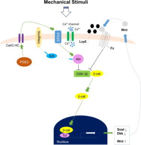
Here’s a diagram of how Calcium signaling may alter a cell.  So Ca2+ is anabolic but it doesn’t really seem to have a direct impact on what lineage a cell traverses(osteo- versus chondro-).

fluid flow stimuli

Interrelation between external oscillatory muscle coupling amplitude and in vivo intramedullary pressure related bone adaptation.

“Interstitial bone fluid flow (IBFF) is suggested as a communication medium that bridges external physical signals and internal cellular activities in the bone, which thus regulates bone remodeling. Intramedullary pressure (ImP) is one main regulatory factor of IBFF and bone adaptation related mechanotransduction. Our group has recently observed that dynamic hydraulic stimulation (DHS), as an external oscillatory muscle coupling, was able to induce local ImP with minimal bone strain as well as to mitigate disuse bone loss. The current study aimed to evaluate the dose dependent relationship between DHS’s amplitude, i.e., 15 and 30mmHg, and in vivo ImP induction, as well as this correlation on bone’s phenotypic change. Simultaneous measurements of ImP and DHS cuff pressures were obtained from rats under DHS with various magnitudes and a constant frequency of 2Hz. ImP inductions and cuff pressures upon DHS loading showed a positively proportional response over the amplitude sweep. The relationship between ImP and DHS cuff pressure was evaluated and shown to be proportional, in which ImP was raised with increases of DHS cuff pressure amplitudes. A 4-week in vivo experiment using a rat hindlimb suspension model demonstrated that the mitigation effect of DHS on disuse trabecular bone was highly dose dependent and related to DHS’s amplitude, where a higher ImP led to a higher bone volume. This study suggested that sufficient physiological DHS is needed to generate ImP. Oscillatory DHS, potentially induces local fluid flow, has shown dose dependence in attenuation of disuse osteopenia. ”

“Direct ImP measurements in rats under DHS over a frequency spectrum indicated that its optimal loading at 2Hz can generate maximum ImP of 14.48±3.10mmHg. On the other hand, dynamic components with large loading amplitudes typically result in pronounced osteogenic responses”

“a micro-cardiovascular pressure transducer was carefully inserted into the tibial marrow cavity through a 1mm drilled hole, and was then tightly sealed within the drilled hole”

” the observed ImP inductions were found to be positively proportional to DHS cuff pressure, namely, which indicates promising potential of DHS loading dose dependency on ImP related bone adaptation.”

“DHS loading at 2Hz between 28.41±10.50 mmHg and 33.75±10.69 mmHg cuff pressures can induce significant ImP increases. Coincided with these observations, our 4-week in vivo study showed that DHS loading at 2Hz with 15 mmHg and 30 mmHg dynamic pressure was able to mitigate disuse trabecular bone loss in a rat functional disuse model.”

“At baseline, normal heart beats generated approximately 1mmHg tibial ImP within the marrow cavity.”

Dynamic hydraulic fluid stimulation regulated intramedullary pressure.

“Physical signals within the bone, i.e. generated from mechanical loading, have the potential to initiate skeletal adaptation. Strong evidence has pointed to bone fluid flow (BFF) as a media between an external load and the bone cells, in which altered velocity and pressure can ultimately initiate the mechanotransduction and the remodeling process within the bone. Load-induced BFF can be altered by factors such as intramedullary pressure (ImP) and/or bone matrix strain, mediating bone adaptation. Previous studies have shown that BFF induced by ImP alone, with minimum bone strain, can initiate bone remodeling. However, identifying induced ImP dynamics and bone strain factor in vivo using a non-invasive method still remains challenging. To apply ImP as a means for alteration of BFF, it was hypothesized that non-invasive dynamic hydraulic stimulation (DHS) can induce local ImP with minimal bone strain to potentially elicit osteogenic adaptive responses via bone-muscle coupling. The goal of this study was to evaluate the immediate effects on local and distant ImP and strain in response to a range of loading frequencies using DHS. Simultaneous femoral and tibial ImP and bone strain values were measured in three 15-month-old female Sprague Dawley rats during DHS loading on the tibia with frequencies of 1Hz to 10Hz. DHS showed noticeable effects on ImP induction in the stimulated tibia in a nonlinear fashion in response to DHS over the range of loading frequencies, where they peaked at 2Hz. DHS at various loading frequencies generated minimal bone strain in the tibiae. Maximal bone strain measured at all loading frequencies was less than 8με. No detectable induction of ImP or bone strain was observed in the femur. This study suggested that oscillatory DHS may regulate the local fluid dynamics with minimal mechanical strain in the bone, which serves critically in bone adaptation. These results clearly implied DHS’s potential as an effective, non-invasive intervention for osteopenia and osteoporosis treatments.”

” bone fluid flow (BFF) with altered velocity or pressure acts as a communication media between an external load and the bone cells, which then regulate bone remodeling. In converse, discontinuous BFF can initiate bone turnover and result in osteopenia”<-Initiating bone turnover could be a good thing if bone structure inhibits anabolism so lowering the frequency may be key.

“DHS was achieved through a costume-made inflatable cuff placed around the right tibia”

” the inflation and deflation of the cuff was driven by a syringe pump with the loading magnitudes and frequencies delivered by a programmable waveform/signal generator”

” 1mmHg tibial ImP and 5mmHg femoral ImP were generated by normal heart beat within the marrow cavities.”

” muscle contraction compresses the blood vessels in muscle, which generates an arteriovenous pressure gradient that further increases the hydraulic pressure in skeletal nutrient vessels and amplifies the capillary filtration in bone”

” using oscillatory muscle stimulation (MS), in which oscillatory MS induced maximal ImP at 20Hz. Oscillatory MS was achieved via two disposable needle-sized electrodes inserted into the quadriceps of the stimulated rats. The electrodes were then connected to the waveform generator with 2V supplies to induce muscle contraction”

“Due to the different physical orientations of how oscillatory MS and DHS contact the loaded tissue, as well as the different material densities and viscosities within hard and soft tissues, maximal DHS-induced ImP may result at relatively lower frequency compared to MS.  Direct hydraulic coupling may influence bone adaptation in a more physiological frequency range, where normal heart rate is 360 times per minute.”

“Changes of MSC number in response to DHS bias their differentiation towards osteoblastogenesis, leading to bone formation even under disuse conditions”

“increased bone fluid pressure and bone fluid flow regulation are strongly correlated, our current results of DHS-driven ImP induction suggest a possible mechanism that the induced ImP may subsequently enhance bone fluid flow.”

Quantifying bone adaptations then and now: Breakthrough from 1920 study

Is some of the recent scientific advancements hiding key features that may give the secrets to growth?  Compare this recent study to a 1920s study that provides an insight about adult longitudinal bone growth related to bone softening.

Development of a protocol to quantify local bone adaptation over space and time: Quantification of reproducibility.

In vivo micro-computed tomography (µCT) scanning of small rodents is a powerful method for longitudinal monitoring of bone adaptation{May be helpful to measure LSJL?}. However, the life-time bone growth in small rodents makes it a challenge to quantify local bone adaptation. Therefore, the aim of this study was to develop a protocol, which can take into account large bone growth, to quantify local bone adaptations over space and time. The entire right tibiae of eight 14-week-old C57BL/6J female mice were consecutively scanned four times in an in vivo µCT scanner using a nominal isotropic image voxel size of 10.4µm. The repeated scan image datasets were aligned to the corresponding baseline (first) scan image dataset using rigid registration. 80% of tibia length (starting from the endpoint of the proximal growth plate) was selected as the volume of interest and partitioned into 40 regions along the tibial long axis (10 divisions) and in the cross-section (4 sectors). The bone mineral content (BMC) was used to quantify bone adaptation and was calculated in each region. All local BMCs have precision errors (PE%CV) of less than 3.5% (24 out of 40 regions have PE%CV of less than 2%), least significant changes (LSCs) of less than 3.8%, and 38 out of 40 regions have intraclass correlation coefficients (ICCs) of over 0.8. The proposed protocol allows to quantify local bone adaptations over an entire tibia in longitudinal studies, with a high reproducibility, an essential requirement to reduce the number of animals to achieve the necessary statistical power. ”

“in rodents like mouse, bone growth spans across the animal׳s life time”

Here’s a 1926 study regarding the regeneration of bone:

THE REGENERATION OF BONE

An early theory of the bone: “the “juice” of bone is released by a fracture,
fills the gap between the ends and there solidifies.”

“The excision of a necrotic bone (the modern sequestrectomy) was more than once reported with complete reconstitution of the affected bone.”

“bone regeneration depends upon the periosteum.  the thickening of this membrane and its later consolidation into bone in experimental fractures and drill holes.”

The four main elements of adult bone: bone cells; periosteal cells; endosteal cells; calcified matrix.

The marrow cavity of a bone(that which contains the juice) is broken up by the trabeculae of spongy bone.

“the bony shell or cortex, we find a definite architecture. On the inner and outer surfaces, the matrix appears in layers lying parallel to the surface. Further in, these layers lose the parallel arrangement and are there grouped in a series of concentric circles. In the center of each of these circles is an opening, the haversian canal, containing a blood vessel and lined with a membrane.  Throughout the substance of the matrix between these canals and in the
parallel layers are numerous smaller openings, the lacunae, in which lie the bone cells.”

“Enveloping the outer aspect of the bone is the periosteum, a membrane consisting
of several layers of cells resembling connective tissue cells, with an enlongated nucleus and abundant fibrillar intercellular substance.”

“solid bone will not grow in length from within by expansion. The calcified matrix cannot
expand. Whatever growth takes place must take place by accretion.”

“Completely calcified bone will not grow interstitially but soft bone will grow interstitially. The experiment just described, if done on a young bone not yet hard, will result in separation of the nails. This is an observation which has a curious reference to adult pathology. Bones in certain pathologic states that render them soft, will occasionally increase in length long after the epiphyseal cartilages are united. This growth is undoubtedly a genuine interstitial addition of new bone, a true expansion of the matrix exactly comparable to such growth in young bones. The commonest conditions in which this phenomenon is encountered are osteomyelitis, Paget’s distlase of bone (osteitis deformans) and Recklinghausen’s disease (neurofibromatosis).
all of which result in a certain degree of decalcification, that is a softening of the bone”

“bones assume a shape adapted to the function they are to perform.”

“Suppose a small drill passed through the cortex of a bone into the marrow cavity. The first effect, of course, is hemorrhage. On withdrawing the drill, the defect fills with blood. Within a few hours this clot begins to be replaced by a plug of fibrin containing polymorphonuclear
leucocytes. Fibrin is the culture medium, so to speak, of young tissue. Then appears a proliferation of the periosteal and endosteal cells, the production of periosteal and endosteal fibroblasts which differ from connective tissue fibroblasts only in their inherent capability of producing bone. These proliferating cells heap up at the edges of the defect, producing a macroscopic thickening of the periosteum and endosteum, and then grow down and up respectivcly into the fibrin that fills the defect, until it is completely replaced. The
periosteal and endosteal reaction extends on the surfaces of the bone over a considerable distance from the point of injury.”

“Before the process of fibrosis is complete, ossification begins at the points where the periosteal and endosteal proliferation began, namely over the surfaces of the bone at the edges of the defect.”

“when a foreign body is brought into contact with bone, one must anticipate that the regenerative processes will be diminished and retarded and that actual bone destruction may occur.”

A discussion attached to the end of the paper suggests that periosteums function merely is a membrane that constrains the bone and that it’s the fluid within the bone that results in bone regeneration.  The periosteum also serves as a mechanism of communication with other tissues.  The commenter stated that the periosteum only appears after the bone has taken place.

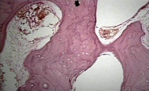
Here’s the bone histology for Paget’s disease:

paget's disease bone histology

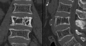
Here’s Paget’s disease in an x-ray of the spine:
pagets disease spine

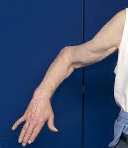
Here’s someone with paget’s disease in the arm:
pagets disease arm

Paget’s disease can make bones longer but not in a real symmetrical way so it’s not really appealing.

Paget’s disease is a disease where bone undergoes break down and rebuilding much faster than normal(so basically faster bone remodeling cycle) and this results in weaker bones as bone does not have time to develop strength.  If there was a way that Paget’s disease was found to increase height we could mimic the effects somewhat by increasing the amount of bone remodeling that occurs.

Osteomylitis seems caused by infection which would be hard to manage.

Neurofibromatosis can cause overgrowth of specific extremeties related to NF1 and NF2 mutations.

Why Basketball Great Michael Jordan Has A Height Complex

For the longest time I had a lot of respect and admiration for the great basketball player Michael Jordan. I watched him in the 90s play and reign supreme. For many guys who grew up in the 90s, MJ represented a hero that you looked up to and aspired to be like. I had thoughts of being a professional basketball player but I just wasn’t good enough. I wasn’t fast, I wasn’t tall, I wasn’t strong. Of course that is my short personal story on basketball.

What makes me think that MJ, who is often considered to be the greatest basketball player in history, has a height complex?

A few key, stories that are mentioned over and over again.

  1. If you ever listened to MJ’s Hall of Fame acceptance speech, you can hear him still talk to his high school basketball coach about cutting him in the varsity team the sophomore year. MJ still had huge grievances against people who supposedly failed him when he was younger from even as back as 30 years ago. What was the real reason Jordan didn’t make it to the Varsity team his Sophomore year? – His height. At the time, MJ was around 5′ 10- 5′ 11. That position in the High School Varsity basketball team if I remember correctly went to MJ’s friend who was 6′ 8″ (named Leroy??)
  2. When MJ first got into the league, he wanted Adidas to sponsor him. He had worn Adidas his whole basketball career before making it to the NBA. He wanted to be in the Adidas crowd but they actually rejected him. Instead, a little company from Oregon named Nike somehow got him to join their team, and the rest is history. Only years later did the truth come out on why Adidas skipped over him. He was not tall enough. At the time, Adidas wanted a “big man” aka center to represent them. They wanted someone like Hakeem Olajuwon. Jordan did not get the company he wanted because he was too short.
  3. MJ was a extremely skilled shooting guard in UNC. He was drafted in the 1984 NBA Draft behind Olajuwon and Sam Bowie. Based on his skill alone, he should have gone at least number 2 (I personally give Hakeem respect on this point). The only reason Portland chose to go with Sam Bowie over MJ was over height again. Bowie being 7′ 1″ was a the main thing that made Portland to choose him. His size was his main attribute and what made Portland think that he was the type of player that could instantly change a franchise and make them title contenders. Portland at the time already had Clyde Drexler. They couldn’t see any point in getting another 6′ 6″ shooting guard on their team. Portland wanted a center, someone to guard the paint in the post. Portland choose height over skill that time. They would do it again in 2009 with Greg Oden with the #1 overall draft pick over the just 2 inch shorter Kevin Durant, and be bitten in the ass again for believing that height was the most important thing.
  4. There is a famous documentary on MJ in the early 90s called “Come Fly With Me”. In that documentary you can hear Jordan talk about the fact that he always wanted to be 7 feet tall when he was little. He would talk about how he would hang onto the pullup bars in the playground in the backyard each day to try to grow taller. His parents saw him doing that and thought he was crazy.
  5. When he was younger, he would be playing basketball against his older brother Larry, who would always beat him in the games. Larry would later only grow to around 5′ 9″. His late father James Jordan would talk about how Larry would beat Michael so often in basketball that Michael would go crazy over that.
  6. MJ’s biological family is not that tall genetically. He has a sister who is only 5′ 5″ and his late father James Jordan has previously been the tallest in the family at just 6′ 0″. It was asked in the documentary how it was possible that Michael grew to be so much taller when the rest of the family was just average height. The joke the family came up with was that the milkman was tall. However, genetics came back to reveal itself when MJ’s sons from his 1st marriage to Juanita turned out to be shorter and closer to the average. One of his sons was just 6′ 2″ and the other was supposed to be 6′ 3″, as listed on their basketball player profiles at the time.
  7. There is a rumor or story told from a former Bulls assistant or someone associated with the association that back in the 1995 year, Chicago was playing against Charlotte in a best of 5 game series. It was said that in game 5, when Muggsy was getting to shoot a free throw with Charlotte down just 1 point, MJ said to Muggsy “Shoot it you F**king midget”. That one sentence was the catalyst that supposedly caused the entire career of Bogue to be destroyed. The problem with this story turns out that there was no Game 5 that year. However, a recent youtube video did reveal that there was a very similar incident in game 4, where Bogue was at the free throw line and missed a very bad shot, and it does appear that MJ said something to him before that shot. Is that Muggsy Bogues story true? It is extremely possible. Let me explain. Back in the pre-season time of 2014, there was an exhibition game between the Los Angeles Lakers and Golden State Warriors in Shanghai China at the Mercedes Benz Arena. Yao Ming showed up. Muggsy Bogue did as well. Kobe did not play that game but Pau Gasol was there. Muggsy Bogue did was well. There was a famous picture taken later after the game where Yao is pictured with Muggsy and that picture is of that night.
  8. A year later around Oct of 2015 at the same Arena there was another game between Charlotte and the Los Angeles Clippers.  There is a famous picture of the current MJ getting his picture taken with Patrick Ewing and Yao. MJ is standing in the center (google those pictures). In the videos that happened at the same time as that picture, MJ started to stand on his tippy-toes to correct for the height differences between him and the former centers. He might have done that as a joke but it does reveal that MJ has a huge height insecurity when he is around taller people.
  9. MJ is a notorious trash talker, and it has been revealed by many people who have played against him that if the opposing guy guarding him is shorter, he would start trash-talking them about them being short, and calling them names associated with having short stature. This supposedly happened to John Starks of the Knicks. Horace Grant who is 6′ 10″, who is taller than MJ revealed in a recent podcast that MJ would be very hard on his teammates in practice. Grant admitted that MJ didn’t harass the taller guys as much, and if MJ did try to get physical with Grant like he did with Steve Kerr, Grant would have probably have gone very physical on Michael. This reveals that MJ has a very long history of harassing shorter basketball players.

It is my opinion that the basketball great Michael Jordan who lead his team to 6 NBA champions back in the 90s has always had a height complex which started from a very young age which he never has fully gotten over.

It is my personal believe and theory that one of the main reasons why he was always so competitive was because at some level MJ suffered from the “Napoleon Complex”. It might not make sense to us normal sized people since MJ is 6′ 6″ but you have to realize that when you are a professional basketball player, your height is a very critical part of your identity. If you look at the starting players on the Bulls team during this reigning era, Michael was often playing the position of Point Guard since the Shooting Guard position was often played by the equally tall 6′ 6″ Ron Harper.

Michael never got over what it felt like, the frustration that you feel, when you are playing against a person who is just taller than you , and thus can just overwhelm you based on his size alone. You sort of see this exact thing in the 1995 playoffs when the Bulls played against the Orlando Magic, who had Shaq at the time. If you look closely that the play on why the Bulls lost to the Magic in the playoffs back in 1995, you see that they just couldn’t get over the size and dominance of Shaq. There was no way that.

It doesn’t seem to matter to MJ that he was objectively tall himself because his job was to be surrounded by guys who are often much taller than him. Based on the fact that the average height of NBA players is supposed to be 6′ 7″, it would be reasonable to say that MJ would in fact be considered “short” or at least below average in height for a NBA player.

Of course, MJ would over time be able to overcome his lack of height with his disproportionately large hands.

New study shows LSJL induces Bone Deformation

Bone Deformation is change in the bone shape or structure.  This deformation can be compression of various cavities, stretching of the bone, twisting, and so son.  This is important as bone deformation is one way to increase hydrostatic pressure by decreasing the cavity size.  Hydrostatic pressure is the pressure exerted by a fluid at rest.   Compressing the bone laterally inhibits the fluids ability to move thus increasing hydrostatic pressure.   If there is more fluid within a smaller space than it follows that hydrostatic pressure increases. Hydrostatic pressure has been consistently shown to induce chondrogenic differentiation.  Chondrogenic tissue is the key for longitudinal bone growth as traditionally chondrogenic tissue is capable of interstitial(growth from within) whereas bone is not.  Only interstitial bone growth has been shown traditionally to induce significant longitudinal bone growth but there are potentially other ways to stimulate longitudinal bone growth.

Knee loading inhibits osteoclast lineage in a mouse model of osteoarthritis.

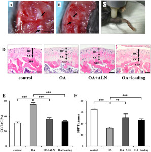
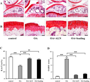
“Osteoarthritis (OA) is a whole joint disorder that involves cartilage degradation and periarticular bone response. Changes of cartilage and subchondral bone are associated with development and activity of osteoclasts from subchondral bone{Since osteoarthritis does affect the subchondral bone that does affect our ability to say that LSJL affects bone deformation in a normal bone but there is no reason why it shouldn’t}. Knee loading promotes bone formation. Knee loading regulates subchondral bone remodeling by suppressing osteoclast development, and prevents degradation of cartilage through crosstalk of bone-cartilage in osteoarthritic mice{This “crosstalk” may stimulate chondral tissue within the bone as well}. Surgery-induced mouse model of OA was used. Two weeks application of daily dynamic knee loading significantly reduced OARSI scores and CC/TAC (calcified cartilage to total articular cartilage), but increased SBP (subchondral bone plate) and B.Ar/T.Ar (trabecular bone area to total tissue area). Bone resorption of osteoclasts from subchondral bone and the differentiation of osteoclasts from bone marrow-derived cells were completely suppressed by knee loading{Knee loading affects the differentiation of bone marrow-derived cells which is the first step in proving that it causes chondrogenic differentiation}. The osteoclast activity was positively correlated with OARSI scores and negatively correlated with SBP and B.Ar/T.Ar. Furthermore, knee loading exerted protective effects by suppressing osteoclastogenesis through Wnt signaling. Overall, osteoclast lineage is the hyper responsiveness of knee loading in osteoarthritic mice. Mechanical stimulation prevents OA-induced cartilage degeneration through crosstalk with subchondral bone. Knee loading might be a new potential therapy for osteoarthritis patients.”

“Daily dynamic knee loading was applied at 1 N, 5 Hz, 5 min/day for 2 weeks”

joint loading on subchondral bone

Compare the OA+ loading to the control bone.  The subchondral bone plate looks much more dynamic.  There are three bone marrow regions rather than two(bone marrow is the blue dots).

You’ll also note that loading+OA increased the ratio of calcified cartilage out of total articular cartilage(but not above statistic significance.  It did not fully restore the thickness of the bone plate.  Alendronate is an anti reobsorption agent.  Given that the ALN and loading group is different we can say that change in subchondral bone shape is likely not related to inhibiting osteoclast activity and is something unique to the loading group.

loading affect on subchondral bone2

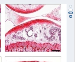
Compare the joint capsule region of control and OA+loading group.  The Joint Capsule is the region that’s not inside the bone.  The cells are a lot more spread out.  There’s a dense redness in the control group which is not present in the OA+Loading group

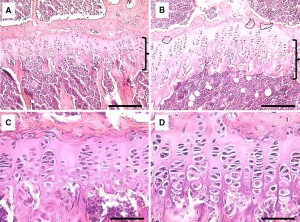
We can see that the loaded group again has distinct characteristics and we can also see the growth plate.  The growth plate of the LSJL group is distinct and there does seem to be signs of cellular migration.  I’ll have to blow it up.

cell migration

Circled is the region of possible cell migration.

LSJL-growth-plates

Similar signs of migration in earlier in LSJL studies(above taken from Lengthening of Mouse Hindlimbs with Joint Loading).

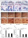
“the expression of Wnt3a was significantly increased by knee loading. However, the protein and mRNA levels of NFATc1, RANKL, TNF-α, and Cathepsin K were significantly suppressed by knee loading”

“Female C57BL/6 mice (~14 weeks of age)”

If you look at this image of bone marrow derived cells extracted from the loaded group and the other groups you can see that the cells are more condensed and condensation is a prerequisite for chondrogenic differentiation,

Loaded cells are more condensed

This is an image of what mesenchymal condensation looks like:

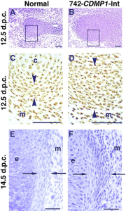
mesenchymal condensation