Monthly Archives: July 2014

Updates For Regenerative Tissue Engineering Using Stem Cell For New Growth Plates – How Much Longer?

Updates For Regenerative Tissue Engineering Using Stem Cell For New Growth Plates – How Much Longer?

So the most recent relevant post I had written had said that we would need to wait 30 or even 50 years before the limb lengthening surgery alternative would be finally be available. That made many people very upset that this idea which I had claimed to be “almost there” would still take that long. Let me try to give you guys a better estimate on what is reasonable to expect to happen within the 21st century in terms of biotech.

The short answer is this. If we were to be extremely optimistic, It would take 10 years for the stem cell technique to be viable for for humans to use, but only for real medical issues. Within 5 years, the physicians performing the technique would realize that there is a large financial incentive in using the stem cell approach on people for cosmetic reasons. So being very optimistic, it would take 15 years before we can finally have the chance to use the new growth plate method. On the opposite end, it would take maybe 30 years for the stem cells to have all the technical details worked out for them to be for use for humans, and still another 10 years before doctors are willing to put the technique for use to lengthen bones. So the worst we can do is wait 40 years. Yes, it sucks that it could take almost half a century, and maybe some of us might not even live that long, but biological science is not like computer science, where you have dedicated people who can pump out a new product in a dorm room with a computer working 24 hours a day.

Here is something else which we need to consider. Exponential Growth. When we remember Ray Kurzweil’s Law of Exponential Growth/Returns which is supposed to signal the point known as the Technological Singularity, it suggest that maybe, just maybe the technology may come even faster, if the exponential growth of technological advancement is true. That might mean that at the earliest, the idea of implanting new lab grown growth plates into bones to lengthen the bone might be available for at least young kids suffering real orthopedic limb angular deformities within as close as 5 years, and have it available for adults for cosmetic application within 10 years.

Let’s give you guys some perspective. If you guys typed into Google right now the term “new growth plate”, you would find as the first link a article/reference to Dr. Robert Ballock’s research at the Cleveland Medical Clinic. That shows that Ballock is very serious on getting the technical details on lab grown growth plates to work out. His desire is to use his knowledge from Harvard to help young children to correct bone grown irregularities. The article you find #1 in Google Search is called “Regrowing Growth Plates: A Fix for Kids’ Injuries? Promising research to mend growth-stifling plate damage” was written back in Feb of this year, 2014. The website by Cleveland Clinic showed that he is focused on the research, and his grant (which we linked to on the previous post) revealed that the project would end around August-Sept this year.

Quote… “In test tube studies, Dr. Ballock’s research team developed a 3-D cell culture model that replicates the key features of what appears to cause growth plate cartilage to develop in the column-like shape that leads to lengthening of the long bones in children.”

At the end, Ballock stated in typical research fashion that his research would be years away from being used for real application. This is disheartening to many teenagers and young people who want to be taller NOW and want something as soon as possible, but it takes time. Even when you want to use the stem cell approach for cosmetic use, other entities can resist you and your research.

The recent reveal that Alexander Teplyashin’s team in Mocow, Russia in getting bones to lengthen by implanting culture grown “growth plates” and trying them out on ram/sheep shows that in theory and concept, the stuff would work. However, Teplyashin is still a researcher (actually has a Ph. D in plastic surgery) and he must follow the guidelines and protocols that the academic and corporate rules set for him.

Here are some problems that have caused the whole stem cell research to be resisted. When George Bush was president (the 2nd one) he imposed laws which made it not possible for USA based researchers to use embryo derived stem cells. Because of Bush, the research into stem cells was halted for his entire 2 terms, which was basically a full decade. While the USA was held back, other countries in the world like Japan, South Korea, Germany, and Russia rushed forward and was willing to take the chance the Americans were not. It wasn’t until 2009, when Obama came to office did the law against stem cell research (at least the embryo derived ones) get lifted. (source: Obama Lifts Bush’s Strict Limits on Stem Cell Research).

The 2nd problem is that Teplyashin’s research has also been halted by the government branched in Russia, one source we found stated. His team is written up grants, petitions, and proposals to move forward in their research, but the government seems to be blocking his team from further progress.

All of this may sound like really bad news, but there is a LOT of good news which I have found recently which shows that the endeavor is definitely being pushed forward.

The Good News

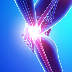
Good News #1 – On the problem on cartilage material strength and whether it can handle the upper weight loading – It seems that this is not a problem. I do read a lot of reddit threads and on one reddit thread, where people were talking about the possibility of getting lab grown cartilage transplanted into their knees for pain management and osteoarthritis treatment ( http://www.reddit.com/r/science/comments/24gghv/for_the_very_first_time_engineers_have_grown/) it was revealed that cartilage that is created in the lab synthetically can withstand the weight load, with the high enough equilibrium compressive strength/modulus. Refer to the study Comparison of the equilibrium response of articular cartilage in unconfined compression, confined compression and indentation for more information.

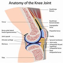
Good News #2: The problem that the people who want to fix their knee articular cartilage degeneration have cleared up is that cartilage implants like autologous chondrocyte implantation has not worked too well to the defects on the surface of articular cartilage. The problem is that the neo-cartilage tissue doesn’t seem to be able to fused properly with the older, natural cartilage. The cartilage then turns into the fibrocartilage tissue type. Of course, these guys on the Reddit Science Thread wants to add cartilage to an area of the body where there is already cartilage. As we know, cartilage is an anareobic environment aka avascular, where the cells inside the ECM of the cartilage can’t get nutrients through blood vessels. Of course then it would make sense that plopping new cartilage on top would not work out too well, since traditionally you need vascularization to get the transplants to fuse with the rest of the body. Our desire is instead to add cartilage to a place where there is no more cartilage (fully fused) and only bone. Bones are very nicely vascularized. In fact, we reported news recently that the vascularization issue was figured out by Teplyashin’s team in a paper that they published (How Svetlana Zagirovna Sharifullina Developed A Vascularized Fully Functioning Growth Plate)

(Side Note: Here are some places which I regularly read up on and make sure I keep up with the most up to date information on reconstructive tissue engineering, stem cells, and such. http://www.reddit.com/r/science, http://www.reddit.com/r/stemcells, http://www.reddit.com/r/tissueengineering, http://www.reddit.com/r/Futurology/, http://www.reddit.com/r/orthopaedics, http://www.reddit.com/r/cancer . In addition, I also read up on the works for the dozens of researchers cited http://www.beckersspine.com/spine/item/14329-10-spine-surgeons-researching-disc-regeneration)

Conclusion:

So when it comes to the technical problems of getting the synthetic lab grown growth plates to meld with the surrounding bones, which is usually just a vascularization problem as Hakkers stated in our interview years ago, that seems to have been resolved.

And when you look at how strong the lab grown cartilage really are, looking at the values of the equilibirum compressive modulus, you find that the major issues that I have come up with seems to be almost all gone.

The few problem that I can think of is that the scaffold derived epiphyseal hyaline cartilage is not producing enough ECM organic compounds, most specifically Collagen Type II, the GAGs, and proteoglycan. However, I would suspect that the technical issue would be a rather easy one to figure out. The other thing is how would the surgeon make the right type of cut into the epiphysis-metaphyseal connected region to implant the cartilage. If you look at how real cartilage is layered in the human body (I saw off the end of a dead young pig’s leg), they are not flat, but curved and bumpy, which is probably due to the different rates of chondrocyte columnar activity.

Then when you look at how easily it is for people with a 3-D printer to print out layers of hydrogel and scaffolds these days, with maybe dozens of people doing similar research, things seem to be looking very good.

I don’t want to make it sound too positive, since there will always be huge issues. The article I found written by someone at Regenexx over the fact that Mesoblast spent hundreds of millions but still couldn’t get their human subjects to have similar results as their lab rats show that sometimes the best results on lab animals can not be translated to humans. The article back in May of 2013 MAJOR STUDY UPDATE: STEM CELLS EASE BACK PAIN revealed that while the subjects did indeed decrease back pain, the disc height did not increase like what happened in the lab with the rats.

Harald Oberlander who I have been in contact with referenced Mesoblast’s research to the people on the Make Me Taller forums but I don’t think even he is aware of the most up to date releases that is coming out of the company. Progress is not easy.


Theories On The Causes And Treatments For Cold Knees and Cold Feet

Theories On The Causes And Treatments For Cold Knees and Cold Feet

Cold KneesNote: This post does not deal with the endeavor of height increase, but is related. I have stated multiple times before that I planned to expand the scope of the research of this website to include bone and cartilage related medical conditions. Height Increase research is for cosmetic reasons, but real medical problems like osteoporosis and osteo-arthritis should always be dealt with first. 

Recently my Asian girlfriend started to notice that her feet and knees was getting unusually cold even though the temperature is quite warm in this Summer weather. She has had a history of dealing with cold knees and cold feet, as well as cold hands.

However, this recent episode has made me start to suggest that there might be some deeper connections and physiological causes to her condition which even most medical professionals have never been able to decipher. Having lived in Asia and seen the bone physiology of the East Asian ethnic groups, and done meta-statistical analysis to compare various racial groups, I feel that I might be able to give a completely new and rather original spin on this rather common and annoying little symptom.

Here is what I do know.

  • East Asian females on average, in general seems to have a lower than average bone mineral density (BMD) than other races.
  • It has been contributed to the fact that while still an embryo, the developing child did not get the high enough level of calcium needed for the prenatal baby to grow. (refer to Bone Health in Chinese American Women). it seems that based on the diet of East Asian cultures, the females of those cultures just wasn’t getting enough calcium (maybe high level of Lactose Intolerance??) , which translated to their developing baby, which grew up with low levels of bone density as well.
  • Not only that, since pregnancy means that a high level of calcium will be transferred from the mother’s body to the babies, the mother will have an increase chance of osteoporosis.
  • Anecdotally, my mother who has multiple Asian female friends have recently told me that they were getting knee replacements as early as their 50s. Knee replacements is one brutal surgery, where a prosthetic is placed where there used to be cartilage and bone before.

So what does this all have to do with cold knees and cold feet?

Based on my research of the past 2 years, cold knees and cold feet, which seems to predominantly effect East Asian females and Caucasian females, is the result of the individual who has a low level of bone mineral density, which has a ripple effect on the other types of tissue connected to the osteo tissue (aka bones).

The cold knees is a sign that they have weaker than average bones and joints. However, what do we mean when we use the word “weaker”?

I know from personal experiences that my girlfriend has never been able to donate blood because the blood donating centers say that she has a very low level of Iron in her blood, which has led to her having anemia. The result is that she has had to go see her personal gynecologist to get ferrous fulmarate intravenously dripped inside her on a regular basis.

In addition, she has a horrible habit of drinking very low levels of water consumption. Her incentive in changing her water consumption level has resulted in the cold knee symptoms disappearing for a long duration, until recently when she moved and changed her eating habits.

It was also found that she has a slight thyroid issue when I took her for a full medical exam more than a year ago. In addition, her menstrual cycle is much larger than average, resulting in more blood loss, which might also contribute to the low level of iron in her blood.

So here is what I am willing to outline for the medical condition of Cold Knees and Cold Feet…

Symptoms…

  • Cold Knees
  • Cold Feet – Sometimes Cold Hands is another symptom

Causes…

Note: All the following causes are theories and guesses based on my personal experience and research.

  • Lack of Iron – the person might be anemic
  • Hypothyroidism – the symptoms of hypothyroidism include 1) increased sensitivity to cold temperature, heavier or irregular menstrual periods, joint pain, and depression. (source)
  • If not hypothyroidism, then some type of thyroid issue (don’t quote me on that one)
  • Habitual Low Consumption of Water 
  • Low Bone Mineral Density – This suggest that the person has a much higher chance of getting osteoporosis later in life.
  • Decreased Circulation 

Update 8/13/2014: It seems there there might be 2 other medical conditions which might be linked to cold knees. They are 1) celiac disease/gluten sensitivity and 2) unusual period types, specifically heavy thick bleeding periods, which I suspect is causing the onset of the cold feet and knees.

Treatments…

Note: Everything I will recommend will be based on personal, amateur level research.

  • First, go to one’s GP (Primary/Family Doctor) to get a full blood work done to see if they are low on any vitamins or minerals. – Check for hypothyroidism and anemia.
  • Start Taking a MultiVitamin – I Suggest a Centrum Silver For Women at the local GNC or Walgreens
  • Take Vitamin D3
  • Take Iron Supplement – this is based on my experiences with a gf with anemic symptoms.
  • Take Vitamin K2 – This is critical. I can not stress how important MK-2 and MK-7 are.
  • Get the recommended daily level of Magnesium – (source: Magnesium Is Crucial for Bones). Magnesium is critical for Vitamin D to work properly.
  • Drink at least 2 liters of water each day – stay hydrated. This will improve on the circulation problem and help with the thyroid issue a little
  • If the person recently went through pregnancy ie a woman, add Calcium to one’s diet to replace for the loss of calcium which got transferred to the baby.
  • Get deep tissue massages ie Swedish – for increased circulation
  • Yoga – for increased circulation

Low Term Considerations.

Here is what my research has told me after many older females I’ve known in my life who had to go through full knee surgery has suggested to me.

If you are in your 20s or 30s and experience cold knees or cold feet (and hands) it is a sign that the bones (and thus also the cartilage) in your body is getting weak.

The way that humans anatomically work, being bipedal, and walk upright, means that structurally speaking, the knees are the weakest area on the human body. Knees and the lower back are the two most common locations to result in pain and problems with people, whether they have an active life or not. (Have anyone else noticed just how many chiropractors practices and offices have been popping up in American towns, even though the theory behind chiropractor work is based on Subluxation, which has no scientific basis? Based on my guesses, chiropractors is nothing more than modern bone setting (which was called Manipulation Surgery at the turn of the 20th century) combined with deep tissue massage physical therapy. The theory of chiropractors is just pseudoscience. From one source (available here) it is said that  “Back pain is the leading cause of disability in Americans under 45 years old. More than 26 million Americans between the ages of 20-64 experience frequent back pain”.  )

Cold knees and cold feet is a sign that the person is at a much higher level of risk for osteoporosis (aka low bone mineral density) and osteo-arthritis (aka degradation of articular cartilage structure) later in life, as quickly as even 10-15 years into the future. 

They will be the ones getting the total knee replacements and they don’t even realize yet in the future. No regular/family doctor with their limited experience and knowledge on the physiology of the bones and cartilage would be able to explain what is going on.

Physiologically speaking, it is caused by three main things…

  1. Decreased circulation in that area
  2. A specific mineral is being transported out of the region (currently, I am not sure which one). (Note: From a physics point of view, it would not make any sense how it is possible that within the human body, a certain local area is cooler than the surrounding area on a Summer Day (I measured the temperature on the skin using a Thermocouple) unless there is a biological process acting similar to an air conditioner going on i.e. a heat transfer and/or a type of chemical endothermic reaction.)
  3. Some form of hormonal imbalance is triggered at some level in the pathway upstream.

This post is for my fiance/future wife, and all of the rest of the people in the world who experience this condition and don’t know what it means. I’ve consulted internal medicine and orthopedic specialists and they have never been able to provide for me a decent and solid sounding answer to the causes and potential treatments to this condition, which I believe is a symptom and sign that something much worst is going to happen later in life.

A Suggested Idea On How We Can Theoretically “Reopen” The Closed Growth Plates

A Suggested Idea On How That We Can Theoretically “Reopen” The Closed Growth Plates

I have said multiple times that it would be next to impossible within this century to create any type of technology to ‘reopen’ fully closed growth plates, at least completely non-invasively. That has been my opinion for a long time since I really started to understand how human bones really work and are organized. Unless there is some type of radical biomedical technology I am not aware of that is coming down the pipeline in the next few decades, I would not be looking for a completely non-invasive way to get the growth plates back.

However, I did recently read an article which seems to suggest at a very theoretical level that it might be possible to kind of “reopen” the closed growth plates, but it would still not be completely non-invasive. Refer to the paper “Roles of neutrophil-mediated inflammatory response in the bony repair of injured growth plate cartilage in young rats“. The PDF for the article is free for download.

I was made aware of the paper when I was trying to find relevant research that Cory Xian had done, since Tyler said that Xian was one of those people we should be following. This paper I did take the time to really look over extensively, and it just slightly hints at the idea that we might be able to change the progenitor cell lineage to something which it is not naturally supposed to go into.

Here is what you need to understand about the microbiological processes that seems to occur after the growth plates in a rat model were intentionally fractured and then analyzed.

Process #1: Inflammatory – This would be the initial injury induced inflammatory response, which is the automatic process that is activated when almost every part of the body is injured. What happens is that inflammatory cells, which comes in the types known as neutrophils, monocytes, lymphocytes, starts being rushed to the local area of injury. In terms of time, at least on the rat models, the first process lasts around 3 days, the maximum effect being around 1 day.

  • Process #2: Fibrogenic
  • Process #3: Osteogenic
  • Process #4: Bone Bridge Maturation

More Details On the Initial Inflammatory Response – The researchers have identified at least 5 different types of chemicals which will be stimulated to go towards the injury area. They include a chemokine, pro-inflammatory cytokines, and fibrogenic growth factors.

  • 1 type of neutrophil Chemotactic Chemokine – CINC-1
  • 2 types of Pro-inflammatory Cytokine – TNF-Alpha and IL-1Beta
  • 2 types of Fibrogenic Growth Factors – TGF-Beta1 and PDGF-B

What the researchers did was add a type of chemical which will decrease the number of neutrophil cell count. Quote: “Using an antirat neutrophil antiserum, a significant 60% reduction in neutro- phil count was achieved at the growth plate injury site”

(Side Note: The most interesting part of the neutrophil to pro-inflammatory cytokine stimulation is that while the antis-serum reduced the neutrophil count, it did not decrease the expression and numbers of the TNF-Alpha or the IL-1Beta (or the Fibrogenic Growth Factors like TGF-Beta1 and PDGF-B) . The reasoning is because besides the neutrophil expression increasing in the bone fracture site, there is two other types of cells which are rushed there, the monocytes and the lymphocytes, and the monocytes, which are also known as macrophage, are the other type of cells which really increase the expression of the Interleukin (IL-1Beta) and the tumor necrosis factor (TNF-Alpha). The results is that the number of MSCS which rushed to the local fracture sight was not not decreased. That suggested that the number of neutrophils in the growth plate injury sight had no real link to the amount of MSCs which will go to that growth plate injury area.)

The result of adding the chemical was that in the injury area, there was more bone tissue than cartilage tissue by ratio that had developed. This suggests that neutrophil cell types has the effect of at least shifting the osteogenic/chondrogenic ratio the other way. The researchers tested the osteogenic and chondrogenic gene expressors the SOX-9 and the COl-Type IIa and saw that the gene expressions were reduced. In reverse, the expression of bone cell differentiation transcription factor cbf-alpha1 and bone matrix protein osteocalcin, which create osteogenic tissue was increased.

If there is an injury aka fracture in the bones, the neutrophils would be able to help the fibrogenic growth factors to develop more cartilage progenitor type tissue and cells.

I quote the last sentence that is in the paper…

“These results suggest that an injury- induced, neutrophil-mediated inflammatory response may be involved in regulating downstream chondrogenic and osteogenic events, leading to bony tissue formation at the growth plate injury site; it appears to play a role in suppressing mesenchymal cell osteoblastic differentiation and increasing chondrogenic differentiation in the repair of injured growth plate cartilage”

It seems that one might be able to block the formation of bone bridges and keep the MSCs in the chondrogenic lineage if we could around the Fibrogenic or Inflammatory stage find some type of chemical to block the injury response processes from ever reaching the osteogenic stages.

It would be the pro-inflammatory cytokines the TNF-Alpha and IL-1Beta that we need to suppress while at the same type increase the number of neutrophils. The researchers did notices from their previous studies that if they did a local induction of chemokine IL-8 (or CINC-1 in rats) the neutrophil numbers would increase, but from past research, interleukin types are not good for cartilage tissue generation. The result of using the suggested IL-8 seems to be an increase in the macrophages in releasing the TNF-Alpha and IL-1Beta. So another chemical besides the IL-8 for neutrophil number increase would be needed. The researchers did say that The TNF-Alpha and the IL-1Beta are what will stimulate the fibrogenic growth factors to be stimulated so we might need to find a third compound to stimulate the fibrogenic growth factors while at the same time we decrease the levels of cytokine inflammation.

What ever chemical has that duo effect, when injected into the fracture area of a bone, might mean that cartilage type tissue, even if it is fibrocartilage in nature, would develop. Sure, we are talking about the growth plate response mechanism in rat models, but there is still some type of knowledge we can transfer for human application. If we do that, I believe that we might be able to create some type of pseudoepiphyseal cartilage layer, which can be slowly changed over time to be hyaline in nature, and then expanded for lengthening of the bones.

Growth Plate Regeneration Update – Robert Ballock and Eben Alsberg Working Together – Getting Very Close!

Growth Plate Regeneration Update – Robert Ballock and Eben Alsberg Working Together – Getting Very Close!

Almost ThereI am sure that the regular readers of the website has already seen this grant that I have talked about at least half a dozen times already and linked to but this is the big news I wanted to say.

Tyler have said that there are two people who would be very helpful in our quest and they were Cory Xian and Robert Ballock. I fully agree, and I have wrote multiple article on Ballock. This guy was given an award more than a decade ago for being able to fully grow a functional growth plate.

(Source: http://projectreporter.nih.gov/project_info_details.cfm?aid=8458506&icde=21045832)

I also said that people like Dr. Warren Grayson of JHU (with his 3-D Bone grafts) and Jean Welter of Case Western University (with his knowledge on differentiating MSCs into the chondrogenic lineage) would also be critical in pushing the effort forward. The recent news that maybe Alexander Teplyashin’s team based in Moscow might have already put their own created growth plates into human legs to lengthen them showed that some people have already taking it out of just the lab and into human clinical trials.

However, from certain russian sources I found on the russian search engines, it seems that Teplyashin’s team is being legally prevented from moving forward on their goals to get that stem cell tissue engineering technique to be fully researched. The government seems to have implemented certain legislative or legal rules to keep the stem cell research from being implemented to the full capacity.

I am happy to inform the readers that the grant that I had referenced before on Ballock had a key critical bit of information which I did not see in the previous times in looking at them.

It seems that Dr. Robert Tracy Ballock’s partner in the grant is a Dr. Eben Alsberg.

This Alsberg is the same Alsberg which wrote the seminal paper which got me super excited last year, entitled “Engineering Growing Tissue“. Refer to the year old post “Engineering Growing Cartilage Tissue In Vivo Through Chondrocyte Transplantation (Big Breakthrough!)

Let’s recap some critical facts….

In the grant (Project #: 5R21AR061265-02) , Ballock writes the following…”Successful regeneration of growth plate cartilage architecture in vivo would have a transformational impact on the practice of pediatric orthopaedic surgery, providing for the first time not only the ability to replace growth plates irreversibly damaged by trauma, infection or irradiation, but also the possibility of restoring longitudinal growth in individuals beyond the age of skeletal maturity.”

So Ballock clearly realizes that what he is trying to get to work would have huge implications, and could be used to make people which completely fused growth plates to start to volumetrically grow in bone size again.

If you clicked on the “Details” Tab on of the grant link (Available Here), it is revealed the Alsberg is the other project leader of the project. That means that two of the biggest hopeful professional researchers, both of have successfully grown functional growth plates, have teamed up together to get the technical details and technical hurdles figured out.

Here is probably the most incredible news, which I didn’t share before. The grant was accepted and given money back in April of 2013. The project would be finished by the end of September 2014, which is only two months away the last I checked. So It could be that within 3 months, there will be at least 2 people somewhere in the Western World (not counting the Russians and the Chinese, whose military and government would never reveal their classified research to the rest of the world) who have proven that the regeneration of growth plates is completely viable and would be functional when implanted into humans.

Growth Plate Regeneration

As for anyone who is still hoping that a technology will be created that will “reopen growth plates”, I don’t think that is even theoretically possible, since the basic laws of physics and how matter works would be broken in some ways. I imagine the bone tissue like the tissue of a tree. Once the young, limber tissue of the tree turns old and semi-dead, there is no way to get that tissue back into its younger state. If you want a very specific section of the tree to turn back into its young morphological state, the easiest way is to make a cut into the tree and insert that young piece of tissue. I personally don’t think the technology to “reopen growth plates” will be something that will come about for centuries to come, and the only way for something that crazy to happen is from nano-sized robots which can stimulate each individual cells in the local area to go into transdifferentiation, and at the same time remove the hydroxyapatite calcium crystals and convert the inorganic compounds found in the bone ECM into the organic compounds found in cartilage ECM like collagen type 2 and certain types of proteoglycans.

If you want to find an alternative to limb lengthening surgery in your lifetime, hopefully within the next 30-50 years, this stuff that Ballock and Alsberg is collaborating on is where it is definitely at, which is revolutionary, and I suspect have all the technical kinks figured and worked out already. This is the future of bone remodeling.

Yes, the technique is invasive, and you can’t do that in the privacy of your home, away from prying eyes, judgement by your peers, and embarrassment that you would do something for a cosmetic gain. It would require that a certain part of your bone to be broken, if not sawed in a certain configuration and shape for the cartilage/growth plate implant. That is something which I don’t think we can get around with, at least in the coming century of biotechnology (unless some higher alien civilization is willing to share with us their insane alien medical technology)

However, you would be under the anesthesia. After just maybe 1 week after the growth plates become vascularized and they fuse with the surrounding bone tissue, and the cartilage tissue is tested to be able to handle the weight of the adult person, the surgical patient would be able to go home, and go about their daily lives, with maybe a few injections of MSCs, growth factors, and other chemicals to help modulate the bone growth.

For the perfect analogy, this stem cell & tissue engineering scaffold and graft implant techniques to lengthen bones and make one smaller compared to the older techniques of limb lengthening surgery with metal fixators or internal metal rod which slowly lengthen would be similar to the shift from the older cars using internal combustion engines (wither 4,6,or 8 cylinder) to the new electric, zero-emissions cars like the Tesla Model S (which I got to test drive yesterday). The shift from metal rods which forcefully pull the bones apart to cartilage implants which simulate the natural process of bone volumetric growth is such a shift in paradigm and biotechnological innovation that things will be forever changed, for the good.

LSJL knee method + Progress update Part II

In the previous post I offered proof that my current LSJL was working for my fingers(and reported anecdotal evidence of increasing my wingspan by a couple of inches without photographic evidence) but that the LSJL method for the legs was lacking.  So I’m revising the LSJL method to be more like what I’m doing for the fingers which I’ve gotten gotten results from.  With an emphasis on intensity rather than duration.  If anyone has any ideas on how to get a more intense clamp please suggest them.  This is only for people with closed growth plates, people with open growth plates should clamp similarly but much lighter as there is no need to try to use clamping to create a more favorable microenvironment for cartilage growth.

knee joint anatomy

You want to clamp on the synovial joint between the tibia and femur.    Michael has suggested that you should load lower on the epiphysis rather than above.  However, I think loading the synovial joint is important.  The bone there is weaker and it’s easier to cause deformation of the synovial joint then it is the bone itself.  There might be stimulus from the synovial joint that stimulates neo-growth plate formation.  The synovial joint is connected to the growth plate area after all.  You want to clamp more on the femur than the tibia as the clamp will eventually slip to be closer to the middle.  You always want to start clamping more on the bigger bone so it slips to the middle.  If it slips to be clamping on one bone then restart.  Essentially:  Make sure you are clamping the synovial joint.

20140715_143030Now you want to use the Irwin Quick Grip(Irwin Industrial Tools 512QCN Next Generation 12-Inch Clamp and Spreader).  But any clamp that can generate enough pressure is sufficient as long as it avoids slipping.  Try to clamp as hard as possible.  Use both hands to get harder clamps.  Take breaks with the clamp still on and the pressure still applied and try to clamp harder.  This method is untested so I can not make any guarantees on the optimal pressure nor can I guarantee that you won’t suffer an injury.  Right now my goal is to generate enough pressure such that there’s a visual or feeling of increased blood flow.  The goal is to generate hydrostatic pressure to create a pro-chondrogenic microenvironment to encourage new growth plate formation.  With my fingers I could see visually increased blood flow and I could feel it.  The goal is to generate enough pressure in the synovial joint to encourage a pro-chondrogenic microenvironment.  Any clamping method that does that is sufficient.


Here’s another angle:
20140715_143046

Me clamping between the tibia and talus:


20140715_100809

Me clamping between the talus and calcaneus:


20140715_100819

Please let me know if you have any questions.  I know people will want a video so I’ll work on that for Part III.

Michael: Here is what I would suggest. Get a 2nd clamp to clamp simultaneously for the knee area. We do have two hands, and the clamping area is on the legs. A easy position to get into. To get the frequency correct, squeeze both hands at the same time.

Fit the two clamps along side of each other, one on the angular part while the other is on the sides, which you suggest. Since load is just pressure (Force/area) to increase the load, we just get a 2nd clamp to double the amount. At this point, I would not suggest increasing the amount of force from one clamp, but put the clamps into series on the sides of the tibia.

How Muscle Tissue Is Lengthened – Bone Lengthening Will Not Be Limited – Breakthrough!

How Muscle Tissue Is Lengthened – Bone Lengthening Will Not Be Limited – Breakthrough!

I have read so many posts and thread on the MMT and LLF discussion boards and so many people keep on suggesting that the real reason why bone lengthening over say 7-8 cm become particularly risky is because the muscle tissue can not be stretched beyond that limit before snapping apart. However, in my recent look back at the stuff that Dr. Dror Paley was working on, it seems that he has been putting the effort in since the early 2000s of looking into how to lengthen muscle tissue effectively. It seems that he might have figured out how to get past the major technical hurdles.

Since at least the 1994 year, Paley with a few collaborators have been applying for grants and getting money to do research on muscle tissue lengthening

Refer to his Curriculum Vitae – Source Here

2/94 – 6/01 – Co-Investigator , ASAMI-North America Grant , Muscle Lengthening , $12,500, Tetsworth K, Paley D, Herzenberg JE, Bhave A

7/98 – 6/03  – Clinical Mentor , National Institutes of Health Award , Effects of Passive Stretch in Skeletal Muscle , $438,574 , De Deyne P, Gonzales-Serratos H, Bloch RJ, Randall WR, Paley D, Herzenberg JE

7/00 – 6/01 – Co-Investigator , POSNA Grant , IGF-1 Gene Therapy of Muscle during Distraction Osteogenesis $14,400 , De Deyne P, Paley D, Herzenberg JE
J Orthop Res. 1999 Jul;17(4):560-70. – Muscle regeneration and fiber-type transformation during distraction osteogenesis. – De Deyne PG1, Hayatsu K, Meyer R, Paley D , Herzenberg JE.

It seems that Paley (along with most orthopedic surgeons with their decades of knowledge) seems to realize that the compound seems to have some type of muscle lengthening effect. That sort of makes sense when one thinks about how it is possible that the muscle striations keep up in lengthen with the lengthening of the bones during puberty, and especially when the person suffers from excess growth hormone stimulation and release.

If we remember the basics on IGF-1, it is a compound very similar to Insulin in the body which an activate and bind to the insulin receptor. That receptor supposedly would set off a cascade of molecular biological pathways which eventually causes the compound known as mTOR to stimulate muscle creation.

All those grants that Paley filed for to do research on was more than a full decade ago. The results of those projects would have been read and understood in their implications by Paley for over a decade.

Now, my focus for the last 2 years has been on bone tissue and cartilage tissue. That is what I know the best. I don’t know enough about the other types of connective tissue in the body, like ligaments, tendons, or muscles.

However, what I am taking away from the CV of Paley is that he has been trying to see how effective it is to get injections of IGF-1 to stimulate muscles growth

One might then ask the question, “What exactly do you mean by muscle growth?”

There is only so many strands of muscle tissue in the human body. Exercise makes the strands of muscle thicker. It doesn’t increase the number of muscle strands in any part of the local area of the body. The real question is to ask, “when I am referring to the fact that IGF-1 is injected into the body for muscle growth, does it only mean it makes the strands of muscles thicker? or does it mean that the muscles also get longer too?

Based on Paley’s grants, it seems that IGF-1 is probably just one of many chemical compounds which can make the muscle tissues longer.

At this time, I would say that muscle tissue is much easier to lengthen than bone tissue, even though its intrinsic nature is elastic, which suggests that even though it may be easier to lengthen, it would just as easily contract and become shorter. It is the addition of compounds like IGF-1 which makes the muscles longer.

The main thing I am trying to show is that the limitation set for and stated by so many people over the limitations of distracting bones to lengthen them should in theory be no much of a problem.

As you lengthen the bone using the metal distractors, the muscles are lengthened concurrently by injections of certain muscle tissue stimulating chemical compounds.