Monthly Archives: April 2015

More info about the enthesis

Since the new proposed LSJL modality, involves loading sites where bones attach to each other specifically at the enthesis, due to the similarity of the enthesis of the zone of ranvier which is where the pool of stem cells resides to help formulate the growth plate.

Here’s some images and quotations from the orthopaedics blog:

Note although this article mainly refers to tendon enthesis, a majority of the info should apply to ligament enthesis(which tend to be attached closer to the regions near where the growth plate used to be).

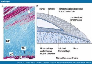
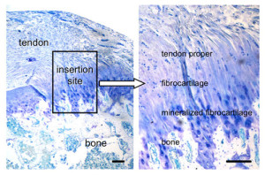
tendon enthesis

This next image shows in much more detail into how the enthesis really integrates into the bone and how enthesis stem cells could be stimulated to form a mini growth plate.  Bone erosion could occur allowing this growth plate to extend across the bone.  In fact, in arthritis which is associated with thickening of the enthesis’ bone erosion does occur.:

detailed view tendo enthesis
Note how the mineralized fibrocartilage integrates into the bone.  The thickening of the enthesis and the erosion of bone in some forms of arthritis, I could only find citations of it occuring in tendons and have not found instances of it resulting in increased length.  But that is likely a result of tendons being more subject to mechanical loading and this new method of LSJL being a novel way to mechanical load the ligaments.  Also ligaments in contrast to tendons are in a better position to contribute to longitudinal bone growth.  In fact, most of the studies I could find on mechanical loading on ligaments referred to loading on the ligament cells themselves rather than loading of the ligaments in the body.

Ligament injuries tend to occur due to heavy impact or overstretching of the ligament.  Pressing of the bones against each other is not a common way of ligament injury indicating that this method of LSJL loading is in fact a novel way of loading the ligaments.

“There is physiological thickening of the fibrocartilage with stress.”<-This tends to happen more with tendons as they are attached to muscle but with LSJL we can encourage it to happen at the ligament fibrocartilage.

” there are other components adjacent to the enthesis proper which also share the stress forces and are termed the “Enthesis organ”. These include the Periosteal fibrocartilage, the Sesamoid fibrocartilage, the Fat pad and Bursa. The Synovial entheseal complex is a concept that the adjacent bursa or joint lining share stress forces, especially compressive forces and are an integral part of the enthesis organ”<-These will be different for the ligament.

Here’s some info from another site that could be pertinent:

Bone Erosion at Normal Insertions

“Bone erosion is a process whereby the surface of a bone (the bone cortex) is degraded or eroded and is most typically seen in the setting of inflammation. However, the normal skeleton appears to be riddled with microscopic erosions. The enthesis is a highly mechanically stressed site which leads to microtrauma to the immediately adjacent bone. This is the basis for small erosions in the normal non-diseased skeleton which likely repair spontaneously.”<-Can we cause sufficient bone erosion as to allow for a new growth plate?

“The early phases of erosion may start to damage or loss of the shock absorbing fibrocartilage that covers the bone. “<-We don’t want this however as this would be the foundation of the neo growth plate.

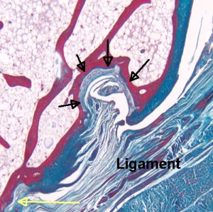
“Normal small joints tend to develop microscopic erosions at sites where the ligament immediately adjacent to the enthesis compresses the bone. This is because the shape of the bone leads to the forces being spread over a wide area that contributes to damage. This occurs at a structure termed a synovio-entheseal complex.”

synovio epiphysis complex

“The black arrows show a microscopic erosion over a knuckle joint. The overlying ligament is shown. The yellow arrow shows the point of ligament attachment closest to the joint cavity. Small blood vessels in the base of the erosion are likely linked to attempted repair. ”

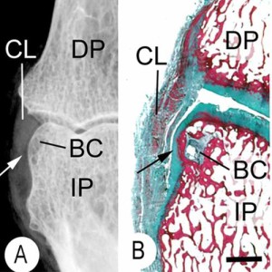
“Sometimes the bone compression by the enthesis organ transmits stresses to the underlying bone and this initially manifests as a small cyst. Later on the roof of this may cave in leading to erosion. ”

bone cyst enthesis

“This is an X-ray (A) and a corresponding tissue section (B). It shows a small bone cyst (BC). This is underneath the cartilage lining the side of the bone. (black arrow). Damage occurs here because the ligament (CL) presses against the bone as it runs between the joints. “<-We’d need more information about the bone cyst to see how promising it is in terms of neo growth plate formation.

Akt1 related overgrowth

Akt1 overgrowth tends to create proportional overgrowth whereas CNP overgrowth tends to create lankier overgrowth

Mosaic overgrowth with fibroadipose hyperplasia is caused by somatic activating mutations in PIK3CA.

“The phosphatidylinositol 3-kinase (PI3K)-AKT signaling pathway is critical for cellular growth and metabolism. Correspondingly, loss of function of PTEN, a negative regulator of PI3K, or activating mutations in AKT1, AKT2 or AKT3 have been found in distinct disorders featuring overgrowth or hypoglycemia. We performed exome sequencing of DNA from unaffected and affected cells from an individual with an unclassified syndrome of congenital progressive segmental overgrowth of fibrous and adipose tissue and bone and identified the cancer-associated mutation encoding p.His1047Leu in PIK3CA, the gene that encodes the p110α catalytic subunit of PI3K, only in affected cells. Sequencing of PIK3CA in ten additional individuals with overlapping syndromes identified either the p.His1047Leu alteration or a second cancer-associated alteration, p.His1047Arg, in nine cases. Affected dermal fibroblasts showed enhanced basal and epidermal growth factor (EGF)-stimulated phosphatidylinositol 3,4,5-trisphosphate (PIP(3)) generation and concomitant activation of downstream signaling relative to their unaffected counterparts. Our findings characterize a distinct overgrowth syndrome, biochemically demonstrate activation of PI3K signaling and thereby identify a rational therapeutic target.”

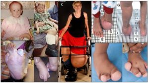
Proteus syndrome, a progressively deforming regional overgrowth syndrome that affects bones, adipose, and other mesenchymal tissues, is caused by a somatic p.Glu17Lys AKT1 mutation which constitutively activates PI3K/AKT signaling”

The Overgrowth can be local:

akt1-overgrowth

Interesting that in one case ” Muscle is replaced by fibrous and adipose tissue with occasional residual muscle fibers (arrow)”.  So muscular tissue basically dedifferentiated.  Maybe the same thing could happen with bone and allow neo growth plate formation.

“somatic occurrence of both AKT2 and AKT3 p.Glu17Lys mutants, paralogous to the Proteus-associated AKT1 mutation, have been described.”

Muscular loading may influence Akt1 signaling in bone:

The masticatory contractile load induced expression and activation of Akt1/PKBalpha in muscle fibers at the myotendinous junction within muscle-tendon-bone unit.

“The cell specific detection of enzyme activation in response to the physiological contractile load within muscle-tendon-bone unit is essential for understanding of the mechanical forces transmission from muscle cells via tendon to the bone{Are these mechanical forces transmitted via fluid flow or by some other means?}. The hypothesis that the physiological mechanical loading regulates activation of Akt1/PKBalpha at Thr308 and at Ser473 in muscle fibers within muscle-tendon-bone unit was tested using quantitative immunohistochemistry, confocal double fluorescence analysis, and immunoblot analysis. In comparison to the staining intensities in peripheral regions of the muscle fibers, Akt1/PKBalpha was detected with a higher staining intensity in muscle fibers at the myotendinous junction (MTJ) areas. In muscle fibers at the MTJ areas, Akt1/PKBalpha is dually phosphorylated at Thr308 and Ser473. The immunohistochemical results were confirmed by immunoblot analysis. We conclude that contractile load generated by masticatory muscles induces local domain-dependent expression of Akt1/PKBalpha as well as activation by dually phosphorylation at Thr308 and Ser473 in muscle fibers at the MTJ areas within muscle-tendon-bone unit.”

“The muscle-tendon-bone unit contains myocytes, fibroblasts, nerve fibers, blood vessels, osteoblasts, osteoclasts, osteocytes, and extracellular matrix.”

“Tendons transmit forces generated from muscle cells at the muscle-tendon-junction (MTJ) to bone cells.”

“Full activation of Akt1/PKBα requires phosphorylation of the enzyme at Thr308 and at Ser473”

“Thr308 is phosphorylated by 3-phosphoinositide-dependent kinase-1 (PDK1). The phosphorylation of Akt1/PKBα at Ser473 is mediated by both mammalian target of rapamycin-rictor complex (mTORC2) and DNA-dependent protein kinase (DNA-PK) depending on type of stimulus.”

“muscle fibers apply forces to the bone cells via tendon cells, which arise from a specialized region called the MTJ. In the MTJ, myofibrils and collagen fibers overlap, forming longitudinal infoldings”

According to The role of Akt1 in terminal stages of endochondral bone formation: angiogenesis and ossification., Akt1 may be involved in regulating MMP14 levels and Akt1 deficient mice had a delay in the formation of secondary ossificiation centers.

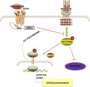
According to Adiponectin inhibits osteoclastogenesis and bone resorption via APPL1-mediated suppression of Akt1., Akt may reulate osteoclastgenesis.  So Akt1 may play a role in growth by increasing bone turnover.

akt1-pathway

So Akt1 increases Osteoclastogenesis and decreases Osteoclast Apoptosis.

Homocysteine may be an Adiponectin inhibitor according to Inhibition of adiponectin production by homocysteine: a potential mechanism for alcoholic liver disease.

Tensile strain and articular Cartilage

If tensile strain can induce endochondral ossification of articular cartilage then it could help you grow taller.

Micromechanical response of articular cartilage to tensile load measured using nonlinear microscopy.

Articular cartilage of 2-5 year old horses were used.  The deep zone was found to be more resistant to tensile strain so it could explain why it’s harder to induce endochondral ossification.

Effects of cyclic tensile strain on chondrocyte metabolism: a systematic review.

“Chondrocytes reorganize the extracellular matrix of articular cartilage in response to externally applied loads. Thereby, different loading characteristics lead to different biological responses. Despite of active research in this area, it is still unclear which parts of the extracellular matrix adapt in what ways, and how specific loading characteristics affect matrix changes. This review focuses on the influence of cyclic tensile strain on chondrocyte metabolism in vitro. It also aimed to identify anabolic or catabolic chondrocyte responses to different loading protocols. The key findings show that loading cells up to 3% strain, 0.17 Hz, and 2 h, resulted in weak or no biological responses. Loading between 3-10% strain, 0.17-0.5 Hz, and 2-12 h led to anabolic responses; and above 10% strain, 0.5 Hz, and 12 h catabolic events predominated{catabolic responses do not necessarily mean a bad thing it could indicate remodeling events that are needed for anabolic responses such as endochondral ossification}. However, this review also discusses that various other factors are involved in the remodeling of the extracellular matrix in response to loading, and that parameters like an inflammatory environment might influence the biological response.”

“after loading with CTS, cells exhibited a more elongated cell shape and aligned perpendicular to the loading direction”<-a more growth plate like organization perhaps?

“Fibronectin connects collagen fibers and other ECM proteins. It is linked to the cell membrane through integrins and might transmit forces from the ECM to the chondrocyte. CTS at 7%, 0.33 Hz and 0.5 Hz, for 4, 12 and 24 h increased the fibronectin mRNA levels in comparison to non-loaded chondrocytes”

“cartilage oligomeric matrix protein (COMP) was increased in response to cyclic tension in chondrocytes”

“a peak hydrostatic pressure of 3.45 MPa occurs in the femoral cartilage during a squat”

“during cartilage compression, the cell´s height (in split line direction) is reduced whereas the cell´s width (perpendicular to the split line) is increased. From the following considerations, one can assume that under physiological cartilage loading this increase in width represents a cell elongation of about 5%”

“in direction of the strain, in uniaxial experiments 79% ± 34% of the strain were transferred to fibroblasts and 63% ± 11% were transferred to tenocytes. In other experiments, 37% ± 8% and 45–60% of biaxial strains were transferred to tenocytes and bone marrow-derived stromal cells”

“in a non-inflammatory environment loading protocols up to 3% cell strain, 0.17 Hz and 2 h could be determined as “low CTS”, between 3–10% cell strain, 0.17 Hz—0.5 Hz and 2–12 h as “moderate CTS” and above 10% cell strain, 0.5 Hz and 12 h as “high CTS”. Loading duration might be the key parameter in triggering gene expression in response to CTS”

“there were no obvious differences between the response of chondrocytes e. g. from the temporomandibular joint and from the knee joints”

Cyclic equibiaxial tensile strain induces both anabolic and catabolic responses in articular chondrocytes.

“Mechanical disturbance is directly implicated in the development of osteoarthritis (OA) but the precise mode for degenerative changes is still largely unknown because of the complexity of the biomechanical and biochemical milieu in the articular joint. To investigate the effects of tensile strain on articular cartilage, cyclic equibiaxial tensile strain (CTS, 0.5 Hz, 10% strain) was applied to monolayer cultures of porcine articular chondrocytes by using a Flexercell strain unit. Overproduction of proinflammatory mediators and imbalanced expression of anabolic and catabolic genes were induced. The cellular secretion of nitric oxide (NO) and prostaglandin E(2) (PGE(2)), as well as the mRNA level of cyclooxygenase-2 (COX-2) were up-regulated in response to mechanical stimuli. Additionally, CTS resulted in an initial peak of anabolic response at 3 h of stretch with respect to the expression of type II collagen and aggrecan. After 12 h of CTS, the expression for these two cartilage-specific matrix proteins fell to control levels. A distinct catabolic response developed after 24 h of stretch with an increase in matrix metalloproteinase-1 (MMP-1). Interestingly, a parallel increase in transforming growth factor (TGF) beta3 was associated with the anabolic changes while an increase in expression of TGF beta1, the predominant isoform of the TGF family, appeared at 24 h. The expression at 24 h of MMP-1, an enzyme that degrades interstitial collagens as well as other cartilage matrix proteins and TGF beta1, may signify a shift towards matrix remodeling and potentially a change in matrix composition as a consequence of continuous CTS.”

The cells did change alignment in response to strain.  6 hours seemed to be the most anabolic stretching time period for cartilage growth but the catabolic genes may be needed for endochondral ossification remodeling.  Since only superficial zone was used it’s difficult to tell the effects of inducing endochondral ossification.

Modelling cartilage mechanobiology.

“The growth, maintenance and ossification of cartilage are fundamental to skeletal development and are regulated throughout life by the mechanical cues that are imposed by physical activities. Finite element computer analyses have been used to study the role of local tissue mechanics on endochondral ossification patterns, skeletal morphology and articular cartilage thickness distributions. Using single-phase continuum material representations of cartilage, the results have indicated that local intermittent hydrostatic pressure promotes cartilage maintenance. Cyclic tensile strains (or shear), however, promote cartilage growth and ossification. Because single-phase material models cannot capture fluid exudation in articular cartilage, poroelastic (or biphasic) solid/fluid models are often implemented to study joint mechanics. In the middle and deep layers of articular cartilage where poroelastic analyses predict little fluid exudation, the cartilage phenotype is maintained by cyclic fluid pressure{tensile strain may disrupt this fluid pressure thus disrupting the cartilage phenotype} (consistent with the single-phase theory). In superficial articular layers the chondrocytes are exposed to tangential tensile strain in addition to the high fluid pressure. Furthermore, there is fluid exudation and matrix consolidation, leading to cell ‘flattening’. As a result, the superficial layer assumes an altered, more fibrous phenotype. These computer model predictions of cartilage mechanobiology are consistent with results of in vitro cell and tissue and molecular biology experiments.”

“In a developing cartilage rudiment, one can recognize the same endochondral growth and ossification processes both at the primary ossification front and around secondary ossification sites. There are regions of quiescence that are characterized by the presence of resting chondrocytes. As growth proceeds, these cells proliferate and then mature as they begin to increase the production of ECM, which is characterized by important cartilage molecules, aggrecan and collagen II. As these mature chondrocytes begin to hypertrophy, they express Ihh an important growth factor that upregulates the expression of bone morphogenetic proteins. In the late stages of chondrocyte hypertrophy, the cartilage ECM septa between the columns of hypertrophic chondrocytes calcify. There is increased expression of MMPs (MMP9, MMP13), vascular endothelial growth factor, collagen X and CTGF, in preparation for angiogenesis and ossification. The hypertrophic chondrocytes then undergo apoptosis, and vascular invasion is initiated through their vacant lacuna. Chondroclasts are recruited to the site and begin to resorb the calcified cartilage, eventually destroying two-thirds of the calcified matrix. Perivascular mesenchymal cells differentiate into osteoblasts and begin to form new osteoid on the remaining calcified septa. The osteoid mineralizes to form primary bone trabeculae and the growth and ossification process is complete. ”

“Although ossification of articular cartilage from the underlying subchondral growth front is greatly diminished, it is not entirely stopped ”

” Because the fluid permeability of cartilage is quite low, it is difficult to squeeze the water out. Consequently, for short static loading periods or for cyclic loading with moderate or high frequencies, the tissue behaves mechanically as a single-phase solid. In these conditions, the simplest constitutive model represents cartilage as a homogeneous, linear elastic, incompressible or nearly incompressible material. ”

” Hydrostatic fluid pressure inhibits cartilage growth and ossification, thereby maintaining the cartilage phenotype. (ii) Tensile strain (or octahedral shear stress) accelerates cartilage growth, ossification and replacement by bone. (iii) Matrix compressive consolidation, with or without fluid pressure, decreases cartilage proteoglycan synthesis and content and results in a more fibrous cartilage phenotype.”

The vascularity and remodelling of subchondrial bone and calcified cartilage in adult human femoral and humeral heads. An age- and stress-related phenomenon.

“A quantitative study of the vascularity and a qualitative study of the remodelling of the calcified cartilage and subchondral bone end-plate of adult human femoral and humeral heads were performed with respect to age. In the femoral head the number of vessels per unit area was found to fall 20% from adolescence until the seventh decade and in the humeral head 15% until the sixth decade. Thereafter an increase was noted in the femur but none in the humerus. More vessels were present at all ages in the more loaded areas of the articular surfaces: 25% more for the femur and 15% more for the humerus. The degree of active remodelling by endochondral ossification declined 50% from adolescence until the seventh decade in the femoral head, and 30% until the sixth decade in the humeral head, rising thereafter to levels comparable to those found at young ages. More remodeling was noted in the more loaded areas at all ages.”

<-this study is cited in the above as one that’s showing that articular cartilage ossification occurs throughout life.
LIPUS can affect face size

There have been conflicting reports on LIPUS for height but ultrasound has been shown to increase condylar growth in rats before.

Effect of nonviral plasmid delivered basic fibroblast growth factor and low intensity pulsed ultrasound on mandibular condylar growth: a preliminary study.

“Basic fibroblast growth factor (bFGF) is an important regulator of tissue growth. Low intensity pulsed ultrasound (LIPUS) stimulates bone growth. [We] evaluate the possible synergetic effect of LIPUS and local injection of nonviral bFGF plasmid DNA (pDNA) on mandibular growth in rats.
Groups were control, blank pDNA, bFGF pDNA, LIPUS, and bFGF pDNA + LIPUS. Treatments were performed for 28 days. Significant increase was observed in mandibular height and condylar length in LIPUS groups{it is reasonable to assume that this increase can be extended to other long bones to which LIPUS was applied}. Significant increase in bone volume fraction in bFGF pDNA + LIPUS group.  Increased cell count and condylar proliferative and hypertrophic layers widths in bFGF pDNA group.  Increased mandibular condylar growth in either bFGF pDNA or LIPUS groups compared to the combined group that showed only increased bone volume fraction.”

“Growth factors like vascular endothelial growth factor (VEGF) and bFGF play an important role in the process of new blood vessel formation”

“blocking of bFGF leads to the prevention of bone formation at the craniofacial suture sites”

The condylar length and ramal height was about 1.5mm longer in the LIPUS group versus the control group.

LIPUS seemed to increase cell size in the growth plate for the hypertrophic zone while also increasing the number of cells in the proliferative zone(by increasing chondrocyte proliferation or causing resting zone stem cells to differentiate into chondrocytes?).

Although the one study “Application of low-intensity ultrasound to growing bone in rats.”, did not find that LIPUS increased longitudinal bone growth it’s possible that there methodology was not correct for inducing longitudinal bone growth.

The mandibular growth in this study indicates that LIPUS may have promise yet for being a part of increasing longitudinal bone growth at least for active growth plates.

Can eating oysters while actively growing increase height?

This study suggests that this may be the case.

Taurine, a major amino acid of oyster, enhances linear bone growth in a mouse model of protein malnutrition.

“we evaluated the effects of Oys or Tau on linear bone growth in a mouse model of protein malnutrition{So there’s no guarantee that it will increase height in those who are not malnourished}. To make the protein malnutrition in a mouse, we used a low protein diet. Growth plate thickness was increased by Oys or Tau. Bone volume/tissue volume, trabecular thickness, trabecular number, connection density, and total porosity were also improved by Oys or Tau. Oys or Tau increased insulin-like growth factor-1 (IGF-1) levels in serum, liver, and tibia-growth plate. Phosphorylations of Janus kinase 2 (JAK2) and signal transducer and activator of transcription 5 (STAT5) were increased by Oys and by Tau.  Oys or Tau may increase growth plate thickness by elevating IGF-1 levels and by promoting the phosphorylations of JAK2-STAT5, and suggest that Oys or Tau are growth-promoting substances of potential use in the food and pharmaceutical industries.”

“When a child is undernourished, circulating IGF-1, and thyroid hormone levels decline and in adolescents, undernutrition causes reductions in sex steroids, and these endocrine changes suppress bone growth “<-However this does not guarantee that overnutrition will stimulate bone growth.

“GH is required for linear growth, and its actions are initiated by its binding to GH receptor (GHR) on cell surfaces. This binding induces receptor homodimerization and activation of GHR-associated tyrosine kinase Janus kinase 2 (JAK2). JAK2 is then phosphorylated and, in turn, phosphorylates GHR and signal transducers and activators of transcription (STAT). Upon phosphorylation, STAT undergoes homo or heterodimerization, translocates to the nucleus, binds to appropriate DNA response elements, and stimulate the transcriptions of GH-regulated genes. IGF-1 is one such gene and acts as a mitogenic factor for various cells and plays an important role in cell growth and survival. The majority of plasma IGF-1 is biosynthesized in liver”

Unfortunately there was no group in this study that had adequate protein and had additional taurine supplementation.

“the mRNA expression of IGF-1 was dose-dependently increased. The effects of 100 µg/mL of Oys and 50 µg/mL of Tau were greatest, and thus, we evaluated the effects of these doses in our in vivo mouse model.”

” Mean lengths of proximal tibial growth plate in the CON and PEM groups were 115.64 ± 3.40 and 84.98 ± 2.70, respectively, whereas growth plate lengths in the Oys and Tau groups were 125.97 ± 8.07 and 123.05 ± 7.52, respectively. Oys or Tau significantly enhanced longitudinal bone growth”

Growth plate thickness was a little bit shorter in the oyster and taurine group versus the control group as were IGF-1 and GH levels.

Mechanical Loading on Tendons(Some evidence for Entheses LSJL)

Considering the alternative LSJL method relies on pushing two bones against one other at ligament and tendon attachment sites, understanding how mechanical loading affects tendons is important.

The effects of mechanical loading on tendons–an in vivo and in vitro model study.

“Mechanical loading constantly acts on tendons. [We] investigate tendon mechanobiological responses through the use of mouse treadmill running as an in vivo model and mechanical stretching of tendon cells as an in vitro model. Mice underwent moderate treadmill running (MTR) and intensive treadmill running (ITR) regimens. Treadmill running elevated the expression of mechanical growth factors (MGF) and enhanced the proliferative potential of tendon stem cells (TSCs) in both patellar and Achilles tendons. In both tendons, MTR upregulated tenocyte-related genes: collagen type I (Coll. I ∼10 fold) and tenomodulin (∼3-4 fold), but did not affect non-tenocyte-related genes: LPL (adipocyte), Sox9 (chondrocyte){this is the gene were are looking for}, Runx2 and Osterix (both osteocyte). However, ITR upregulated both tenocyte (Coll. I ∼7-11 fold; tenomodulin ∼4-5 fold) and non-tenocyte-related genes (∼3-8 fold){so the load has to be sufficient to affect the target chondrogenic gene for our purposes.  If the load is not intense enough it will not upregulate novel genes.}. In the in vitro study, TSCs and tenocytes were stretched to 4% and 8% using a custom made mechanical loading system. Low mechanical stretching (4%) of TSCs from both patellar and Achilles tendons increased the expression of only the tenocyte-related genes (Coll. I ∼5-6 fold; tenomodulin ∼6-13 fold), but high mechanical stretching (8%) increased the expression of both tenocyte (Coll. I ∼28-50 fold; tenomodulin ∼14-48 fold) and non-tenocyte-related genes (2-5-fold){Stretching has to be high to affect the target gene}. However, in tenocytes, non-tenocyte related gene expression was not altered by the application of either low or high mechanical stretching{So mechanical stretching likely cannot induce transdifferentiation or the microenvironment is partially responsible for the upregulation of non-tendon specific genes}. Excessive mechanical loading caused anabolic changes in tendons, it also induced differentiation of TSCs into non-tenocytes.”

So we induce the differentiation of TSCs and Ligament Stem Cells(which are likely similar to tenocyte stem cells) into chondrocytes at the entheses which can then form new growth plates.

“[IGF-1’s] Eb isoform, also known as mechano-growth factor (MGF), may be a key component of the mechanism that translates mechanical loads into cellular biological changes.”

“under mechanical loading conditions, TSC population in the tendon grows, providing progenitors”

““round tenocytes” were observed in the supraspinatus tendon after intensive treadmill running (16.7 m/min) for 12 weeks. Based on our findings in this study, we suspect that these “round tenocytes” could be chondrocytes differentiated from TSCs, because i) TSCs, not tenocytes, are able to undergo non-tenocyte differentiation under high mechanical loading conditions; ii) a round shape is a typical morphology of chondrocytes, and iii) these round cells produce abundant proteoglycans detected around the cells in the tendon”

This study provides evidence for the entheses method of LSJL loading(termed henceforth as entheses LSJL).  Where the bone is clamped at the site where two bones are connected by a ligament.  These ligaments contain entheses which attach to the bone and are similar in attachment to the Zone of Ranvier.  That tendons contain stem cells which can differentiate into chondrocytes suggests that it’s probably highly similar that ligaments contain ligament stem cells which can differentiate into chondrocytes.  The difference primarily being that tendons are constantly subjected to mechanical load by virtue of their attachment to muscle.  The issue now is proving that there are stem cells within the entheses of a ligament.