Monthly Archives: November 2015

Factors that alter the bone microenvironment

Here’s a review paper on factors that alter the bone microenvironment.  There’s a lot of stuff here.  More than is mentioned.

 Biomechanical and biophysical environment of bone from the macroscopic to the pericellular and molecular level.

” Bones with complicated hierarchical configuration and microstructures constitute the load-bearing system. Mechanical loading plays an essential role in maintaining bone health and regulating bone mechanical adaptation (modeling and remodeling). The whole-bone or sub-region (macroscopic) mechanical signals, including locomotion-induced loading and external actuator-generated vibration, ultrasound, oscillatory skeletal muscle stimulation, etc., give rise to sophisticated and distinct biomechanical and biophysical environments at the pericellular (microscopic) and collagen/mineral molecular (nanoscopic) levels, which are the direct stimulations that positively influence bone adaptation. While under microgravity, the stimulations decrease or even disappear, which exerts a negative influence on bone adaptation. A full understanding of the biomechanical and biophysical environment at different levels is necessary for exploring bone biomechanical properties and mechanical adaptation. In this review, the mechanical transferring theories from the macroscopic to the microscopic and nanoscopic levels are elucidated. First, detailed information of the hierarchical structures and biochemical composition of bone, which are the foundations for mechanical signal propagation, are presented. Second, the deformation feature of load-bearing bone during locomotion is clarified as a combination of bending and torsion rather than simplex bending. The bone matrix strains at microscopic and nanoscopic levels directly induced by bone deformation are critically discussed, and the strain concentration mechanism due to the complicated microstructures is highlighted. Third, the biomechanical and biophysical environments at microscopic and nanoscopic levels positively generated during bone matrix deformation or by dynamic mechanical loadings induced by external actuators, as well as those negatively affected under microgravity, are systematically discussed, including the interstitial fluid flow (IFF) within the lacunar-canalicular system and at the endosteum, the piezoelectricity at the deformed bone surface, and the streaming potential accompanying the IFF. Their generation mechanisms and the regulation effect on bone adaptation are presented. The IFF-induced chemotransport effect, shear stress, and fluid drag on the pericellular matrix are meaningful and noteworthy. Furthermore, we firmly believe that bone adaptation is regulated by the combination of bone biomechanical and biophysical environment, not only the commonly considered matrix strain, fluid shear stress, and hydrostatic pressure, but also the piezoelectricity and streaming potential. Especially, it is necessary to incorporate bone matrix piezoelectricity and streaming potential to explain how osteoblasts (bone formation cells) and osteoclasts (bone resorption cells) can differentiate among different types of loads. Specifically, the regulation effects and the related mechanisms of the biomechanical and biophysical environments on bone need further exploration, and the incorporation of experimental research with theoretical simulations is essential.”

“Osteocyte processes are attached to the canalicular wall via transverse tethering elements
(the average spacing between the elements is 32 ± 14 nm) and only directly contact the canalicular wall at specialized collagen fibril protrusions ”

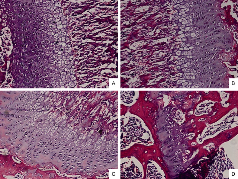
“In trabecular bone, the trabecular rods and plates form three-dimensional structures, and within the trabeculae are less regular arranged lamellae and osteocytes”

“The organic material, which is primarily type I collagen (90%) and nonfibrillar organic matrix
(osteocalcin and osteopontin present in a large proportion) gives the bone toughness. The mineral salts, mainly nanocrystallite apatite materials, permeate the organic matrix
and provide the characteristic rigidity and strength of bone. Common belief holds that the apatite materials are hydroxyapatite [Ca10(PO4)6(OH)2] (HAP). In fact, there is no OH-band in bone mineral detected in a Raman spectrum, therefore the bone apatite is not hydroxyapatite ”

“in two adults tibiae during comfortable walking and strenuous running; the results suggested that the compressive strain ranged from approximately 400 to 1300 µε, and the tensile strain ranged from approximately 380 to 750 µε ”

“The pericellular space in the lacunar-canalicular system (LCS) of cortical bone and the porous
structures within trabecular bone are filled with interstitial fluid. Due to the low permeability of mineralized bone matrix, interstitial fluid flow (IFF) is principally generated during alteration of intramedullary pressurization (ImP) and bone matrix deformation”

“Pulsatile extravascular pressure resulting from blood circulation can also drive IFF, but it is much smaller than that induced by ImP and bone deformation ”

“The IFF generated from ImP usually follows two spatially distinct flow profiles: IFF within the
LCS and at the endosteal surface.  Uniform pressurization [such as that due to decreased intramedullary cavity resulting from elevated bone marrow lipids induced by high level of corticosteroid administration will generate radial flow from the intramedullary compartment to the endosteal surface and into the LCS due to the pressure gradient
from the marrow cavity to the bone matrix. Non-uniform pressure gradients within the intramedullary cavity [such as those due to local heterogeneous permeability or fluid displacement changes in the intramedullary compartment from the interaction
between mechanical loading/oscillatory muscle stimulation and capillary filtration in bone tissue will cause tangential fluid flow to the endosteal surface ”

“Because the mineralized matrix is nearly impermeable{This is partially why it is so hard to grow taller} and the pericellular annular fluid space surrounding the osteocyte cell body and processes is narrow (gap < 1 µm) , the transportation of nutrients (e.g., glucose), oxygen, waste products (e.g., lactic acid), and regulatory signals (e.g., sex hormones, nitric oxide, prostaglandins, cytokines, and growth factors) by simple diffusion is not sufficient to maintain cell viability and function IFF within the LCS serves as the primary transport mechanism between the blood supply and osteocytes”

“hydrostatic pressure is induced by the bone compression that results from physiological loading. Based on the data that walking at a frequency of 1 Hz induces bone strain of 1000 µε and 0–18 MPa cyclic stress{this is surprisingly high},   the maximum hydrostatic pressure in LCS is approximately 0.27 MPa{this is actually within the chondrogenic range of 0.1-10MPa; there must be additional factors going on}, this pressure is estimated to be 12% of the applied axial stress and 40 times higher than that in the vasculature”

” the compressed areas develop negative potentials, whereas the tensioned areas develop positive potentials. These changes are known as the direct piezoelectric effect ”

“The researchers subjected demineralized type I collagen to bending deformation and bathed the collagen in continually flowing physiologic fluid over a period of weeks. Interestingly, the compressed internal surface (negative) of the collagen attracted calcium ions (positive), leading to the subsequent nucleation and crystallization of apatite and an increase in precipitate. In contrast, the tensioned portion (positive) of the collagen showed no difference in precipitate compared with that in the undeformed collagen. ”

“Apatite is piezoelectric by nature ”

“dry collagen is piezoelectric, but fully hydrated collagen is not piezoelectric because the structured water may increase the symmetry of the collagen molecule”

” In the presence of apatite, access of water to parts of the collagen fibril and the collagen swelling to its fully hydrated form can be prevented, thus the collagen can show some piezoelectricity even when the bone is fully hydrated ”

” the negative and positive potentials caused by polarization can be stored in apatite for years ”

“osteons are irregular cylinders always with complex branching and interconnections”

“The strain-induced movement of interstitial fluid through bone porosities can induce another
bioelectrical potential in addition to piezoelectricity; this potential is called the streaming potential ”

“At the interface of the bone matrix and interstitial fluid, negative charges accumulate on the bone surface due to the electronegativity of collagen; these charges attract positive ions in the liquid, and the positive ions concentrate near the interface, forming an electrical double layer.  The electric potential in the interfacial double layer relative to the interstitial fluid away from the interface is the zeta potential (ζ), and when interstitial fluid flows tangentially along the interface under pressure, a streaming potential appears along the flow direction. The zeta potential is mainly generated by deformed collagen and corresponds to the streaming potential ”

” the body fluid shifts[during space flight], and perfusion is diminished in the lower limbs and increased in the upper limbs and head ”

“ultrasound may primarily exert an effect on increasing blood flow and producing micromechanical strains; the actual acoustic intensity experienced by the bone cells at the target site might be lower due to the possible attenuation caused by the overlying soft tissue ”

“[Oscillatory muscle stimulation] at 20 Hz generated a maximum ImP and a relative high bone strain”

Understanding Mesenchymal to Epithelial cell transition may be key for neo-growth plates

This study is huge because we can get adult epithelial cells to potentially form new growth plates.  Since the resting zone is composed of mainly endothelial-like cells and the resting zone is the foundation of the growth plate.  Understanding how to transition adult mesenchymal stem cells into endothelial cells may be the basis for forming a new growth plate.

Epithelial-mesenchymal transition and mesenchymal-epithelial transition response during differentiation of growth-plate chondrocytes in endochondral ossification.

“For linear longitudinal bone elongation, the stem-like progenitor chondrocytes distributed in resting zone (RZ) of growth plate have a capacity to differentiate towards the spindle chondrocytes in proliferative zone (PZ), then towards the columnar and tightly adjacent chondrocytes in hypertrophic zone (HZ). We hypothesized this process of endochondral ossification with cells morphological change was occurred along with the inter-conversion between epithelial to mesenchymal cell types. Consistent with this hypothesis, the chondrocytes highly expressed mesenchymal-like biomarkers and loss of epithelial surface markers in PZ, while converse in RZ and HZ of the growth plate in mice distal tibia in vivo.  The 4-week old male and female mice were treated with estradiol cypionate or oxandrolone, then investigated the response of epithelial- and mesenchymal biomarkers, and demonstrated that estrogen blocked the EMT process from RZ to PZ while androgen promoted MET from PZ to HZ. Our observations supported the hypotheses that the growth plate firstly go through EMT from RZ to PZ, then MET process from PZ to HZ during the epiphyseal fusion. Our results could interpret the different roles of estrogen and androgen in growth plate cartilage [undergoing] endochondral ossification.”

In Epiphelial to Mesenchymal Cell Transition, cells lose cell-cell adhesion and cell polarity properties to become more migratory mesenchymal cells.  Since the growth plate firstly grows via Epipthelial to Mesenchymal Cell Transitiation at the RZ to PZ that is the most important stage for us to focus on as that causes the formation of the growth plate and the formation of the growth plate would be a great help in growing taller.  However, the cell condensation stage to establish the resting cell zone should first require epithelial cell types as cell to cell adhesion would be needed.

“Multiple tissues differentiation and organs formation in embryonic development arise from a
series of conversion from epithelial to mesenchymal cells, through epithelial to mesenchymal
transition (EMT) or mesenchymal to epithelial transition (MET). In primary EMT process,
the primitive epithelia lose their characterization of rounded shape, sequential arrangement and compact junctions to convert a population of spindle, loosely organized but motile mesenchymal cells{hydrostatic pressure may help organize the stem cells and change the structure of these cells} for gastrulation formation and neural crest migration. Then, after a transient epithelial structure condensation through MET, these population in notochord, somites, somatopleure and splanchnopleure derived from mesoderm generate mesenchymal cells which have ability to differentiate into specific cells types of diverse tissues via the secondary EMT

” the neural crest cells migrate to somites of mesoderm following stereotyped pathways and undergo a secondary EMT to generate mesenchymal condensation.  These mesenchymal cells differentiate into osteoprogenitors for intramembranous ossification and chondrocytes for endochondral ossification. ”

” Pluripotent stem cells exhibit epithelial characteristics, down-regulate the epithelial markers such as Cdh1, Cldn6, Epcam and enhance the mesenchymal markers including Snai1/2, Zeb1,
CtnnbIP1″

” estrogen administration maintains the epithelial type genes expression in growth plate particularly in RZ implies that estrogen appears to block EMT process.”

“Not like human or rabbit, the expression of estrogen receptors within HZ of growth plate in mice and rat was extremely low until at the last time point prior to epiphyseal fusion, which also
reflects the less effect of estradiol cypionate to in the HZ in our study. Conversely, Androgen
effectively promotes EMT for  chondrocytes differentiation”

” estrogen may interdict TGF-beta, then further repress Smad3 expression, so that
postpone chondrocytes differentiation via EMT blocking. ”

“Androgen is determined to promote EMT for differentiation. However, androgen improves Smad3 expression but appears to have no response to SIS3, which indicates that androgen may participate in other pathways rather than TGF-beta/Smad3.”

” A notable presence of growth plate was observed in distal tibia of the 4-week but not in 16-week old mice”<-Note that LSJL has worked in 16-week old mice.  Although in the LSJL study they used Sprauge -Dawley rats in contrast to outbred ICR mice.

” The mRNA level of epithelial markers including Cdh1, Cldn6, Col4a1, Krt19, Lamc1 expressed in RZ and HZ were significantly higher than in PZ while the mesenchymal markers such as Acta2, Ctnnb1, Smad3, displayed the converse tendency. The results suggested that a process of EMT occurred in the programming of RZ towards PZ and MET in PZ towards HZ.”

Here’s the 16-week old growth plate, still present but weak:

16-week old mice growth plate

Epithelial cells may already exist in adult bone marrow.

Epithelial cells in bone marrow: do they matter?

“epithelial-like cells can be detected in the bone marrow of many patients not known to have cancer. ”

Inducing a mesenchymal to epithelial transition would be difficult as I haven’t found any studies of it occuring due to physiological stimuli.  Another possibility though would be to have a growth plate without a resting zone as the proliferative zone consists mainly of mesenchymal cells.  The viability of this depends on the viability of a growth plate without a resting zone.  The resting zone may play a role in growth plate orientation which makes sense as epithelial cells tend to be involved in cell polarity and cell adhesion.

Here’s a diagram of the mesenchymal-epithelial transition:

mesenchymal-epithelial transition

LSJL upregulates Pcdhb2(protocadherin beta 2), Cdh13, Ctnna3, Fat1(a cell adhesion model).  It downs regulates protocadherin subfamily A, 4(Pcdhga4), AK002616(a miscellaneous Cadherin related protein), Celsr2, Cdh15, Cdh11.  So LSJL has the definite potential to affect the mesenchymal-epithelial transition although how isn’t clear as it affects a lot of related genes but not in a clear pattern

For the other parts LSJL downregulates Cldn13, Dsp(Desmoplakin isoform 1, the downregulation of this gene suggests that LSJL likely encourages the epithelial to mesenchymal transition but that doesn’t mean that it doesn’t encourage the mesenchymal to epithelial transition as well), and upregulates a gene related to Cldn19, Muc3.  It also downregulates an anti-mucin gene.

This diagram mentions the reverse markers:
EMT

Here’s another diagram:
another EMT diagram

According to Actin stress fibres and cell-cell adhesion molecules in tendons: organisation in vivo and response to mechanical loading of tendon cells in vitro.

“Tendons consist of parallel longitudinal rows of cells separated by collagen fibres. The cells are in intimate contact longitudinally within rows, and laterally via sheet-like lateral cell processes between rows. At points of contact, they are linked by gap junctions. Since tendons stretch under load, such cell contacts require protection. Here we describe the organisation of the actin cytoskeleton and actin-based cell-cell interactions in vivo and examine the effect of cyclic tensile loading on tendon cells in vitro. Cells within longitudinal rows contained short longitudinally running actin stress fibres. Each fibre was aligned with similar fibres in the cells longitudinally on either side, and fibres appeared to be linked via adherens junctions. Overall, these formed long oriented rows of stress fibres running along the rows of tendon cells. In culture, junctional components n-cadherin{this increase is actually not good news for creation of epithelial cells as this is a mesenchymal marker} and vinculin and the stress fibre component tropomyosin increased in strained cultures, whereas actin levels remained constant.  (1) cells are linked via actin-associated adherens junctions along the line of principal strain; and (2) under load, cells appear to attach themselves more strongly together, and assemble more of their cytoplasmic actin into stress fibres with tropomyosin. Cell-cell contacts are protected during stretch, and also that the stress fibres, which are contractile, may provide an active mechanism for recovery from stretch. In addition, stress fibres are ideally oriented to monitor tensile load and thus may be important in mechanotransduction and the generation of signals passed via the gap junction network.”

So according to this in response to load cells may establish more cell-cell contact characteristic of epithelial cells.

Mechanism of the Mesenchymal–Epithelial Transition and Its Relationship with Metastatic Tumor Formation

epithelial characteristics were dramatically associated with increased bone and soft-tissue colonization after intracardiac or intratibial injection.”

“Multiple complex signaling systems are required for the induction of EMT and are also closely related with MET. The FGFR2 gene, which is located at human chromosome 10q26, encodes for FGFR2b and FGFR2c isoforms due to alternative splicing and mutually exclusive use of exon IIIb or exon IIIc. FGFR2b primarily binds FGF10 and FGF7 and is the isoform of choice in epithelial cells, whereas FGFR2c binds FGF2 and is mainly expressed in cells of mesenchymal origin. FGF/FGFR2 signaling governs the EMT that is required for organogenesis in mouse embryos.”

“expression of FGFR2b induced MET [induced cancer in one instance]”

“As for the regulation of FGFR2 isoforms’ alternative splicing, a highly conserved GCAUG element was shown to be required for efficient exon IIIb activation. Afterward, Fox protein family members, especially Fox-2, were shown to regulate the FGFR2 exon choice, and this regulation was absolutely dependent on the GCAUG elements present in the FGFR2 pre-mRNA. Fox-2 induced the FGFR2c to FGFR2b switch, accompanied by molecular and morphological changes consistent with MET”

“2 paralogous epithelial cell type–specific RNA binding proteins, Rbm35a and Rbm35b, which are essential regulators of FGFR2 splicing. Ectopic expression of either protein in cells that express FGFR2c caused a switch in endogenous FGFR2 splicing to the epithelial isoform”<-Note it’s FGFR3 that’s typically associated with dwarfism.

MET

EMT and MET as paradigms for cell fate switching

“Cell fate determination is a major unsolved problem in cell and developmental biology. The discovery of reprogramming by pluripotent factors offers a rational system to investigate the molecular mechanisms associated with cell fate decisions. The idea that reprogramming of fibroblasts starts with a mesenchymal-epithelial transition (MET) suggests that the process is perhaps a reversal of epithelial to mesenchymal transition (EMT) found frequently during early embryogenesis. As such, we believe that investigations into MET-EMT may yield detailed molecular insights into cell fate decisions, not only for the switching between epithelial and mesenchymal cells, but also other cell types.”

“In any given animal tissue, one may find two very common cell types: the epithelial cells that line the surface of a tissue or organ and mesenchymal cells that are embedded in the three-dimensional matrix. “<-Growth plate cells line the bone matrix.

“the epithelial cells are attached to the basement membrane, establish an apical–basal axis of polarity, and communicate with each other through the gap junction. Across and underneath the basement membrane, there is the stroma made of the three-dimensional extracellular matrix synthesized by the resident mesenchymal or stromal cells.”

“Inside the nuclei, Snail genes are considered as the most important downstream targets of the nodal-SMAD2/3 pathway during gastrulation. The Snails may in fact be the guardians of the mesenchymal phenotype by activating mesenchymal genes and suppressing epithelial genes. Indeed, Snails have been shown to down-regulate E-cadherin effectively, which is one of hallmarks for epithelial cells. Snail-deficient embryos could not proceed through gastrulation and form mesodermal cells as they could not down-regulate the expression of E-cadherin in the primitive streak. It is generally recognized that the embryonic EMT process is orchestrated and maintained through the collaboration of extracellular signals and intracellular transcription factors.”

F1.medium

Update On Older Research – Melatonin and Growing Taller

When I first started writing on the website, I remember that people were talking about this supplement combination which was supposed to help you grow taller. M.E.N.S.

That was the acronym used for sleep, exercise, niacin, and melatonin. I wanted to talk about melatonin.

I recently started to take melatonin just to help me sleep and I remembered that I never really did much research on Melatonin. A quick Google Search on the link between melatonin and the possibility that it can help you grow taller reveals many of the older posts written maybe 4–7 years ago, derived from the now dead GrowTallForum.com website.

People believed in this supplement combination. Melatonin was promoted a lot by people of that old forum because the thinking was that since it helps you sleep, and growth hormones are primarily released when you are sleeping, melatonin would mean you have a greater chance of having growth hormones being pushed into your system.

I looked at the link. Is there something there?

I refer to 7 studies, some of them recently published just in 2014.

The 3rd study suggests that Melatonin does have this ability to turn MSCs into the chondrogenic lineage. Other sources (#7) says that for the growing child, over-expression of melatonin in the system can cause abnormal chondrocyte growth. It turns out that young kids who have idiopathic scoliosis actually have longer bones (vertebral column and arm length) than their peers. The curvature of the vertebral is because the anterior region of the vertebral is growing faster than the posterior region.

Out of all of the 7 studies, I felt that study #1 was the most insightful.

Short stature is most often caused by 2 ways.

  1. Idiopathic Short Stature (ISS)
  2. Growth Hormone Deficiency (GHD)

It seems that for children who suffer from GHD, they have GHD because of the overexpression of melatonin.

This means that based on this 1 studies, when we generalize the conclusion, it seems that having too much melatonin in the child’s system is bad for their height. However, this condition is something that can be medically treated, unlike ISS, which most family doctors would say makes the kids “genetically predisposed to be short later in life”.

So while MSCs might be helped slightly by Melatonin, too much Melatonin is a bad thing. Referring to study #5, the conclusion states the following for peri-pubertal children. – “These findings indicated that melatonin could inhibit the proliferation and stimulate differentiation of GPC (Growth Plate Chondrocytes) in human”{Tyler-Actually this could be a thing for a height growth as peak chondrocyte hypertrophy is more important than chondrocyte proliferation for peak height growth}

Taking Melatonin does not work. Try to figure out another type of supplement to take to sleep easier.

Excess Pituitary Stimulated Growth Hormones Increases Male Genitalia aka Penis Size

Something I read from the reddit/r/short threads recently made me remember something I discovered years ago. It seems that male genitalia, (aka Penis Size) can be altered/increased through excess levels of hormones.

I just don't know what the title might be, just please read it, not because of me.

Whoever the poster was, I am guessing that they are feeling the symptoms of a pituitary related tumor, whether benign or malignant. The tumor is stimulating excess HGH release into their system.

Like they said they are already 26, which suggest that their normal growth plates should be closed. This recent events which are indeed unusual for most people doesn’t seem to surprise me. I had guessed that excess HGH can cause even people with closed growth plates to gain 1 full inch in height, just based on the fact that there are still thin slivers of hyaline cartilage tissue in their vertebrate, tibia, femur, etc. which goes through hypertrophy. When you combine all of the hypertrophy the hyaline articular cartilage locations goes through, the total increase in height would often be around 1 inch.

As for height increase, this guy went from 6′ 2″ to 6′ 3″. His height will most likely not increase any further. However, he stated the other fact. His genitalia increased{Tyler-One thing to note is the penis is considered to be like a hydroskeleton}. When I think about this, it all starts to make sense.

Let’s go back a little.

For the normal, average, heterosexual, American men, the most common insecurity might not be their size, in terms of height/stature, but their size as in their “genitalia”. There are in fact entire internet forums dedicated by young men who actively do exercises to increase the size of their genitalia. Since the forums seem to be quite active, I am guessing that these guys who have that pursuit are slightly more successful in their endeavor than ours.

I had proposed in previous posts the idea that stereotypes have in them kernels of truth. I theorized that the stereotype that African American males have larger than average sized genitalia is true. The reason that is the truth is because I guessed that African Americans have higher than average levels of IGF-1 which goes through their system when they are going through puberty.

Higher levels of IGF-1 suggests higher rates of cardiovascular system related medical conditions later in life (diabetes, prostate cancer) which also has a slight correlation to having an earlier than average puberty. I am not sure, but I think I remember seeing studies which show that African American females (at least in urban locations) goes through puberty around 0.5 years earlier than their Caucasian American counterparts. This gives them a slightly less time to grow, assuming all other factors in their growth progression is the same.

Let’s remember our endocrine system. Pituitary gland derived GH goes into the liver, which converts it to IGF-1 which goes to the chondrocyte area (growth plate) of bones making them longer. We also learned that IGF-1 can be created located in the tissue right next to the growth plate. IGF-1 is created in both areas.

If my theory on the link between African Americans and increased IGF-1 levels during puberty is true, it would explain the increase in tissue (size and number) that is not just in bones.

This guy who posted stated that he noticed himself growing again. The obvious guess is that his pituitary gland has gone into overdrive, secreting GH, going to the lier, converting into IGF-1 and that is what is contributing to making his genitalia larger.

Now, this is not the only way chemically. There are a LOT of other options for men to increase the size of their genitalia. (think Relaxin, Prostaglandin ES , Prostaglandin Alpha F2.). In fact, it seems that taking something as simple as Niacin or Nitric Oxide (found in most GNC stores) would be able to do that.

Here is what most people need to realize about growing tissue in their body. Pituitary Gland derived HGH can grow almost all forms of tissue in the human body except for bones, at least interstitially.

If you have ever seen most people suffering from acromegaly, or maybe gigantism when they were younger, they often have wider than average noses, and have wider than average upper torso. The GH going through their system is expanding the costal cartilage in their sternum and the fibrocartilage in their nose even when they are in their 30s, and 40s, of course in small levels.

In fact, analogous to how men in today’s American society worry about the size of their genitalia, American females are too concerned with their mammary gland organs (breasts).

Too many American females get plastic surgery to make their breasts larger, to supposedly give themselves higher levels of confidence/self-esteem. What they don’t understand is that nature (being the generous “mother” that she is) figures out a way to give females who have smaller than average mammary glands larger ones, once they have gone through the process of pregnancy.

In fact, the actress Mila Kunis went on the Conan O’Brien show recently and talked about her experiences in adjusting to the fact that her mammary glands have increased from pregnancy, and now she needs to wear a bra, which she has not had to do throughout her adult life before. So girls, stop going to the plastic surgeon. Just wait for pregnancy to do it naturally. Sure, there is also the added side effect of increased nipple and aereola size, which is for the baby’s mouth. However, the mammary glands definitely increase in size.

Some people might counter and say that the increase is not permanent. I agree on that point, since based on female menstrual cycles, the breasts’ size increases and decreases based on the monthly cycle. However, pregnancy does cause certain chemicals to go often, which have such a dramatic affect on the female body that often the physiological and anatomical changes are completely permanent.

(I am referring to the fact that many caucasian females who had blonde hair before throughout their entire lives had their hair color changed to becoming brunettes because of the release of excess melanin from pregnancy. In fact, you will find many stories of females who had their hair progressively get darker and darker over time after each pregnancy they go through.)

From pregnancy, women gain these changes in their body

  • Feet size increase – Shoe sizes often go up by 1 full size, permanently. This is due to relaxin.
  • Breast size increase – I suspect at least 25% of all women who have gone through multiple pregnancy would notice an increase in cup/bra size that is permanent.
  • Hair color changes – Most pronounced in caucasian females. Light blonde hair turns progressively darker/more brown after each pregnancy.
  • Body hair changes – Increased levels of hair on arms/legs. Also, hair might sprout up in areas of body which wasn’t there before.
  • Hip size increase – Some medical profession might argue with me and say that certain women can never loss the extra 15 lbs they gain from their first pregnancy. They account hip size increase due to fat deposits that become an extra layer. However, I might argue against this and say that hip size increases can be due to realignment of the pelvic/sacroiliac girdle, causing the pubic symphysis to become stretched out.
  • Height increase – Now, this is the phenomena which is most rare. I had guessed that this only happens to maybe 1 our of every 10,000 women.

Interestingly, just today some other woman commented on a previous post “Another Case Of Pregnancy Causing Woman To Grow Taller And Increase In Height”.

Let me show you guys just how often I get messages to this website to that post about this phenomena.

Pregnancy