Monthly Archives: November 2014

The Calcium and Vitamin D In Milk Doesn’t Seem To Make Bones Stronger Either

I was driving home today and heard over the radio that there was news that a certain Dr. Karl Michaelsson in Sweden conducted a very large observational study to try to get the definitive answer on whether drinking milk does help make bones stronger or not. After looking at the carefully tabulated data of more than 100,000 subjects who took careful notes of their life over time, it seems that drinking more milk over a 20 year time span does NOT make the bones less likely to fracture.

This would seem to be going against what we were taught as little kids. The idea was that drinking milk was supposed to make young children taller and make the bones stronger. So far, we have proven that the correlation between young kids drinking milk and them ending up taller as adult is extremely weak. Now it seems that the claim that milk should make the bones stronger has also been sort of disproven.

Refer to the article written a month ago http://www.telegraph.co.uk/health/healthnews/11193329/Three-glasses-of-milk-a-day-can-lead-to-early-death-warn-scientists.html

Other Sources –  http://www.washingtonpost.com/news/to-your-health/wp/2014/10/31/study-milk-may-not-be-very-good-for-bones-or-the-body/

From what I could get out of the radio, drinking 3 glasses of milk a day doesn’t decrease the chances of fracture, and may in fact cause the person to die earlier. For women, the risk of fracture actually increases.

The theory proposed by Dr. Michaelsson on why this is is the following – The two types of sugar in milk, glucose and lactose, seem to cause the human body to go through even more oxidative stress. Oxidative stress is supposed to be one of the main causes for the human body to go through aging/senescence.

It turns out that when we were babies first coming out of our mothers, the first source of food was our mother’s breast milk. That breast milk had a lot of lactose in it. However, the new born baby as the lactase enzyme in the body to break down that lactose sugar. Over time, as the human develops and grows older, the level of lactase in their bodies seem to drop at a dramatic rate. In some countries like Asia, the level of the lactase enzyme is so low that people develop the condition “lactose intolerance”. The just don’t have enough of the enzyme in their body that is specifically used to break down the lactosse sugar found in milk and other dairy products.

What was shown was that instead of milk, hard cheese seemed to be able to decrease fractures. The difference between why hard cheese is effective and milk is not seems to come down to the fact that hard cheese has less levels and concentrations of lactose.

Conclusion

The first thing to realize is that drinking more than average milk does NOT decrease fracture incidences. In fact, it might have the opposite effect. This point is further validated by the PubMed Study “Milk, dietary calcium, and bone fractures in women: a 12-year prospective study.

What has been traditionally believed is that calcium is something that is desired for a developing children who is still growing taller. In the medical textbook that I have been reading, it seems that to have a developing child to be growing at their optimum level, they should be adding around 0.5 grams of extra calcium into their body everyday, and most especially during the puberty years, when they get their huge growth spurts, which should be around 1 gram of extra calcium each day

The reason milk doesn’t work in strengthening the bone is guessed by the researchers to be the negation of the effects of the calcium by the fat content in the milk.

Based on what I do know, it would still not be a good idea to stop giving the developing human child milk. Getting a reasonable amount of milk into a child is still somewhat important.

It might be that bovine derived milk is not completely compatible with the human stomach bacterial ecosystem. However, it does have some type of negligible effect.

Final TIp: Based on our research for the last two years, I am happy to tell the person who is worried about or suffers from low bone mineral density that they should instead look into a much better BMD(Bone Mineral Density) stimulant. –  Sclerostin Inhibitors (Refer to study “Sclerostin inhibition for osteoporosis–a new approach.”)

This Chemical Formulation Injection Will Make You Taller

Chemical Formulation InjectionAfter writing the recent long post about the fact that articular cartilage can go back into growth more from increased HGH stimulation after growth plate closure, I thought I would leave a real formulation which will definitely work to make the interested reader taller.

If the reader is rich enough, I would tell them to get the following three compounds.

  1. Real Human Growth Hormone
  2. Relaxin
  3. Growth Differentiation Factor-5

The last 2 years of my life dedicated to the pursuit of this knowledge has made me realize that this formulation of 3 compounds, when you inject it into your feet area, will cause the bones in the feet to loosen up, and then grow wider through periosteal appositional growth. The end result is that the feet bones now have more layers of bone and when the patient stands back up again, the overall skeletal structure has been “lifted” up slightly.

In addition, one would also have to keep their feet elevated for more than 6 months, while at the same time stretching the feet from the ankle area in a physical therapy type of situation.

The result would be a noticeable few millimeters or even 1 centimeter of increase. This formulation would absolutely work.

As always, all 3 chemicals are extremely hard to obtain, being very expensive. The normal person might be able to get their hands on real HGH these days, but the other two, I have no idea.

Overstimulation Of HGH After Growth Plate Closure Does Cause Slight Height Increase

HGHThis is probably the first really long post I have written in maybe 2 months. However, there have been an accumulation of new evidence suggesting that for some people even in their mid-20s, after full epiphyseal growth plate closure, could notice height increases, but at a smaller level.

So why do I now believe in this theory which goes against everything that the medical literature claims?

The first thing is that I have finally gotten around to reading this book “Bones and Bones, 2nd Edition” by Weinmann and Sicher for the last 4 months on my desk which I only cracked a few times. Tonight I took a really close look at what the medical authors wrote in the section on abnormal pituitary stimulation causing gigantism and acromegaly.

From page 210 to 228, the focus was on the overproduction of hGH from the pituitary gland. The stuff on Gigantism wasn’t too helpful, but the stuff written about Acromegaly was.

If the readers would ever buy the book, on page 214 I quote the following sentences….

“The elongation of the hands and feet is often first noted by the patient since he is forced to buy larger gloves and shoes….Probably the fact that each digit in the hand and foot consists of four segments (three in the thumb and big toe) is responsible for the considerable lengthening of the digits by small increments at the articular ends

“Although increased endochondral ossification of the articular cartilages cannot contribute significantly to the length of the humerus, femur, or similar long bones, an accumulation of small increments on seven sites on the three-phalangeal digits will result in marked elongation…”

The authors would then go on to notice that the excess of HGH in adulthood would cause the thorax area of the chest to become distorted, since in the adult human, the thorax/chest area is still fibrocartilage. HGH can make even fibrocartilage get wider/go into hypertrophy.

As for the irregular vertebral bones, the HGH release causes the bones to grow thicker, growing periosteally, on the anterior and lateral sides.

Along with the bones, the discs between them also get wider.

However, the fibrocartilage next to the discs differentiates, especially at the periphery of the disc and adjacent to the bone, into hyaline cartilage!

As the author, says “Thus, a new site for endochondral ossification is established, which creates a spur and a shelflike hyperostosis at the edges of the vertebral bodies.” Of course, we need to realize that they are talking about bone growth in the horizontal direction, not vertical. However, that doesn’t mean that there is no stimulation for the hyaline cartilage to also push against gravity in the vertical direction.

The last thing to realize is that for people who suffer from acromegaly, their mandible jaw bone over time starts to grow and become longer. That is because of the synovial joint in the area which the zygomatic cheek bone bridge meets the temporal bone with the mandible That is known as the Temporal-Mandibular Joint, or the TMJ. On a related note, it seems that the surgery of limb lengthening is done quite often to the TMJ area to make it longer (why, I don’t know)

So here is what the reader should take away. The following points…

  • Even after the long bones have no “growth plates”, growth will still be going on everywhere else, including even the organs inside the body.
  • Acromegalic people notice that their fingers and feet seem to get wider and longer, similar to how pregnant females notice the same thing
  • The fibrocartilage in their IVD area changes into hyaline cartilage!
  • The slight bit of articular cartilage also starts to go into over-activity.
  • When it comes to fingers, they will continue to get wider and longer
  • The TMJ jaw area of the person which never ossifies slowly makes the person’s skull “longer”

What is most surprising to me is the statement made that the fibrocartilage tissue differentiates into hyaline cartilage! And also remember, that between the fibrous and collagenous type of tissue in the discs and the vertebrate bone, there will always be a very, VERY thin layer of hyaline cartilage tissue, usually only at the thickness of say 3-5 cells across.

Now, let’s take a look at a well known case of a person who became tall due to overactive pituitary gland function. Anthony Robbins. He recently did a video interview with Lewis Howes to promote his first new book in 25 years, dealing with Money, Investing, and how to take control of one’s finances. I clipped one picture from their interview (to see the interview, Click Here).

While I was listening to the interview, I couldn’t help noticing how much bigger Robbin’s head was compared to Lewis’s. Lewis is probably around 6’5″- 6’6″ and Tony is well known for being 6′ 7″. However, Tony doesn’t look proportional. His skull, his upper chest/torso area, and his hands are abnormally large, but the length of his femur/upper leg is disproportionally short. Lewis in comparison looks very proportional.

Grow Taller

This reveals (and validates) something which I have theorized for a long time, but may never be proven.

I don’t know anything about Anthony Robbin’s biological father, but I would say that he was not genetically not pre-programmed to end up super-tall, unlike Yao Ming or Shaq, who had tall parents and grandparents. He said he was only 5′ 2″ as a 15 year old kid. The HGH that caused his body to grow seemed to really make his head, torso, and hands, and feet bigger.

That means that when it comes to the long bones, the effect they have didn’t do as much as to the irregular bones.

I remember once theorizing that the disproportionate torso/leg ratio of Michael Phelps was because of his eating habits. For a person who ate a lot as a kid, their torso/leg length ratio would increase.

Now, compare that to a person who was not a big eater as a kid, but still ended up tall, like most NBA players. NBA players are notoriously well known for being “long” which means that their legs, and their arms are super-long, compared to their height. If you look at an old interview with Yao Ming and Tracy McGrady together, you would notice that Yao is 1 foot taller than Tmac when sitting down. That means that for a person like Yao, his height came from his torso mostly, which explains why Yao was known for abnormally short arms, with a wingspan of only 7’4″-7’4.5″. For Yao, his limbs were short.

So I will reiterate my theory again.

  • People who are genetically pre-programmed to be tall get their height from their long limbs (arms and legs)
  • People can change their pre-programming a little to become taller as adult by eating a lot as a kid, to change the length of their torso relative to the length of their legs, which are more or less set by the height of their “genetics”

Now I am not saying that Robbins was a big eater when he was younger. He probably wasn’t since he lived in a very poor family. His height came from this “gift from god” or stupid luck.

Of course, when it comes to Gigantism, I could play Devil’s Advocate and show another case, Elisany Da Silva, who would attribute her height from the lengthening of her limbs, not her torso. However, I would counter-argue that the proportionality is determined by the age at which the onset of overactive HGH started.

Getting back to the subject, I once wrote a post here on Maurice Tillet, the French Angel, who was actually quite short (5’6″ maybe) but developed acromegaly. The argument I made back then was that Tillet didn’t get any taller even though HGH was overactive in his system, to show that HGH stimulation after growth plate closure was not possible. Now, I am reversing my opinions, but only to show that there are exceptions to the rule. However, I suspect that his acromegaly started later in his life. There is a time limit for when HGH is no longer effective, and it is not at the point of growth plate closure, but some time after it. 

Why would I claim this idea? Well, the body of a 30 year old and a 23 year old is very different. I suspect that even at the age of say 24 or 25, even after the growth plates are supposedly “closed” a person who gets a high enough HGH stimulation would find that they would be able to increase their height, but only by as much as 1-1.5 inch though.

The human body is not a fixed entity, similarly to a bone. It is possible to shake the body to made certain biochemical physiological processes to accelerate enough to cause dramatic changes in the body. The very fact that my own girlfriend of the last two years would say that she grew 1 cm makes me wonder whether the human body is capable of making much more dramatic changes in a short period of time.

If the person is young enough, a high enough HGH injection into the body would shock the body into vertical growth for the last time.

It would stimulate the following areas of the body to grow…

  1. The fibrocartilage in the region of the IVDs would differentiate into hyaline cartilage.
  2. The 2 very thin layers of hyaline cartilage sandwiched between the discs and the vertebrate bone will start to proliferate just a little.
  3. The irregular shaped bones in the feet will grow periosteally, have the macroscopic effect of  pushing the overall skeletal system upwards, thus grow taller.
  4. The layer of articular cartilage in the hip joint will increase as well, as well as the articular cartilage of the tibia and femur in the knee joint.

All of the following physiological processes would accumulate in millimeters until the person gets maybe an overall 1 inch of extra height. That is not a lot, but it does prove a concept. The articular cartilage layer will indeed start to get thicker if you get the growth hormone into the system. 

If the person needs even more indication that articular cartilage growth is going to contribute to bones lengthening, realize this…

Mandibular Condyle
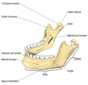
The mandible grows by endochondral ossification at the condyle and by surface apposition in certain areas. The condylar growth increases the height of the ramus and the overall length of the mandible!!

What that means is that for the human who has his/her normal growth plates closed, and they are have GH overstimulated, thus develop the medical condition known as acromegaly, the condylar growth, which is only from an articular cartilage layer, is enough to increase the length of the ramus, which is just the outer posterior lateral section of the mandible!!

This would suggest that if we could get off of our feet, and into an environment of lower gravity, HGH would work in increasing the length of the much bigger long bones like the femur trough articular cartilage layer deposition. If we were able to lie down on a horizontal bed for 24 hours for maybe 3-4 years, or live in a spaceship with 0 G, our long bones would grow longitudinally, at least up to a noticeable amount.

Concluding statement – Finally, let me reference page 220 of the book, the first paragraph “In acromegaly, growth of the mandible can again be initiated and continued even at a time when growth has normally ceased because of the peculiar histologis structure of the condyle. Here, the bone in younger individuals is covered by a cap of hyaline cartilage, which in turn, is covered by a thick layer of fibrous tissue. Remnants of the hyaline cartilage, which serves as a site of growth in the same way as the epiphyseal cartilage of the long bones, persist even in old individuals. As long as this hyaline cartilage is present, its proliferation can again be set in motion by a hyperactive pituitary gland, and it will then assume its function as a growth center of the mandible where it left off at the termination of normal growth. But even in cases in which an eosinophil adenoma or the hypophysis develops after the disappearance of the cartilaginous cap, a differentiation of hyaline cartilage from the fibrous covering of the condyle is not only possible, but also highly probably. If a new layer of hyaline cartilage has developed, endochondral growth can again set in after resorption of the terminal plate. As in other bones, the periosteal appositional growth is stimulated by the growth hormone; but this growth does not keep pace with the endochondral condylar growth, and the effect is a gradual increase in the mandibular angle.”

What the above paragraph shows is that you can make a bone longer even with just a cap of articular hyaline cartilage, similarly to how the antlers of deers grow out, which I did research more than 2 years ago. The key is to not be pushing down on the cartilage cap, which is not possible since we humans must walk. If we were upside down or had our overall weight lifted from the knee cap cartilage, the tibia bone will most likely start to slowly get longer.

Tyler’s Notes:

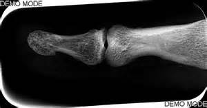
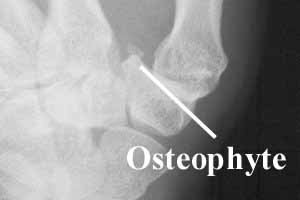
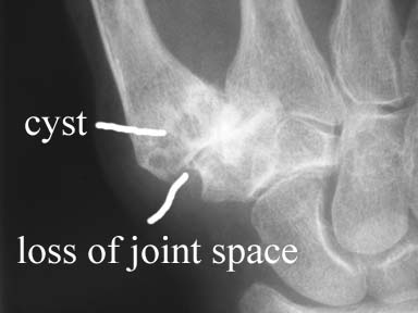
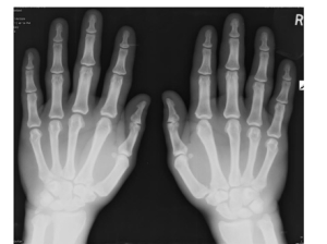
Here’s some hand xrays from an acromegalic hand:


Acromegaly: Hand

First, notice how white it is indicating very high levels of bone density.  The epiphysis is much wider than normal.

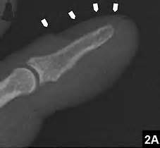
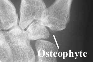
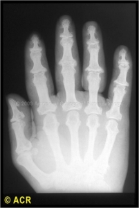
Here’s another acromegalic hand xray:

Acromegalic_hand_x-ray 2The bones are again much whiter than normal indicating increased density but the increase in epiphyseal width isn’t there.

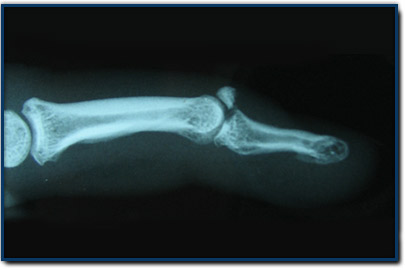
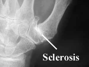
Here is another acromegalic hand xray more like the first:

Acromegalic_hand_x-ray 3

There is epiphyseal widening but not as extreme as the first xray.

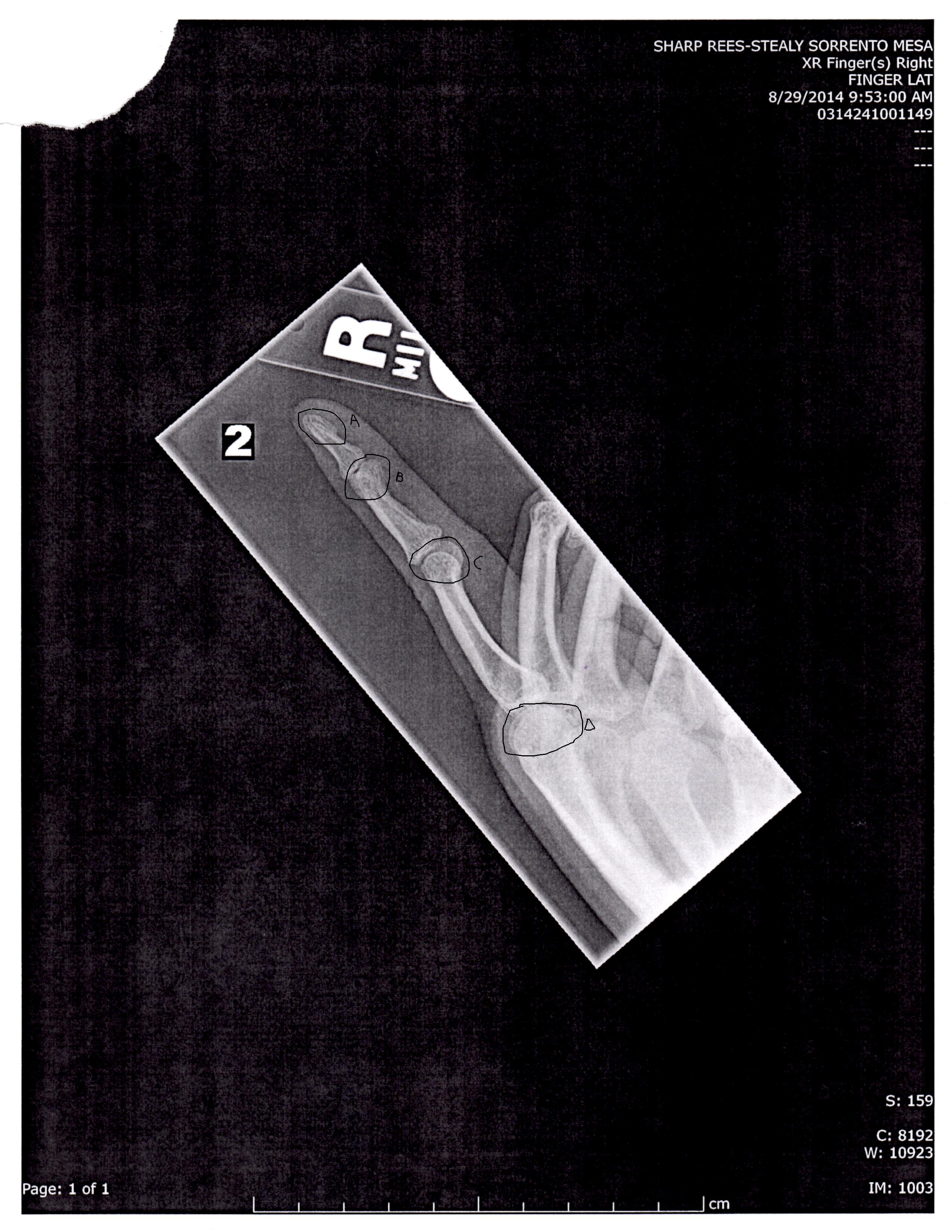
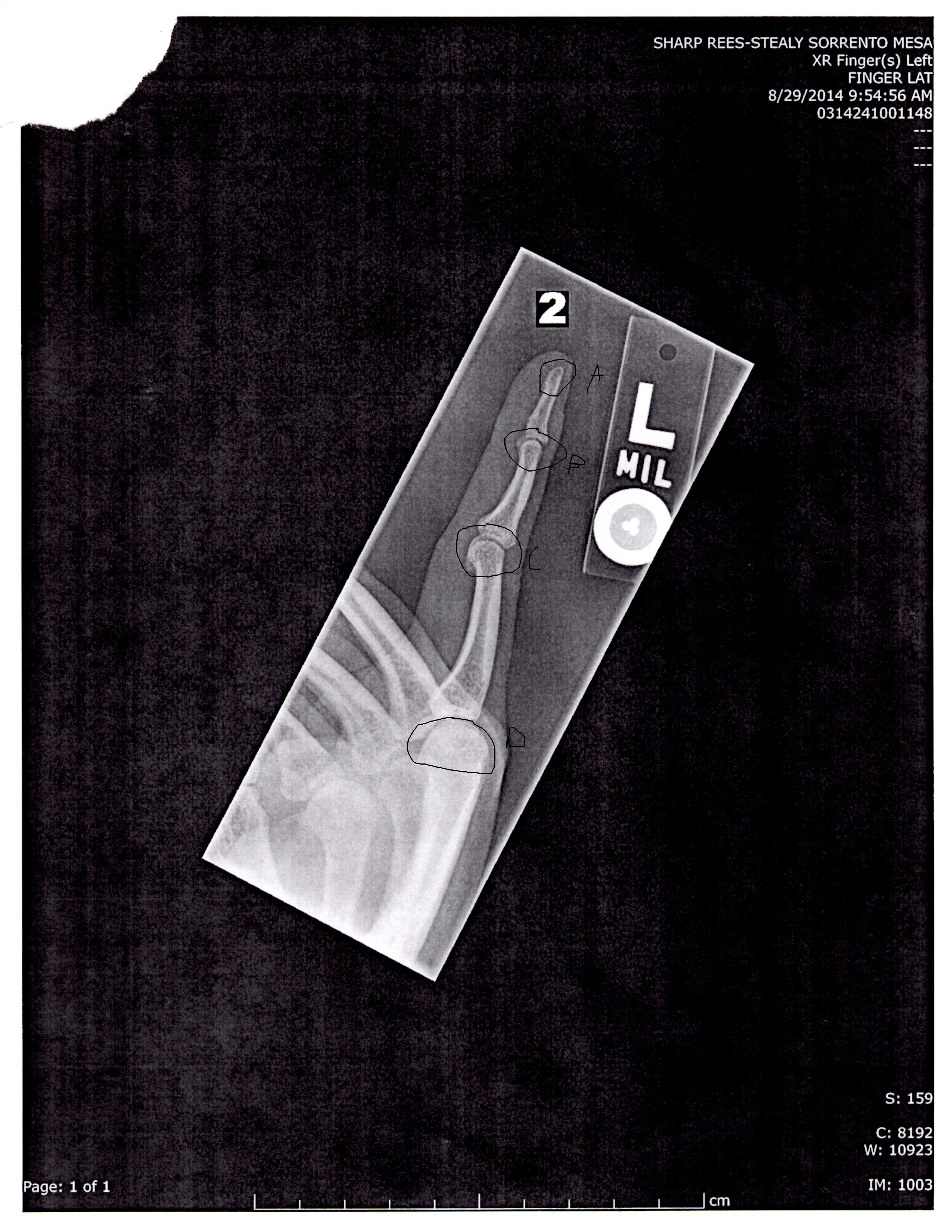
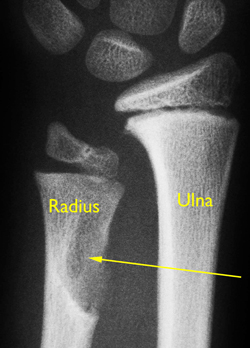
Let’s look at some developing fingers:
finger growth plates

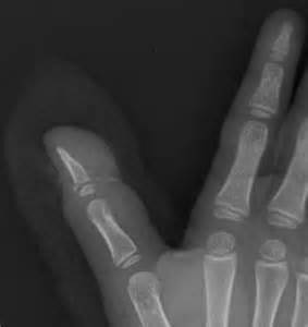
You can see that not only is the growth plate region not fully developed but the articular end without the growth plate too.  It is possible that this development which could occur with physical stimulation or with elevated HGH due to acromegaly could continue into adulthood and contribute to longitudinal bone growth.

My Girlfriend Claims She Grew 1 cm

I had said in a few posts back that I would not be doing too much posting on this website for the next 4-5 months as I am building up another project of mine. However, something interesting did happen today which made me start to rethink some theories I have had ever since last year.

I was on Skype with my girlfriend, and she told me that she found out from a recent Medical Exam that she was measured at 1.62 meters in height. Before that, she had always claimed that she was just 1.61 meters in height. Now, 1 cm is only about 0.394 inches, but since the rest of the world bases the measure of length on the meter, and thus the centimeter, that 1 extra centimeter in height is noticeable.

How was I supposed to take that type of news, since I am probably one of the only researchers in the world who has been actively looking for this type of phenomena??.

She is 26 now, with a birthday in January. It makes no sense for a girl of her age to ever increase in height.

However, females increasing in height in their late 20s is NOT completely unheard of.

She almost completely validates a theory I had about a year ago.

It seems that females who have been abnormally thin and with low bone density seem to have height increasing capabilities later than most other women.

In addition, she has been suffering from cold knees and knee pain for the longest time.

Recently she went to get 3 of her wisdom teeth removed, almost a dozen cavities filled, and had to see multiple medical specialists over medical problems.

In addition, she has been complaining for more than a year that she feels a strange feeling of pressure on her eyelid. A trip to the eye doctor suggested that she might very likely develop glaucoma. When the likelihood of glaucoma was ruled out, she went to see a Neuro-Specialist, who suggested that she might have a tumor developing in her brain, causing the optic nerve behind her eyeball to become pinched, causing that feeling of pressure on the eyelid. She did go get a CT Scan. Was it possible that she has some type of tumor in her pituitary causing pituitary adenoma?

So let me list the types of medical conditions she has been suffering from

  • Childhood history of being abnormally thin
  • She has the most common symptoms of a person with low bone density
  • She suffers from having knees which are always “cold”
  • Her knees have been developing pain, which has started to affect also her lower back.
  • Her blood test shows that her iron level is low, suggesting maybe anemia.
  • Has a long history of extremely heavy bleeding during menstration.

In response to all this, she has being taking the following supplements

  • Iron
  • Multivitamin
  • Magnesium
  • Vitamin D3
  • Glucosamine HCL (not the Sulphate type)
  • Lexapro

I don’t really know what to think right now since someone very close to me has gone through the small big of transformation which we have been searching for the last two years.

Could her claim to be a fluke, maybe a measurement error, or she just hasn’t measured herself in the last 10 years, ever since she noticed that she leveled off in height at the end of puberty?

If I was to be a sceptic, I would say that she has been at 1.62 meters in height for a long time, but she just never realized it. I remember more than a year ago, at a Zoo in Everland (South Korea), there was a height scale on the side of an exhibit to compare ourselves to the giraffe. You walk by it to get an idea of just how much taller a giraffe is to you. She walked under that height scale and stated that she was just 1.61 meters in height. Where I was standing, it looked like she was closer to the 1.62-1.63 meters range, and she was wearing TOMS shoes, the ones which only have less than 1 cm of heel height. If I was to guess, she was already 1.62 meters in height already.

However, let’s say that she did miraculously grow by 1 cm in the last few months. How would I even be able to validate that idea?

The only thing that I can think of is to maybe wait another 6 months or 1 year to see if she has pushed up in height again, to say 1.63 meters. If that happens, then I know that what I have been searching for the longest time may be already right next to me.

If she is growing, then it is time to ask myself, as probably the worlds most extensive amateur height increase researcher, how is it that my own girlfriend has grown at this late stage of her life?

Tyler’s Notes:  Due to being abnormally thin in childhood, it could be catch up growth due to lack of nutrients.  Remember there’s a proliferative capacity of the resting stem cells.  During the lack of nutrients this proliferative capacity is maintained, but there does some to be some height loss permanently that could not be caught up.

Very promising evidence that LSJL induced longitudinal bone growth in my finger

The coloration of regions of an area in an xray reflect their density.  If a region of bone is grayer than normal that bone is less dense.  If a bone is less dense that could mean that it is a region of new growth or that the bone is naturally less dense.  But even though some regions of less density are natural, if these regions of decreased density are expanded it is very possible that this could be indicative of increased growth.  If this expansion occurs in the longitudinal direction then it is indicative of longitudinal bone growth.  If you compare my right and left lateral finger xrays below you can see that my right bone has much greater expansion of these gray regions than the left and the regions extend into the longitudinal direction thus being indicative of longitudinal bone growth.

I need your help to confirm or deny this theory.

I statement I found on white versus black regions in x=rays: “On x-ray, bone and dense materials such as barium are white. Black would be air (lungs or air in the GI tract). Other tissues are in shades of gray. You can see the outline or shadow of the kidneys or certain muscles, for example. But you cannot see these things in detail. X-ray does not show the difference between normal vs. “dead” tissue. A CT scan, or even better, a MRI would be better suited for that type of diagnosis. ”

The darker regions of the bones are more likely to be less dense or possibly new grown bone.  Some bones are naturally less dense than others.  So we have to compare the bones also to standard bones.  If there are more gray areas than normal then it’s likely that there was new bone growth.

Here’s one lateral index finger view:
right lateral index finger

Which my finger looks more like.  Here’s one that my left finger looks more like(this is also a right index finger):
left proximal index finger

Have to compare color intensity of areas of right versus left bone.

Here’s a finger with some gray areas.  There is a large gray region on the tip of the proximal finger and some on the bottom part of the distal region of the medial index finger.  Note this is a fractured finger so we’d expect there to be some anabolic growth.

xray-mallet

So, it’s normal to have those regions of darker bone.

There are other potential reasons why one part of the bone can be much darker than the other but one strong possibility is that it is new LSJL induced bone growth.  Since LSJL involves lateral loading it makes sense that much of the growth can be better observed than a lateral angle(the differences in coloration can not be seen as well from an overhead view).  From the overhead view the right index finger looks to be whiter than the left index finger infact.  Click on the images below to enlarge them.

LSJL-right hand illustrated

LSJL left hand illustratedEach of the circled points A-D represents a region of bone that is much lighter than the other.  On the left side these regions are present but the are much lighter.  At point A it seems to blend better with the whiter region of the bone in contrast to the right side where it is more a bulbous head.  The bone at B looks totally different.  C and D are similar but the right(LSJL loaded side the darker region of the bone is much bigger).

Percentage growth based on darker area versus lighter area:

Proximal Head(Tip of dark region to tip of light): 23.4 pixels

(bottom of proximal index finger bone to top of light region): 170.4 pixels

13.7% growth.

However, I’d guess that my left bones are about 1% longer than my right.  If my right finger did grow longer than 10% my right proximal finger would measure longer than my left which is not the case.  You can see from the above images of other images that the normal finger has these darker regions, those regions are just expanded above normal.  So maybe the the proximal finger bone didn’t grow the entire gray region but the gray region just expanded in response to the stimulus.  In fact a study has shown that the finger bones naturally grow into adulthood.  So these darker regions of bone could be a sign of adult finger growth.  The same study stated that metacarpals tend to decrease in length so the fact that my right metacarpal grew longer is a good sign that this growth could extend to other bones.

What’s a bit unfortunate is that the growth plates are on the proximal(bottom) end of the finger bones and the gray regions are on the distal(top) ends of the finger bones.  However, the distal regions of my right finger bones do seem to be a bit more gray than the left bones so it is possible that the growth plate region was stimulated by LSJL.

Below is some more information about x-rays.

Here’s something I found describing xrays:

” The bone along the joint is usually whiter (called “sclerosis”) and may have little points of bone growing out (called “osteophytes”). There may be holes in the bone ( called “cysts”) and the bones may be starting to slide out of alignment (called “subluxation”).”<-None of these images quite explains the growth in LSJL.

Here’s a comment I found from Yahoo! Answers:

“Bones always appear as white images in regular xrays. However they arent really white because the film is clear, the plastic on the view box is white. But on xray film it is coated with a metalic layer. The xrays make the metalic layer stick to the film. So the black part of the film is where all the xrays reach the film. If the area on the xray is gray, then that means that some of the xrays are getting through implying that the structure is more dense than the black part. Soft tissue appears gray. The bone is a very dense structure allowing minimal to no xrays getting through. The film is then put through the processor and the film runs through chemicals that make the picture “stay” on the film. It is then rinsed and dried. Since the xrays make the metalic layer stick, none stick to the area of the bone because no radiation got through and the layer was washed off during processing. This is why bones appear white, they are the densest structure.”

Here’s another response from Yahoo! Answers regarding why one part of the bone is darker than the other:

“When a bone X-ray is taken, radiation is momentarily irradiated on the area being examined. X-rays pass through the body to produce an image on film. Structures like bone that are dense and have a high atomic number absorb a lot of X-rays, so less X-rays reach the film and the bone appears white on the film. Muscle, fat, tumour, and fluid absorb less X-ray than bone, and appear darker than bone on the film. Air distributed in various pockets within the body (usually due to an abnormality) has a low atomic number and density, and consequently appears black, because most X-rays pass through without being absorbed. You would be advised to consult the doctor or radiologist dealing with this matter and enquire as to the suspected cause of the description that you describe. You may find that further tests/examinations will result from the X-Ray result which will determine or assist the diagnosis.
The information provided here should not be used during any medical emergency or for the diagnosis or treatment of any medical condition. A licensed physician should be consulted for diagnosis and treatment of any and all medical conditions. “<-I don’t think air pockets could explain the larger gray area on the right side bone.

On this page is an image of tissue types in regard to whiteness: http://radiologymasterclass.co.uk/tutorials/physics/x-ray_physics_densities.html#top_first_img.  The coloration is consistent with soft tissue.

Here’s an image of a periosteal chondroma:

periosteal chondroma“In this x-ray of the wrist, the saucer-like hollow in the radius bone is a classic indication of a periosteal chondroma. “<-That’s not really consistant with what’s going on in my bone.

As for my ganglion cysts according to here, they are usually caused by myxoid(mucous) degeneration of collagen and usually lined with flattened mesothelium.

I can’t really find any adequate explanation as to the brighter regions of my finger.

Update by Michael:

From what I remember from being in some medical derivative courses back in my university days, I do remember the idea of X-rays penetrating into human tissue. White light means that the X-rays did not completely go through the tissue, while the black color is an indicator that the X-rays went through less dense tissue, like fat and tendons. 

I don’t have the enlarged X-rays pictures with me anymore but I don’t remember the left or right index fingers having any major differences in color. I think the important thing to remember is that we are looking for bone length differences, not bone density variations.

Now it is theoretically accurate to say that if we can show that LSJl caused bone density changes, causing the X-ray to cause a darker band on a metatarsal bone then LSJL might lead to bone longitudinal growth. However, on a much more practical level, even if the BMD is lowered, that doesn’t seem to translate to bone length increases, even after half a decade of doing joint loading on a consistent basis. I don’t think that it is worth trying to validate the LSJL theory based on a slight difference in color/density from comparing the left and right X-rays of the index fingers. That is too much of a stretch even for me.

As for the fingers growing longer, that actually makes a lot of sense. The human nose gets longer over time, the human ears droop lower and lower, and even the human mandibular joint area grows longitudinally if you have a pituitary problem.

For the old time regular readers, remember my study on why deer antlers can fall off, and regrow back in length every season? It is because of the occurrence of a laceration/osteonomy at the beginning, causing an open wound. A blastema develops in that region. That blastema eventually grows into a pseudo “growth plate”. The top part of the horn is where the mesenchyme is, and it is covered by a layer of perichondrium, NOT periosteum. That means that the top roof surface that is encapsulating the “rest zone’ type cells in the mesenchyme is flexible. It is only being pushed down by air and gravity, not an entire body, like the grow plates in the tibia of an adult human, which can be over 200 lbs of weight. Similarly, the human fingers have the same type of structure, a human anatomical peninsula, like the human tibia-knee area, which is more like an isthmus.

What I am trying to say is that fingers is similar to the horns on the deer. I have already shown a few times that some stem cell powder can regrow fingers. It is not that a big of a stretch to then conclude that human fingers, like noses, and ears, and pregnant female’s feet can grow throughout one’s life.


Spinal height gained by bedrest could be related to GAGs

This study shows that the increase in spinal height after bed rest and space flight may be due to an increase in GAG content in the intervertebral disc.

Assessment of Lumbar Intervertebral Disc Glycosaminoglycan Content by Gadolinium-Enhanced MRI before and after 21-Days of Head-Down-Tilt Bedrest.

“During spaceflight, it has been shown that intervertebral discs (IVDs) increase in height, causing elongation of the spine up to several centimeters. Astronauts frequently report dull lower back pain that is most likely of discogenic origin and may result from IVD expansion. It is unknown whether disc volume solely increases by water influx, or if the content of glycosaminoglycans also changes in microgravity. Aim of this pilot study was to investigate effects of the spaceflight analog of bedrest on the glycosaminoglycan content of human lumbar IVDs. Five healthy, non-smoking, male human subjects of European descent were immobilized in 6° head-down-tilt bedrest for 21 days. Subjects remained in bed 24 h a day with at least one shoulder on the mattress. Magnetic Resonance Imaging (MRI) scans were taken according to the delayed gadolinium-enhanced magnetic resonance imaging (dGEMRIC) protocol before and after bedrest. The outcome measures were T1 and ΔT1. Scans were performed before and after administration of the contrast agent Gd-DOTA, and differences between T1-values of both scans (ΔT1) were computed. ΔT1 is the longitudinal relaxation time in the tissue and inversely related to the glycosaminoglycan-content. For data analysis, IVDs L1/2 to L4/5 were semi-automatically segmented. Zones were defined and analyzed separately. Results show a highly significant decrease in ΔT1 (p<0.001) after bedrest in all IVDs, and in all areas of the IVDs. The ΔT1-decrease was most prominent in the nucleus pulposus and in L4/5, and was expressed slightly more in the posterior than anterior IVD. Unexpected negative ΔT1-values were found in Pfirrmann-grade 2-discs after bedrest. Significantly lower T1 before contrast agent application was found after bedrest compared to before bedrest. According to the dGEMRIC-literature, the decrease in ΔT1 may be interpreted as an increase in glycosaminoglycans in healthy IVDs during bedrest. This interpretation seems contradictory to previous findings in IVD unloading.”

“Average ΔT1 value of all intervertebral discs was 104.87 ms (sd 7.64 ms) pre-bedrest and -20.20 ms (sd 4.70 ms) post-bedrest.”

“It seems to be well established that increased GAG concentration within the IVD will result in a decrease in ΔT1. A high GAG-concentration causes a small ΔT1 during dGEMRIC-measurements because only small amounts of contrast agent shift into the IVD. A low GAG-concentration in turn leads to a high ΔT1. Increased ΔT1 after an intervention (as compared to before) has been interpreted as degeneration process”<So T1 has nothing to do with the height it has to do with the GAG content.  So a decrease in deltaT1 means that GAG content likely increased.