Monthly Archives: January 2013

A Personal Interpretation And Explanation Of Principles Of Biology And Genetics For The Purpose Of Height Increase

I had once tried to explain what I know about the principles of biology and genetics to a reader of the website so that they would be able to understand what I am talking about when I get into the details of the research.

This will be a rather lengthy post on my own personal interpretation and explanation of the biological, genetic, and biomolecular principles that are being used in the website and which I will be using when I read studies and articles from PubMed.

If we remember from our high school biology classes, we were taught about the idea of genes, hereditary, alleles, and we might have done a few allele charting. As a reminder, we must start from basics.


Biology

From high school (and some college level) biology basic principles…

1. From a macroscopic to microscopic direction approach, we can say that the human body consist of about a dozen organ systems.

From the Wikipedia article on Biological Systems

There is the circulatory, respiratory, lymphatic, digestive, reproductive, nervous, skeletal, endocrine, muscular, and a few others I don’t remember.

For our study on height increase, we are focused on the endocrine system and the skeletal system.

The specialized medical study of the endocrine system is called endocrinology and the doctors which focus on this sytem are known as endocrinologists (ex. Dr. Sanjay Gupta)

The specialized medical study of the skeletal system is called orthopaedics or orthopedics. From the wikipedia article on Orthopedic Surgery

“Orthopedic surgery or orthopedics (also spelled orthopaedic surgery and orthopaedics in British English) is the branch of surgery concerned with conditions involving the musculoskeletal system. Orthopedic surgeons use both surgical and nonsurgical means to treat musculoskeletal trauma, sports injuries, degenerative diseases, infections, tumors, and congenital disorders.”

2. At a lower level, each organ system is made up of individual organs.

An example is the fact that the digestive system is not just the stomach, but also the small intestine, large intestine, pancreas, gall bladder, liver, rectum, and throat area.

For the endocrine system, we study the hypothalamus, the pituitary gland, especially the anterior region, the adrenal glands, the thyroid glands. The glands all are for production of certain hormones that will be used to regulate the function of the body to reach some form of equilibrium/homeostasis.

For the skeletal system, we study the lower limbs, vertebrate, and specific cartilage regions mainly. We focus our attention in the lower limb area on the femur, the tibia/fibula combination which make up the lower part of the human leg, the patella, the calcaneus, and the cartilage on the ends of these bones. For the vertebrate parts, we are looking at all the areas, the cervical, thoracic and lumber regions, focusing on the intervertebral disks on possibly how to manipulate the annulus fibrosus and the  nucleus pulposus. These areas are notorious for being places where serious painful injuries can develop like bulging disks, herniated disks, pinched nerve, and lower back pain so if we plan to try any techniques or methods on the back area, we will be looking for something that doesn’t cause a major disturbance in the skeletal system of that region.

3. At a lower level, each organ is made of specific types of tissue.

From the Wikipedia article on tissue, “Tissue is a cellular organizational level intermediate between cells and a complete organism. A tissue is an ensemble of similar cells from the same origin that together carry out a specific function. Organs are then formed by the functional grouping together of multiple tissues.”

There is 4 types of tissues.

  1. Connective – Connective tissues are fibrous tissues. They are made up of cells separated by non-living material, which is called extracellular matrix. Connective tissue gives shape to organs and holds them in place. Both blood and bone are examples of connective tissue. As the name. It supports and binds other tissues. Unlike epithelial tissue, connective tissue typically has cells scattered throughout an extracellular matrix (Wiki)
  2. Nervous – N/A
  3. Muscle – N/A
  4. Epithelial – N/A

Since our study is looking at the skeletal system, we will not be going into any more detail on the nervous, epithelial or the muscle tissue. Now it is true that technically for us to do anything on the bone, we will have to possibly make a cut/incision through other tissue like the muscle or the epithelial but for now, our focus is on the connective tissues.

The connective tissue we will be focusing on are the cartilage and the bone, and how to remodel them into the form we are looking for. Ultimately, our goal is to figure out how to lengthen long bones (and maybe irregular bones) but to do that, we may also indirectly be affecting the other types of bones around it. To avoid any harm or injury to a subject, we want to also learn about the tissues that wrap themselves around on on the bones.

Cartilage is mainly made of a intercellular matrix filled with collagen and proteoglycans. As for bones, we want to focus on the non-living organic and non-organic materials that gives bones their hardness quality. The main component that forms this non-living matrix of the bones is called hydroxyapatite. It is calcium derived, and if we remember the calcium buildup we find in our bathrooms or certain areas in our body which are so hard to remove being stuck, we would remember that the hardness comes from calcium phosphates, which when in the human body seems to interact with water molecules to add the hydroxide group (-OH) and it gets attached turning it into the hydroxyapatite.

Something to note is that from my personal study on the tissue of cartilage, we can say that in the natural growing process of endochondral ossification, cartilage tissue turn into bone tissue by going through the process of “calcification”.

As for bones…

It is nearly concluded at this point that we should not be focusing on bone growth or bone regeneration but actually cartilage regeneration. Bone growth and regeneration is relatively easy. The bones after fracture automatically and relatively quickly heal themselves. You can increase that healing process by using LIPUS, PEMF, capacitative electrical field application, dynamic mechanical loading, or adding certain osteogenic growth factors. Bones regenerate very quickly and easily.

So what I am implying is that when we are analyzing tissues, at that level, we will be focusing almost all of our effort into looking at cartilage creation and regeneration either inside the bones or through in vitro methods and then do an implantation. Bone tissues we will look at sometimes but the focus will not be on that tissue.

4. At a lower level each tissue is made of a certain type of cell.

For our research we will be only looking at two types of cells, cartilage cells, chondrocytes and chondroblasts, and bone cells, osteocytes, osteoblasts, and osteoclasts.

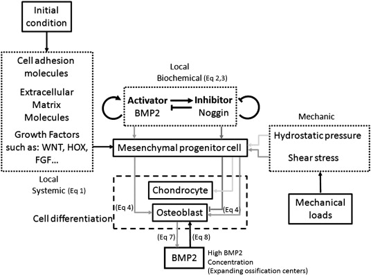
Chondrocytes will be intensively researched here, especially looking for ideas and techniques on how to get the progenitor cells to differentiate into chondrocytes, how to keep the chondrocytes in that state without going into apoptosis or differentiating further into osteocytes and osteoblasts. Chondrocyte Proliferation, Chondrocyte Hypertrophy, and stacking into the correct direction for chondrocyte columns is the key and we will keep looking to find ways to manipulate the genome, nucleotides, or even the microRNA to try to get the chondrocytes to stay in the small region of function we are looking for to increase longitudinal growth.

As for bone cells, osteoblasts is the bone cells which create bone material. Osteoclasts is what break down bone material. We want to focus more on osteoclasts and try to get the osteoclasts to function at a higher rate in a certain region of the skeletal system, like in a band on the long limbs so that bone resorption is increased. If bone resorption is increased, we might be able to get the long bones to become weaker for tensile loading or get the hard bone material to be replaced at a reasonably slow rate to be replaced by chondrocytes, and then cartilage tissue.

5. At a lower level each cells is controlled by certain proteins, kinases, signals, enzymes.

Proteins provide the basic structures for every organ system in the body. The human body is made of many things, including sugars and lipids in the form of adipose tissue. There is also the nucleic acids, and minerals like calcium, phosphorous, and iron which form hard deposits in the extracellular matric of bones, but overall, proteins are what really matter since the very goal of the genes in our genome is to make proteins. Proteins have so many functions that it would be really hard for me to list all of its functions.

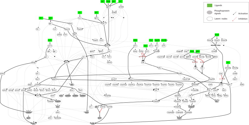
For our purposes, I’ll say that proteins are what will eventually make up the components that are a part of all the signal pathways we find in the intracellular matrix (within the cytoplasm). This includes previous posts about the Wnt/Beta-Catenin Signal Pathway, the MAPK/ERK pathway, and the PI3K/AKT/mTOR pathway. Proteins will act as the signals which send signals from the outside of a cell into a cell, and proteins will be what is send out of the nucleus to the outside of the cell to direct the process of the body.

Our research is specifically focused on seeing which proteins have either a direct or indirect effect on chondrocyte proliferation, hypertrophy, and differentiation in the natural growing process of endochondral ossification in postnatal humans. At some point we are hoping to map out the signal map of the myriad of proteins which effect the growth plate. Once that is done, we hope that  we can pinpoint the 4-5 most influential proteins which either goes into the cell nucleus or leave the cell nucleus which effects the growth process. We can then use external proteins to be injected to either inihibit the proteins which are slowing down the growth process or to up-regulate the proteins which make the growth process still possible.

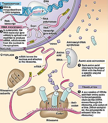
6. At a lower level each protein is produced by genetic mechanisms like transcription, reverse transcription, translation, as well as post-translational processes.

In the nucleus of a cell (sometimes the mitochondria will also have its own DNA/RNA stuff) there will be DNA that need to be duplicated or manipulated on. Go to the Genetics section for more details on what each process or element in genetics will mean.

What I am going to say here is our most important research probably will eventually come down to the genetics, specifically to the microRNA and how each of the microRNA effects each specific gene of the body Sciences have concluded that the human genome has only around 25,000-30,000 genes, much less than expected. However there is so much phenotypical variation in the human species. No two people in our 7 billion population looks alike, and yet it was found that humans in general are 99.99% alike in our genes and differences. Even a dark skinned male from tanzania is still 99.95% similar in the types of genes they have to a light skinned female from norway so it is clear that the master regulators of the human trait of height is probably the mRNAs and that the phenotypical differences we see can not be just from gene differences.


Genetics

From high school (and some college level) genetics basic principles…

The individual human is composed of around 100,000,000,000,000 (that’s 100 trillion!!) cells more or less. In almost all of the many different types of cells in the human, except for say red blood cells, there is a nucleus in the cell. The types of cells that have nucleus include fat cells (adipose), nerve cells (neurons), bones cells (osteoblasts and osteocytes), and many other types. In each of these cells in the nucleus, there is a copy of the entire human genome inside. The human genome has (barring people with strange genetic deformities) 46 individual chromosomes. Some resources would reinterpret the 46 number to say that there is 23 pairs of chromosome pairs. The main exception for the 46 pairs is the human sex cells, the sperm and eggs (aka gametes), which have a 23 individual chromosomes. When there is 46 individual chromosomes total in a cell, that is known as a diploid. When there is 23 chromosome in a gamete, that is known as a haploid.

In one human gemete with the 23 chromosomes, there is about 3 billion DNA base pairs. Base pairs is where two of the 5 main types of nucleotides attach to each other. There is the A,T,G, and C for the nucleic acid known as DNA (deoxyribonucleic acid). For the other main type of nucleic acid, known as RNA (ribonucleic acid), there is the A,U,G, and C.

transcriptionDNA seems to have two main function that that is for it to replicate or to make RNA for some reason.

There is 4 main types of process that is talked about in genetics a lot

  • Replication – replication seems to be exactly what it sounds like. both the DNA and RNA have processes that allow them to replicate (reproduce) an exact copy of themselves
  • Transcription – the process where the DNA is cleaved/cut in the middle breaking the bonds the A (adenine )made with the T (thymine) and the G (guanine) made with the C (cytosine) as a nucleotide base pair. This results in only one half of the double helix strand forming a RNA.
  • Reverse Transcription – the process where the single strand RNA becomes a double helix strand DNA again.
  • Translation – the process where the RNA becomes a protein. For a more detailed description of the process, check out the Wikipedia article on Translation.

Transcription-TranslationThere is 4 main types of RNA (Ribonucleic Acid) that are really important.

  • tRNA – transfer RNA – 
  • mRNA- messenger RNA – 
  • rRNa – ribosomal RNA – 

the 4th type of RNA which our research may have to go into is which is known as microRNA

At this point, the research will be on the microRNA and how it affects the genes that are being produced by the other RNAs.

Conclusion:

As any biology or genetics university major can see, my own personal knowledge on human biology and physiology is very limited at this point. My genetics is very weak and I need to read and study far more to get further into the research. There are at least 100 more published papers I would have to go through before I can come up with anything of real value.

What is the biomechanical method of bone formation?

If we understand how biomechanical bone formation is induced from mesenchymal stem cells we can induce new bone growth to reinvigorate longitudinal growth.

A computational model of clavicle bone formation: A mechano-biochemical hypothesis.

“Clavicle development arises from mesenchymal cells condensed as a cord extending from the acromion towards the sternal primordium. First two primary ossification centers form, extending to develop the body of the clavicle through intramembranous ossification. However, at its ends this same bone also displays endochondral ossification.  [These] embryological events [occur in] two serial phases: first formation of an ossified matrix by intramembranous ossification based on three factors: systemic, local biochemical, and mechanical factors. After this initial phase expansion of the ossified matrix follows with mesenchymal cell differentiation into chondrocytes for posterior endochondral ossification. Our model provides strong evidence for clavicle formation integrating molecules and mechanical stimuli through partial differentiation equations using finite element analysis.”

chondrocyte formation

“areas of low octahedral shear stress and high hydrostatic stress promote bone formation by endochondral ossification. On the contrary, areas of octahedral shear stress result directly in osteogenic induction.”

hydrostatic stress promotes mesenchymal cell into a cartilaginous differentiation pathway, whereas octahedral shear stress stimulates mesenchymal cells to differentiate into bone.”

Signaling pathways in normal chondrocytes versus LSJL gene expression

Modeling of signaling pathways in chondrocytes based on phosphoproteomic and cytokine release data.

“The signaling pathways downstream 78 receptors of interest are interrogated. On the phosphoproteomic level, 17 key phosphoproteins are measured upon stimulation with single treatments of 78 ligands. On the cytokine release level, 55 cytokines are measured in the supernatant upon stimulation with the same treatments. Using an Integer Linear Programming formulation, the proteomic data is combined with a priori knowledge of proteins’ connectivity to construct a mechanistic model, predictive of signal transduction in chondrocytes.
We were able to validate previous findings regarding major players of cartilage homeostasis and inflammation (E.g. IL1B, TNF, EGF, TGFA, INS, IGF1 and IL6). Moreover, we studied pro-inammatory mediators (IL1B and TNF) together with pro-growth signals for investigating their role in chondrocytes hypertrophy and highlighted the role of underreported players such as INHBA, DEFB1, CXCL1 and Flagellin, and uncovered the way they cross-react in the phosphoproteomic level.”

“up-regulation of the SOX9 transcription factor induced by TGFB or FGF stimulation leads to collagen synthesis. On the other hand, over-activation of NFKB induced by several pathways (E.g. Inflammation related pathways or bone development processes leads to the release of MMPs and collagen degradation.”

” a large number of stimuli raised a significant response in chondrocytes, activating at least one phosphoprotein signal. As positive control observations, well known players such as IL1B, TNF, EGF, TGFA, INS, IGF1 and IL6{up in LSJL} responded as expected from previous studies. The pro-inflammatory mediators IL1B and TNF activated IKB, HSP27{note this is reduced by Lithium}, MAPK14 (p38) and JUN{up in LSJL}, already known to promote inflammation in cartilage, together with pro-growth signals such as CREB, ERK, GSK3, IRS1 and MAP2K1 (MEK12), validating their role in chondrocyte hypertrophy”

” pro-growth stimuli such as EGF, TGFA and INS activated only anabolic pathways, leaving inflammation related signals unaffected.”

Stimuli also found to affect chondrocytes include “INHBA (Inhibin beta A), ADIPOQ (Adiponectin), DEFB1 (Defensin beta 1), BTC (Betacellulin){up in LSJL}, CXCL1{highly upregulated by LSJL}, HBEGF{up}, IL19, CXCL10, ODN2006 (TLR9 ligand), NOG (Noggin) and Flagellin.”

chfigure2_v2

“Major inflammatory mediators such as IL1B and TNF, signal through their receptors to IKB, MAPK14, HSP27 and JUN. IL1B also activates CREB and MAP2K1 (growth related signals) via TRAF6″

” Pro-growth stimuli such as TGFA, BTC, EGF, IGF1, INS and FGF2 signal through GRB2 to SOS, RAS and from there either to MAP2K1 (MEK12) via RAF1, or signal through PI3K to AKT and to CREB. IL6 activates mostly STAT3 via JAK1.”

“CXCL1, a small cytokine of the CXC family, binds to CXCR2 and activates RPS6KA1. HBEGF, a ligand of the EGFR, signals via the same pathways as EGF, BTC and TGFA. DEFB1, a TLR ligand, signals through TLR4 to RAC1 and from there to the MAPKs and finally activates HSP27 demonstrating pro-inflammatory action. Flagellin, also a TLR ligand, signals through TLR5 to MYD88 and then merges with the IL1 pathway activating major inflammatory signals, CREB and MAP2K1. INHBA, a ligand of the TGFBR, signals via the MAPKs to activate JNK and P53.”

Could what’s in the drinking water affect height?

Since drinking water contains Arsenic and Fluoride which can affect gene expression, it’s conceivable that drinking water could affect height growth.

Arsenic and fluoride co-exposure affects the expression of apoptotic and inflammatory genes and proteins in mononuclear cells from children.

“Humans may be exposed to arsenic (As) and fluoride (F) through water consumption.  Herein, the expression of cIAP-1, XIAP, TNF-α, ENA-78[also known as CXCL5 which is downregulated by LSJL], survivin, CD25, and CD40 was evaluated by RT-PCR. Additionally, the surface expression of CD25, CD40, and CD40L on peripheral blood mononuclear cells were analyzed by flow cytometry, and TNF-α was measured by western blotting. This study examined 72 children 6-12 years old who were chronically exposed to As (154.2μg/L) and F (5.3mg/L) in drinking water and in food cooked with the same water. The urine concentrations of As (6.9 to122.4μg/L) were positively correlated with the urine concentrations of F (1.0 to 8.8mg/L). The CD25 gene expression levels and urine concentrations of As and F were negatively correlated, though the CD40 expression levels were negatively correlated only with the As concentration. Age and height influenced the expression of cIAP-1, whereas XIAP expression was correlated only with age. Additionally, there was a lower percentage of CD25- and CD40-positive cells in the group of 6- to 8-year-old children exposed to the highest concentrations of both As and F when compared to the 9- to 12-year-old group (CD25: 0.7±0.8 vs. 1.1±0.9. CD40: 16.0±7.0 vs. 21.8±5.8). PHA-stimulated lymphocytes did not show any changes in the induction of CD25, CD69, or CD95. In summary, high concentrations of As and F alter the expression patterns of CD25 and CD40 at both the genetic and protein levels. These changes could decrease immune responses in children exposed to As and F.”

“Studies of As-exposed populations have demonstrated unusual gene expression profiles for cell cycle control-related factors, transcription factors, and inflammatory molecules”

“Using microarrays, our group determined that the IL-6, IL-1β, TNF-α, TGF-β, CD40, IL-2RA, CD40L, CD25, ENA78, SURVIVIN, XIAP, and IAP-1 genes are differentially expressed in adults chronically exposed to high concentrations of As (22.5–148.9 μg/L) and F (2.3–5.4 mg/L) compared to the control group (As: 0.3–1.4 μg/L; F: 0.1–0.7 mg/L)”<-Not strong height altering genes except possibly TGF-Beta.

“cIAP-1 expression was positively correlated with age and height”<-Can clAP-1(also known as clasp1) affect height?

XIAP and TNFalpha were also associated with height but not above statistical significance but closer than the rest of the genes.  p<0.15.  TNFalpha definitely affects height but there is it is likely that there’s an optimal quantity of TNFalpha for height growth rather than it just being good or bad.

This study could link Clasp1 to height growth:

Association of TALS developmental disorder with defect in minor splicing component U4atac snRNA.

“The spliceosome, a ribonucleoprotein complex that includes proteins and small nuclear RNAs (snRNAs), catalyzes RNA splicing through intron excision and exon ligation to produce mature messenger RNAs, which, in turn serve as templates for protein translation. We identified four point mutations in the U4atac snRNA component of the minor spliceosome in patients with brain and bone malformations and unexplained postnatal death [microcephalic osteodysplastic primordial dwarfism type 1 (MOPD 1) or Taybi-Linder syndrome (TALS); Mendelian Inheritance in Man ID no. 210710]. Expression of a subgroup of genes, possibly linked to the disease phenotype, and minor intron splicing were affected in cell lines derived from TALS patients. Our findings demonstrate a crucial role of the minor spliceosome component U4atac snRNA in early human development and postnatal survival.”

” The TALS phenotype includes marked intrauterine and postnatal growth retardation; short, bowed long bones with severe delay in epiphyseal maturation”

“The U4atac snRNA is located within intron 2 of the CLASP1 gene, –682 base pairs (bp) to –556 bp upstream of exon 3. Because of this, TALS mutations could, in theory, alter CLASP1 splicing and/or expression. In silico splice site predictions, real time–quantitative polymerase chain reaction (RT-qPCR) analysis of CLASP1 mRNA levels, and an RT-PCR study of CLASP1 exon 3 splicing in TALS patients revealed no effects of TALS mutations on CLASP1 expression or splicing”

However, the study seems to show a lack of connection of CLASP1 to dwarfism.

Idursulfase

Can Idursulfase affect normal individuals who want to grow taller?

The Effect of Recombinant Human Iduronate-2-Sulfatase (Idursulfase) on Growth in Young Patients with Mucopolysaccharidosis Type II.

“Mucopolysaccharidosis type II (MPS II; Hunter syndrome) is an X-linked, recessive, lysosomal storage disorder caused by deficiency of iduronate-2-sulfatase. Early bone involvement leads to decreased growth velocity and short stature in nearly all patients. [We] investigate the effects of enzyme replacement therapy (ERT) with idursulfase (Elaprase) on growth in young patients with mucopolysaccharidosis type II. Analysis of longitudinal anthropometric data of MPS II patients (group 1, n = 13) who started ERT before 6 years of age (range from 3 months to 6 years, mean 3.6 years, median 4 years) was performed and then compared with retrospective analysis of data for MPS II patients naïve to ERT (group 2, n = 50). Patients in group 1 received intravenous idursulfase at a standard dose of 0.58 mg/kg weekly for 52-288 weeks. The course of average growth curve for group 1 was very similar to growth pattern in group 2. The average value of body height in subsequent years in group 1 was a little greater than in group 2, however, the difference was not statistically significant. In studied patients with MPS II, idursulfase did not appear to alter the growth patterns. ”

So idursulfase therapy only seems to have an impact when used before 6 years of age.

“Mucopolysaccharidosis type II (MPS II, Hunter syndrome, OMIM# 309900) is caused by the deficiency of the enzyme iduronate-2-sulfatase (I2S; EC 3.1.6.13) that is responsible for breaking down heparan and dermatan sulfate (HS and DS) within the cells”

I don’t think idursulfase will have an impact on “normal” individuals.  And it’s only having above a certain threshold of the enzyme affects height.

Increase Height And Grow Taller Using Back Bridging Exercises To Stretch The Torso And Vertebrate

At some point I had found profiles on the old GiantScientific.com forum suggesting that maybe the stretching method known as Back Bridging can help a person increase and grow taller since they are stretching out the torso and vertebrate.

At this point I can’t find the thread or posts which does suggest this form of exercise. However there are quite a few yoga websites which suggest that the idea is feasible. or at least some weak content Internet Marketer created pages which suggested this idea. So I wanted to do a little bit more research to see whether the idea had any chance for effectiveness.

From the MMA website Sherdog.com, I would learn that the backbridging exercise can increase flexibility and build upper body strength.

From the website…

“Bridging works your entire posterior chain, while simultaneously stretching your hip flexors, shoulders and upper back (places where guys tend to get tight). Back bridging can also strengthen your vertebrae and increase spinal circulation.”

This section talks about something called a “hip flexors” when I would google and find out is the muscle that connects the main torso body to the legs, around the same region as where the leg hip joint attaches to the pelvis. Everything else is normal biology language. The other thing that I noticed was that the vertebrate can be strengthened, but it says nothing about stretching or elongating the vertebrate. When a body part is strengthened, it usually suggest that it got thicker or wider, not longer. From this quick review from a MMA based website, there is nothing to suggest that a person has any chance of growing taller from doing the backbridging exercise. The fact that it can increase flexibility gives a slight chance, but the posterior region is stretched in the opposite direction from the normal stretching.

The other website or reference suggesting this would work is from HERE. Unfortunately there is not a lot of information that it is giving except this excerpt below…

“…lie on your back and put arms on the floor beside your legs. After this lift your abs and pelvis towards the ceiling”

They talk about lifting one’s abs and pelvis but that would not change the internal body structure alignement. We could hope that the hip flexors and the ligaments that hold the total hip bone to become slightly moved away from the socket, but that would lead to injury. Lifting the pelvic suggest that maybe the lumbar vertebrate area can be stretched out, but we find out that the lower  vertebrate are almost fused together.

backbridgingSo is it possible that the backbridging pictured to the right can make a person grow taller at least temporarily by stretching out their vertebrate or spine? 

At this point I would say no. It is true that the stretching of the human vertebrate may cause temporarily elongation, but most of the human torso’s weight and organs are on the anterior side of the vertebrate (in front of). When a person does a normal stretch like getting into a pair of yoga pants, and trying to touch their toes while going into a downward dog position, The vertebrate will be slightly bent in the anterior direction and that will cause a little bit of increased separation and width in the intervertebral disks but the vertebrate physiologically designed to be able to bend in that direction. After the person gets back up to the up-right position, The disks may stay in that expanded volume state for a short while.

But one could argue that the backbridging is just the opposite of the position described where the vertebrate are bent in the opposite direction. However my argument is that the human vertebrate is not designed to bend in that direction. If a person did more of the back bridging, they would strengthen their back, but I would guess that the disks between the vertebrate are more likely to be compressed than expanded. This is due to the fact that the most of the torso weight is in the anterior of the vertebrate. When you are backbridging the weight of the front side of the body pushes down ward, causing the disks to be squeezed even more between the vertebrate.

arch-romanThis is similar to the architecture design known as the roman arch. Notice that with the roman arch, the arch stays in placed being squeezed into stability due to the weight from above it. That is similar to what the human body’s intervertebral disks do when they are bend in in the opposite direction and the weight of the body pushes downward on the vertebrate.

On the other hand, when you are stretching out in the normal direction hands touching the toes, the vertebrate is not being pushed down by the anterior torso part, giving it a better chance in expanding, than being compressed.