
Modeling of signaling pathways in chondrocytes based on phosphoproteomic and cytokine release data.
“The signaling pathways downstream 78 receptors of interest are interrogated. On the phosphoproteomic level, 17 key phosphoproteins are measured upon stimulation with single treatments of 78 ligands. On the cytokine release level, 55 cytokines are measured in the supernatant upon stimulation with the same treatments. Using an Integer Linear Programming formulation, the proteomic data is combined with a priori knowledge of proteins’ connectivity to construct a mechanistic model, predictive of signal transduction in chondrocytes.
We were able to validate previous findings regarding major players of cartilage homeostasis and inflammation (E.g. IL1B, TNF, EGF, TGFA, INS, IGF1 and IL6). Moreover, we studied pro-inammatory mediators (IL1B and TNF) together with pro-growth signals for investigating their role in chondrocytes hypertrophy and highlighted the role of underreported players such as INHBA, DEFB1, CXCL1 and Flagellin, and uncovered the way they cross-react in the phosphoproteomic level.”
“up-regulation of the SOX9 transcription factor induced by TGFB or FGF stimulation leads to collagen synthesis. On the other hand, over-activation of NFKB induced by several pathways (E.g. Inflammation related pathways or bone development processes leads to the release of MMPs and collagen degradation.”
” a large number of stimuli raised a significant response in chondrocytes, activating at least one phosphoprotein signal. As positive control observations, well known players such as IL1B, TNF, EGF, TGFA, INS, IGF1 and IL6{up in LSJL} responded as expected from previous studies. The pro-inflammatory mediators IL1B and TNF activated IKB, HSP27{note this is reduced by Lithium}, MAPK14 (p38) and JUN{up in LSJL}, already known to promote inflammation in cartilage, together with pro-growth signals such as CREB, ERK, GSK3, IRS1 and MAP2K1 (MEK12), validating their role in chondrocyte hypertrophy”
” pro-growth stimuli such as EGF, TGFA and INS activated only anabolic pathways, leaving inflammation related signals unaffected.”
Stimuli also found to affect chondrocytes include “INHBA (Inhibin beta A), ADIPOQ (Adiponectin), DEFB1 (Defensin beta 1), BTC (Betacellulin){up in LSJL}, CXCL1{highly upregulated by LSJL}, HBEGF{up}, IL19, CXCL10, ODN2006 (TLR9 ligand), NOG (Noggin) and Flagellin.”
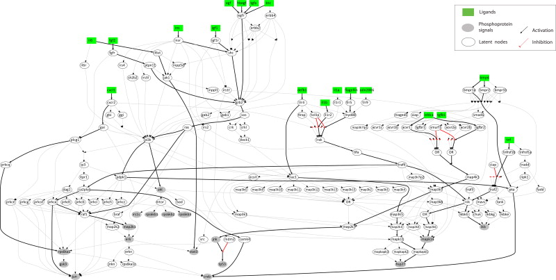
“Major inflammatory mediators such as IL1B and TNF, signal through their receptors to IKB, MAPK14, HSP27 and JUN. IL1B also activates CREB and MAP2K1 (growth related signals) via TRAF6″
” Pro-growth stimuli such as TGFA, BTC, EGF, IGF1, INS and FGF2 signal through GRB2 to SOS, RAS and from there either to MAP2K1 (MEK12) via RAF1, or signal through PI3K to AKT and to CREB. IL6 activates mostly STAT3 via JAK1.”
“CXCL1, a small cytokine of the CXC family, binds to CXCR2 and activates RPS6KA1. HBEGF, a ligand of the EGFR, signals via the same pathways as EGF, BTC and TGFA. DEFB1, a TLR ligand, signals through TLR4 to RAC1 and from there to the MAPKs and finally activates HSP27 demonstrating pro-inflammatory action. Flagellin, also a TLR ligand, signals through TLR5 to MYD88 and then merges with the IL1 pathway activating major inflammatory signals, CREB and MAP2K1. INHBA, a ligand of the TGFBR, signals via the MAPKs to activate JNK and P53.”