
This can provide benefit to the theory of microgrowth plates.
CASE REPORTS: Enlargement of a Calcaneal Osteochondroma after Skeletal Maturity
“Growth or radiologic modification of an osteochondroma after the epiphyseal plate closes suggests the diagnosis of malignant transformation to a chondrosarcoma. However, extensive growth of an osteochondroma in a skeletally mature patient whose tumor proved benign has been reported. We report a similar case in an adult who had a solitary osteochondroma of the calcaneus. The lesion showed marked growth and was removed. Histologic examination showed no evidence of malignancy, and there was no recurrence during the 4-year followup.”
The man was 36 years old although it’s alluded that the osteochondroma was developed during skeletal development.
” bulky 2-cm thick cartilaginous cap, an irregular and indistinct appearance of the surface of the calcified tissues beneath the cap, scattered calcifications in the soft tissue component of the tumor, radiolucent foci in the lesion, and a large low-density soft tissue mass. However, no destruction of the adjacent bone was visible.”
The images tend to not be informative. Just a white mass amonst the calcaneus bone.
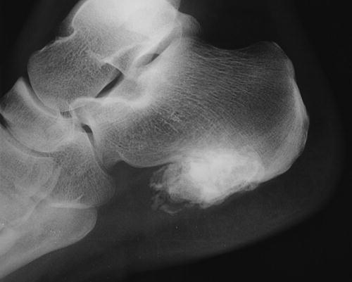 Looking at that image it seems possible that the growth could provide a height and length increase but seems mostly lateral to the region that would increase foot height and length.
Here’s a normal calcaneus:
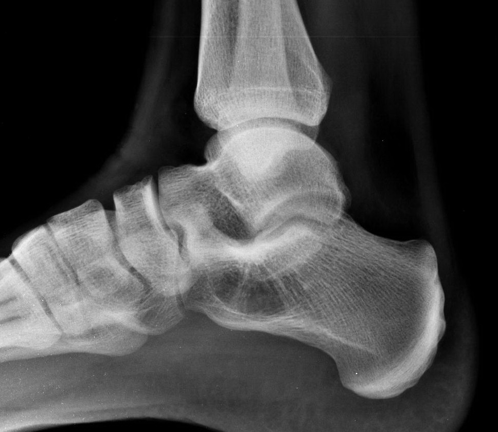 Although this foot seems much larger than the normal.
“the cartilaginous cap appeared hyperplastic and had foci of increased cellularity. The chondrocytes were organized in clusters and had no significant atypia.”
“An osteochondroma develops when growth plate tissue is extruded laterally and proliferates into an exostoses. Therefore, an osteochondroma can arise in any bone that undergoes endochondral ossification, and it usually stops growing when the physes close.
“