
This is the most significant LSJL study to date.
Three key takeaways from this study:
1) LSJL increases bone length in existing growth plates via traditional mechanisms(chondrocyte hypertrophy)
2) LSJL increases bone length in non-traditional mechanisms as shown by the fact that LSJL also stimulates the reserve zone. Reserve zone cells being the chondrocyte precursor cells and the ones able to form new growth plates.
3) LSJL dramatically alters the microenvironment of the bone(as shown by the histological slides). It’s unclear exactly what changed but the decrease in bone trabeculae and the increase in bone marrow means that an LSJL loaded bone is more permissive to growth plate formation. Osteomy is essential for renewed longitudinal bone growth. As cartilage is capable of interstitial growth which induces longitudinal bone growth whereas bone is not.
Lengthening of mouse hindlimbs with joint loading
“Loads were applied to the left hindlimb (5-min bouts at 0.5 N[at 5Hz) of C57/BL/6 mice (21 mice, ~8 weeks old). Compared to the contralateral and age-matched control groups, knee loading increased the length of the femur by 2.3 and 3.5%, together with the tibia by 2.3 and 3.7%, respectively. In accordance with the length measurements, knee loading elevated BMD and BMC in both the femur and the tibia. Histological analysis of the proximal tibia revealed that the loaded growth plate elevated its height by 19.5% and the cross-sectional area by 30.7%. Particularly in the hypertrophic zone, knee loading increased the number of chondrocytes as well as their cellular height along the length of the tibia.”
3min/day for 5 days/week for 10 days total was LSJL applied. Bone was harvested 18 days after the last loading.
“Femoral length was defined as the maximum distance from the distolateral condyle to the
most medial and proximal position on the femoral head. Tibial length was defined from the most proximal position of the tibial plateau to the most distal position of the medial malleolus.”<-this is important as changing where and how femoral length is measured would effect total femur length. It is hard to tell the ramifications of this length setting for sure without more data though.
“The height of the growth plate (GP) was defined from the apical[apex] border of the reserve zone to the lower border of the mineralized cartilage”<-So the measurement of growth plate height would likely include not just growth plate chondrocytes but chondrocyte progenitor cells. And you’d need chondrocyte progenitor cells to form new growth plates.
According to Artificial selection sheds light on developmental mechanisms of limb elongation, an increased number of proliferative chondrocytes is likely the cause of increased height.
“the upper boundary of the hypertrophic zone was identified at the margin of the
chondrocytes that increased their size relative to those in the proliferative zone, whereas its lower boundary was at the terminal intact chondrocytes next to the metaphysis”
“At the cellular level, the numbers of proliferative and hypertrophic chondrocytes were counted and the total number of chondrocytes was calculated as their sum. The height of hypertrophic chondrocytes was determined using at least 20 cells in each slice”
“During knee loading, no apparent damage was detected at the site of loading or injection.”
“the longitudinal length of the femur was increased by 2.3% (14.19 ± 0.28 mm in contralateral control; 14.51 ± 0.28 mm in knee loading)”
“the longitudinal length of the tibia was increased by 2.3% (16.68 ± 0.23 mm in contralateral control; 17.06 ± 0.21 mm in knee loading)”<-interesting that the percent increase is so comparable(both 2.3%).
“Compared to the age matched control, knee loading increased the longitudinal length by 3.5% in the femur and by 3.7% in the tibia”<-Also a very similar percentage.
In the elbow loading study, “humerus was elongated by 1.2% compared to the contralateral and age-matched controls, while the ulna had become longer than the contralateral control (1.7%) and the age-match control (3.4%)”. In 16 week mice(see same link above), the increase in length was 1.6% in the tibia.
Here is the growth plates under LSJL(I provide a more detailed analysis here):
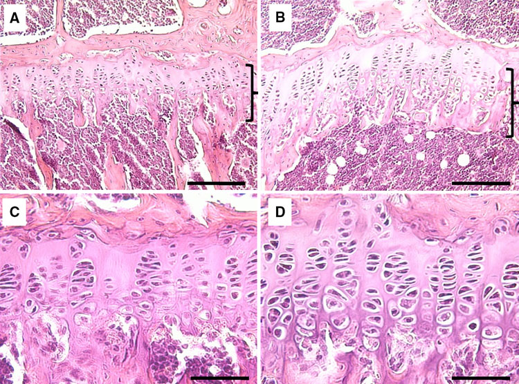
“H&E-stained sections of the growth plate in the proximal tibia. a Growth plate of the contralateral control. The bracket denotes the growth plate. b Growth plate (bracket)
of the loaded tibia. c Proliferative and hypertrophic zones of the contralateral control. d Proliferative and hypertrophic zones of the loaded tibia. Bars a, b 100 micro-m; c, d 200 micro-m”
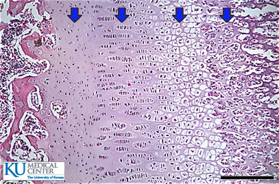
Here’s a baseline growth plate with similar colors:
It’s difficult to say exactly what is going on in the growth plates of the control and LSJL-loaded version but what is clear is that the micro-environment of the two bones is dramatically different. The LSJL loaded growth plate has much more bone marrow and many more osteoclasts(the white spots; although those spots could also be adipose tissue). The increase in bone marrow and loss of bone trabeculae would be more enabling for micro-growth plates. Thus, LSJL could create a more favorable microenvironment for micro-growth plates.
“Histological analysis revealed that knee loading increased the height and the cross-sectional area of the growth plate in the proximal tibia. First, the total growth plate height was increased by 19.5% (175 ± 25.6 micro-m in contralateral control; 210 ± 18.1 micro-m in loading) including the heights of the proliferative zone and the hypertrophic zone. In particular, the height of the hypertrophic zone was extended by 33.6% (48 ± 4.6 micro-m in contralateral control; 65 ± 3.4 micro-m in knee loading). Note that the height ratio of the hypertrophic zone to the growth plate (HZ/GP) was significantly increased, whereas the ratio for the proliferative zone (PZ/GP) was not altered”
“the cross-sectional area of the growth plate was increased by 30.7% (0.263 ± 0.108 mm2 in contralateral control; 0.344 ± 0.095 mm2 in knee loading)”
“At the cellular level, the numbers of chondrocytes were increased in the total growth plate and the hypertrophic zone by 28.5% and 46.3%, respectively. In the proliferative zone, however, no statistically significant difference in the numbers of cells was detected”
“the height of individual chondrocytes in the hypertrophic zone was elevated in the loaded side (16.3 ± 1.67 micro-m) compared to the control side (13.0 ± 1.45 micro-m)”
“oscillatory loads laterally applied to the knee not only induce anabolic responses but also lengthen the femur and the tibia.”<-Interesting that they do not state the necessity of an existing growth plate in this statement although admittedly this is not strong evidence.
“The total length increase in the growth plate was more than the sum of the increases in the proliferative and hypertrophic zones, indicating that other regions such as the resting and calcifying zones were also affected“<-This is huge as the resting zone is where chondrocyte progenitor cells are derived. If LSJL can induce mesenchymal stem cells to become chondrocyte progenitor cells than it can create new growth plates.
“Because the cross-sectional area of the growth plate is significantly increased with knee loading[the growth plate is wider], the data support that the bone-lengthening effects are not limited only to the lateral or medial loading site. At the cellular level, the number of chondrocytes in the hypertrophic zone was increased together with their cellular height. Our results are consistent with the notion that dynamic tensile and compressive loads stimulate and suppress longitudinal growth, respectively”
“In knee loading, the rate of lengthening with 0.5 N loads (peak-to-peak) was 0.1% per bout (femur) and 0.1% per bout (tibia) for 5-min loading per day.”
“both loaded and contralateral hindlimbs increased in length in the tibia.”
Yay it’s up again! I was really worried that the site was done for! I really ought to collect these studies for offline reading – they are really promising.
Pingback: Cilia's involvement to load on the growth plate | Natural Height Growth
Pingback: New study suggests osteocytes can modify height growth - Natural Height Growth
Pingback: How growth plate structure affects longitudinal bone growth - Natural Height Growth
Pingback: Understanding Mesenchymal to Epithelial cell transition may be key for neo-growth plates - Natural Height Growth