
Bone Deformation is change in the bone shape or structure. This deformation can be compression of various cavities, stretching of the bone, twisting, and so son. This is important as bone deformation is one way to increase hydrostatic pressure by decreasing the cavity size. Hydrostatic pressure is the pressure exerted by a fluid at rest. Compressing the bone laterally inhibits the fluids ability to move thus increasing hydrostatic pressure. If there is more fluid within a smaller space than it follows that hydrostatic pressure increases. Hydrostatic pressure has been consistently shown to induce chondrogenic differentiation. Chondrogenic tissue is the key for longitudinal bone growth as traditionally chondrogenic tissue is capable of interstitial(growth from within) whereas bone is not. Only interstitial bone growth has been shown traditionally to induce significant longitudinal bone growth but there are potentially other ways to stimulate longitudinal bone growth.
Knee loading inhibits osteoclast lineage in a mouse model of osteoarthritis.
“Osteoarthritis (OA) is a whole joint disorder that involves cartilage degradation and periarticular bone response. Changes of cartilage and subchondral bone are associated with development and activity of osteoclasts from subchondral bone{Since osteoarthritis does affect the subchondral bone that does affect our ability to say that LSJL affects bone deformation in a normal bone but there is no reason why it shouldn’t}. Knee loading promotes bone formation. Knee loading regulates subchondral bone remodeling by suppressing osteoclast development, and prevents degradation of cartilage through crosstalk of bone-cartilage in osteoarthritic mice{This “crosstalk” may stimulate chondral tissue within the bone as well}. Surgery-induced mouse model of OA was used. Two weeks application of daily dynamic knee loading significantly reduced OARSI scores and CC/TAC (calcified cartilage to total articular cartilage), but increased SBP (subchondral bone plate) and B.Ar/T.Ar (trabecular bone area to total tissue area). Bone resorption of osteoclasts from subchondral bone and the differentiation of osteoclasts from bone marrow-derived cells were completely suppressed by knee loading{Knee loading affects the differentiation of bone marrow-derived cells which is the first step in proving that it causes chondrogenic differentiation}. The osteoclast activity was positively correlated with OARSI scores and negatively correlated with SBP and B.Ar/T.Ar. Furthermore, knee loading exerted protective effects by suppressing osteoclastogenesis through Wnt signaling. Overall, osteoclast lineage is the hyper responsiveness of knee loading in osteoarthritic mice. Mechanical stimulation prevents OA-induced cartilage degeneration through crosstalk with subchondral bone. Knee loading might be a new potential therapy for osteoarthritis patients.”
“Daily dynamic knee loading was applied at 1 N, 5 Hz, 5 min/day for 2 weeks”
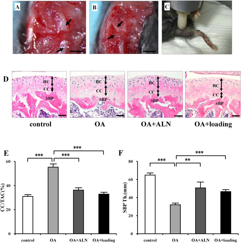
Compare the OA+ loading to the control bone. The subchondral bone plate looks much more dynamic. There are three bone marrow regions rather than two(bone marrow is the blue dots).
You’ll also note that loading+OA increased the ratio of calcified cartilage out of total articular cartilage(but not above statistic significance. It did not fully restore the thickness of the bone plate. Alendronate is an anti reobsorption agent. Given that the ALN and loading group is different we can say that change in subchondral bone shape is likely not related to inhibiting osteoclast activity and is something unique to the loading group.
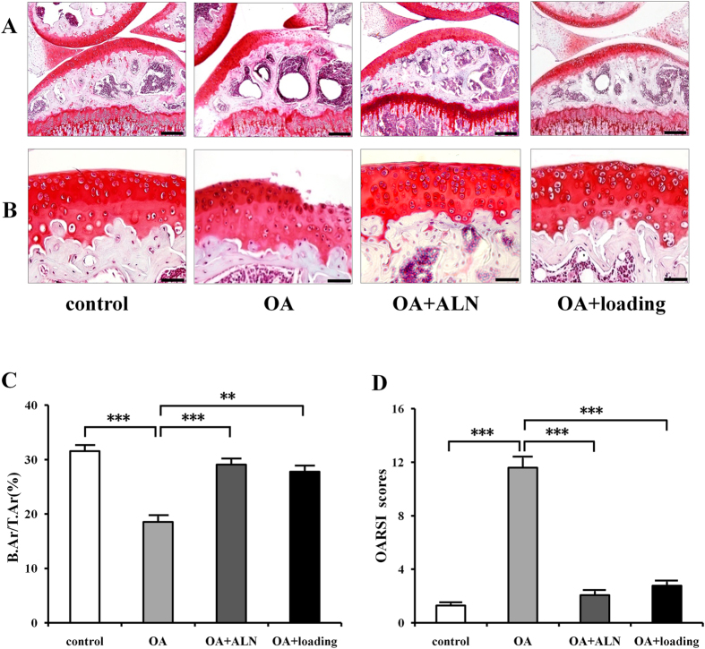
Compare the joint capsule region of control and OA+loading group. The Joint Capsule is the region that’s not inside the bone. The cells are a lot more spread out. There’s a dense redness in the control group which is not present in the OA+Loading group
We can see that the loaded group again has distinct characteristics and we can also see the growth plate. The growth plate of the LSJL group is distinct and there does seem to be signs of cellular migration. I’ll have to blow it up.
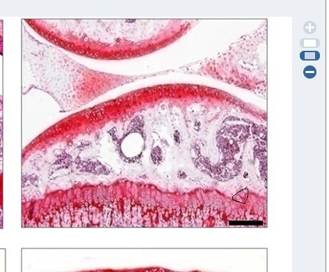
Circled is the region of possible cell migration.
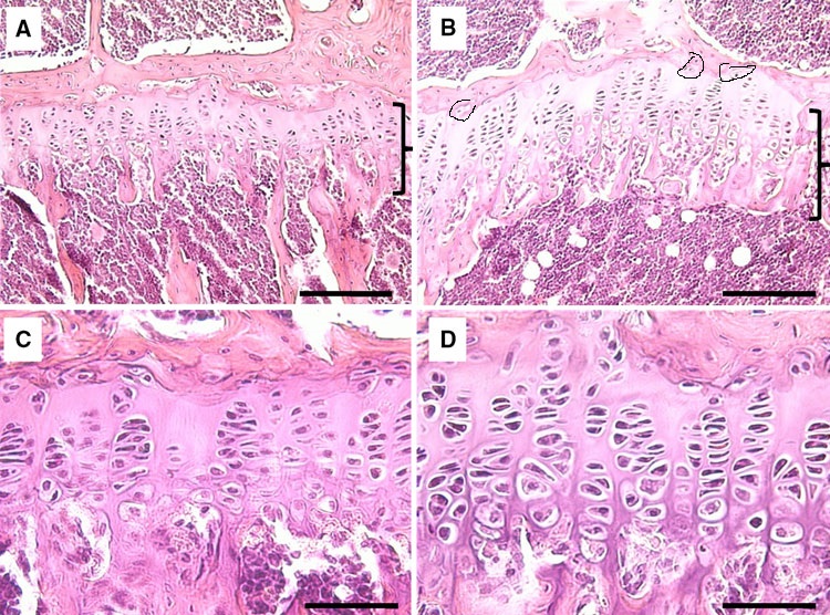
Similar signs of migration in earlier in LSJL studies(above taken from Lengthening of Mouse Hindlimbs with Joint Loading).
“the expression of Wnt3a was significantly increased by knee loading. However, the protein and mRNA levels of NFATc1, RANKL, TNF-α, and Cathepsin K were significantly suppressed by knee loading”
“Female C57BL/6 mice (~14 weeks of age)”
If you look at this image of bone marrow derived cells extracted from the loaded group and the other groups you can see that the cells are more condensed and condensation is a prerequisite for chondrogenic differentiation,
This is an image of what mesenchymal condensation looks like:
This is incredible… Congratulations on finding solid substantial evidence for what was once thought of as wishful thinking. There’s one thing left to consider though, is the method we are using really the optimal one for longitudal growth? I’m now confident that the solution is much closer than previously taken to be. Cheers.
I would recommended also supplementing igf-1 sprays under the tounge. That would speed up LSJL method because creating bone deformation, focuses the igf-1 on the the “wound” instead of organs. Calcium, Zinc, and Magnesium would make sense with the igf-1 promoting substances like the 5 amino and also betaine which would increase igf-1 receptors. The focus should be naturally made igf-1 by the liver aimed at bones only.
Pingback: New study shows LSJL induces Bone Deformation Black media plus
Hey Tyler, I was wondering if you could give and update on your LSJL progress?