Monthly Archives: January 2013

If A Person Has Open Growth Plates, How Much Are They Going To Grow Each Year On Average?

chartThis post is really for the concerned and worried parents out there who might be interested in knowing just how fast their son or daughter should be growing at the age that they are at.

Note: This post is also a key post which will be used to show that if I propose a technique or method for increasing the growth rate of a person who still has open epiphyseal growth plates, for the serious, sceptical, cynical person to see that there is a high reasonable percentage of a chance that the technique probably did do what I had said it would.

All that would need to be done is to compare the average rate of height increase for the time period of a person from say a medical textbook chart to the increases seen from implementing a technique or idea I propose.

First, I would like to cite the Wikipedia article on “Growth Chart” which has a link to the World Health Organization websiteThere is a link to a PDF entitled “WHO Child Growth Standards” from the www.who.int/ website. This massive book is over 300 pages long so I clearly will not try to read the entire PDF.

Analysis #1: For the WHO child growth standards

I would like to refer the reader to pages to 3 main sections of the PDF

  1. pages 32-33 which talks about the growth of boys
  2. pages 59-60 which talks about the growth of girls
  3. pages 74-77 which compare the growth of girls to boys.

whochart
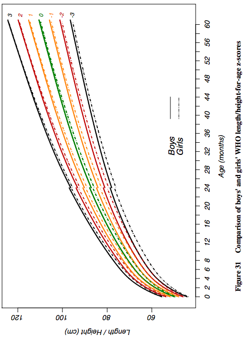
There are charts for all of the three sections. what is interesting is that in this World Health Organization PDF they are comparing the numbers and measurements of the WHO to also the CDC and the NCHS. The study only is looking at the growth rate of very young kids. between the 0-60 months years old group. As we can see from the chart pic on the left there is a non-linear growth pattern for young children between the ages of 0-5 years old. If we look at another picture looking at the growth rate progression of people in general from ages 0 until the ends of puberty, ages 0-22, we see that the entire growth progression chart is also nonlinear.

The graphs like the one (taken from the section 74-77) to the left all show for a y->x axis chart looking at measured height to determined age, in units of cm to months. For the page section comparing the growth of girls to boys, you can see that the heights are all slightly lower for girls compared to boys. This is expected since humans when comparing the sex have a sort of dimorphic distribution of height where the girls and boy height are always diverged apart from each other, but don’t seem to ever converge in the middle.

Note #1: As it is well known, for each different demographic, ethnic group, or nation the growth rate and final adult height of a subset of the entire human population will be different from another. This means that the final numbers or the chart that might be posted at the end of this post will almost always be slightly off from what a real parent would see. What the growth/ height increase progression rate of a child will be different in Vietnam compared to say the Netherlands.

So what does the small chart I have copy and pasted from page 75 actually show about the growth progression in young children from young age, specifically ages 0-5 years old?

If we take a ruler and tried to measure the magnitude of the difference of the y axis versus each year we see that for the first year of life, the growth is the strongest (months 0-12) with going about 40-> 75 cms, a 25 cms increase, which is exactly 1 entire feet in growth. For the 2nd year for average (50 percentile) boys they go from 75 –>90 cm, which is a 15 increase, or 6 inches. For the 3rd year they increase from 90–>97 cm, or a 3 inch increase. This continuation in decrease of growth rate is seen in the 4th and 5th year too. For the issue with girls, they seem to start at about 5 cm shorter than boys at birth and but have a similar growth pattern trend, if not a little less steep growth trend in the first 2-3 years. Since the graph does not continue on for ages 6-18, I had to resort to another graph and other resources to find out just how much a child should grow in later years when their epiphyseal plates are still open.

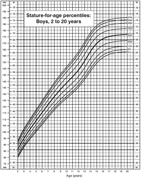
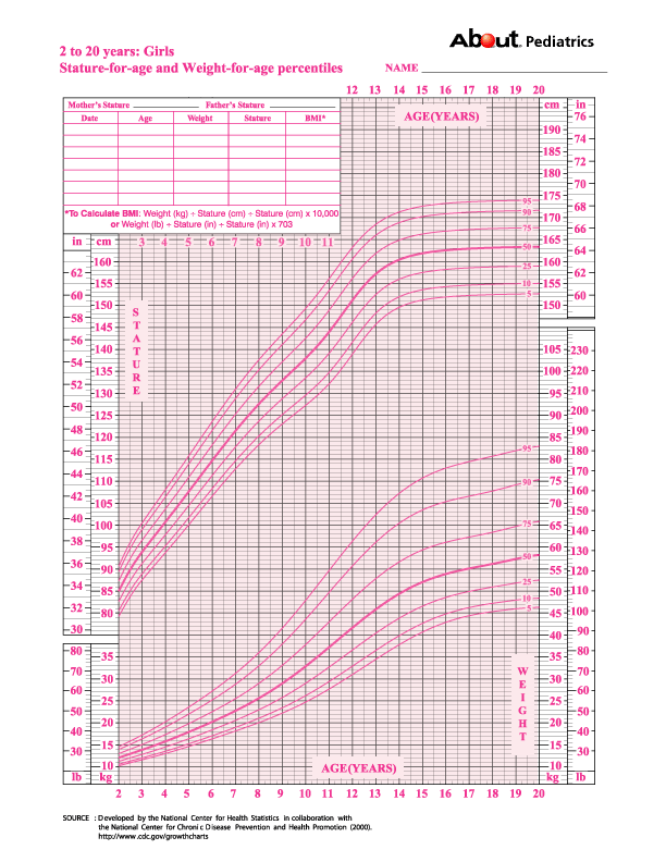
When I try to type in the phrase “growth progression chart” into google what I mostly get are charts for very young children, babies and infants in the 0-5 year range. Most of the image results reveal the two charts (for boys and girls) belows

Male_Growth_Chart

girlstwoyears

Before we look at the charts for the average height growth progression I would like to cite a few PubMed articles.

The first is “Evaluation of growth rate in height over periods of less than one year

Abstract

The stature of 260 well-nourished children aged between 7 and 10 years was measured at intervals of approximately 1 month over 13 months. The growth rate of each child was calculated over periods of 3 months and 6 months ending in each month of the year. Children who missed one or more measurements, due to absence from school, were excluded from the calculations

Centiles of growth rate for these periods are presented. A growth rate of 3-4 cm/yr is well within normal limits for a period of 3 or 6 months ending in December or January, but is below the 10th centile for periods ending between March and June.

Most children reach their maximal 3-monthly rates in the periods of either 3 or 6 months ending between March and July, and their slowest in the periods ending between September and February.

A child’s growth rate over the 3 months of fastest growth is most frequently 2 to 3 times his slowest rate, but may be 7 or more times the slowest if the latter was very low. There may be no measurable growth during a single period of 3 months in a normal child, but maximal rates of up to 10 cm/yr are not necessarily abnormal. An individual’s maximal 6-monthly rate may be up to 3 times his minimal.

A satisfactory assessment of a child’s growth cannot be made over a period of less than one year.

The 2nd study cited is “[WHO growth standards for infants and young children].” which was written in 2009 in French

Abstract

The growth pattern of healthy breastfed infants deviates to a significant extent from the NCHS/WHO international reference. In particular, this reference is inadequate because it is based on predominantly formula-fed infants, as are most national growth charts in use today. The WHO multicentre growth reference study (MGRS), aimed at describing the growth of healthy breastfed infants living in good hygiene conditions, was conducted between 1997 and 2003 in 6 countries from diverse geographical regions: Brazil, Ghana, India, Norway, Oman and the United States. The study combined a longitudinal follow-up of 882 infants from birth to 24 months with a cross-sectional component of 6669 children aged 18-71 months. In the longitudinal follow-up study, mothers and newborns were enrolled at birth and visited at home a total of 21 times at weeks 1, 2, 4 and 6; monthly from 2-12 months; and bimonthly in the 2nd year. The study populations lived in socioeconomic conditions favorable to growth. The individual inclusion criteria for the longitudinal component were: no known health or environmental constraints to growth, mothers willing to follow MGRS feeding recommendations (i.e., exclusive or predominant breastfeeding for at least 4 months, introduction of complementary foods by 6 months of age and continued breastfeeding to at least 12 months of age), no maternal smoking before and after delivery, single-term birth and absence of significant morbidity. Term low-birth-weight infants were not excluded. The eligibility criteria for the cross-sectional component were the same as those for the longitudinal component with the exception of infant feeding practices. A minimum of 3 months of any breastfeeding was required for participants in the study’s cross-sectional component. Weight-for-age, length/height-for-age, weight-for-length/height and body mass index-for-age percentile and Z-score values were generated for boys and girls aged 0-60 months. The full set of tables and charts is presented on the WHO website (www.who.int/childgrowth/en), together with tools such as software and training materials that facilitate their application. The WHO child growth standards were derived from children who were raised in environments that minimized constraints to growth, such as poor diets and infection. In addition, their mothers followed healthy practices such as breastfeeding their children and not smoking during and after pregnancy. The standards depict normal human growth under optimal environmental conditions and can be used to assess children everywhere, regardless of ethnicity, socioeconomic status and type of feeding. The standards explicitly identify breastfeeding as the biological norm and establish the breastfed child as the normative model for growth and development. They have the potential to significantly strengthen health policies and public support for breastfeeding. The pooled sample from the 6 participating countries allowed the development of a truly international reference that underscores the fact that child populations grow similarly across the world’s major regions when their health and care needs are met. It also provides a tool that is timely and appropriate for the ethnic diversity seen within countries and the evolution toward increasingly multiracial societies in the Americas and Europe as elsewhere in the world. The WHO standards provide a better tool to monitor the rapid and changing rate of growth in early infancy. They also demonstrate that healthy children from around the world who are raised in healthy environments and follow recommended feeding practices have strikingly similar patterns of growth.

The 3rd PubMed article cited is “New growth standards for the 21st century: a prescriptive approach.”

Abstract

Breast-fed babies have been shown to grow at a substantially different rate from the current international reference curves, with greater growth rates in height but with smaller body weight increases and substantially less variability in the growth patterns of a group. On this basis, the World Health Organization concluded that there was a need to undertake new studies to establish on a global basis the appropriate growth curves for exclusively breast-fed babies, their growth curves then being potentially seen as optimum standard curves rather than an arbitrary set of reference charts. The Multi-Country Growth Reference Study was therefore carried out from July 1997 to December 2003 as a population-based study covering the cities of Davis, California, USA; Muscat, Oman; Oslo, Norway; and Pelotas, Brazil, together with selected affluent neighborhoods of Accra, Ghana and South Delhi, India. These centers were considered conducive to a study of babies and children under optimum breast-feeding and weaning and early feeding conditions. These studies, to be reported shortly, confirm previous observations on breast-fed children, but also show that the greatest differences are within each population group rather than being international differences.

The last study or article I would like to cite is written by Dr. Robert H. Shmerling entitledCan We Predict Height?“. A part of the article he wrote is copy and pasted below…

What Determines Height?

One’s ultimate height is determined by a complex interaction of many factors including nutrition, genes, and overall health. Growth rates vary over a lifetime:

  • From infancy, when average length is 20 inches, to age 2 there is initially rapid growth, then slowing, with about 14 inches in height added.
  • From age 2 to puberty there is slow, steady growth at about 2½ inches per year.
  • As one enters puberty, a growth spurt of 3 to 5 inches in a year is common.
  • By ages 40 to 50, height actually may begin slowly to decline, even in healthy adults.

Final Conclusion & Analysis Made By Me

So what do the charts and numbers say about the growth rates in children or adolescent who still have open growth plates?

From the first pubmed studies we learn that for children at least in the age range of 7-12, their growth rate is far higher between the months of March and july than the other half of the year.  The growth rate during the fastest 3 months can be as high as 7 times as fast as during their slowest time but is on average 3 times as fast as the slow months between december and february. Growth is usually 3-4 cm/year if one was only accounting the time of growth during the bad time range but can be as high as up to 10 cm/year

The 2nd pubmed study shows that if mothers decided to breastfeed their babies exclusively or more than just usual formula very quickly in the babies development, their height growth progression would be far more different. The modern nations have mothers feeding their babies formula much more now that this causes the World Health Organization height progression charts to be very off from what might be more “natural”. For the study, all the other factors which might make the issue of comparing breastfeeding vs formula to height progression was removed where the mothers were advised to continue to breatfeed beyond a certain month and to refrain from smoking and many other behavior to negate other factors which might make the data useless.

The 3rd pubmed study is written by the same authors so basically only validates the 2nd study. The growth charts for breast feed babies are very different from formula feed babies. The growth rate for the babies show higher height increase in terms of relative to the gain of weight. This implies that if the health organizations like WHO are hoping to get a far more natural and most accurate reference growth chart of babies from around the world, they need to put the babies on a breastfeeding diet for much more than their mothers might be doing currently.

The last resource I cited is from a doctor He states that from ages 0-2, the baby gains about 14-15 inches in height, which is about what the other chart indicated. For the age range of 2-13, or whenever puberty starts, the growth rate is about 2.5 inches per year. Puberty causes the growth rate to increase up to 3-5 inches per year.

So from all the resources I have found, but especially the WHO, CDC, and NCHS the amount of height increase is sort of like this

Age range

  • 0-1 – 12 inches
  • 1-2 – 5-6 inches
  • 2-3 – 3 inches
  • 3-4 – 2.5-3 inches
  • 4-11 – 2.5 inches
  • 11-13 – 3-5 inches
  • 13-17 – 1.5-2 inches
  • 17-18 – 1-1.5 inches

So the number values are based on averages but can still be used to compare one’s children’s growth rate to other kids their age to see if they are growing property and within the aceptable growth rate percentile.

Is Using Microscopic Engineered Nano-Bots To Increase Height And Grow Taller Even Reasonable Or Possible?

On the idea of using microscopic engineered nanobots to increase height and grow taller

urlThere have been a few people who have contacted me through email to ask over this idea. They talked about building and releasing nanobots which can swim around our bodies through the blood streams and be able to focus on the old growth plate areas to somehow regrow the growth plates again. They of course never go into detail on how they would be able to convert the nonliving organic hard calcium phosphate and hydroyapatite into the collagenous fibers or how to take into consideration the fact that the human structure can break apart if too much of the hard element which forms bones is removed.

The main problem is that to be able to recreate something similar to what we found in our younger days would require at least 4 step process to do that.

  1. The first thing is to remove the inorganic hard calcium reinforced matrix that makes up the bone on the inside, the trabecular bone.
  2. The 2nd thing is to remove the inorganic hard material that makes the even harder material, the cortical bone.
  3. The 3rd is to be able to direct the blood vessels and marrow in the empty cavities of the long bone long enough for step 3.
  4. The last step is in creating the actual epiphyseal hyaline growth plate. This in itself is already hard enough to do, due to the fact that the chondrocytes in the growth plate are stacked in columns, which are not seen in any other type of tissue found in the human body.

Overall, when I hear this type of idea I start to roll my eyes and can not believe that since it seems either too fantastic to believe or too far into the future to make it even remotely realistic for people like us who are in our teens or 20s, maybe 30s to use in our lifetime. It is hard enough for computer scientists today to build robots which have any type of Artificial Intelligence capabilities currently and to extrapolate the technology we have currently to make them microscopic for biomedical or cosmetic reasons seems very fantastic.

The current level of robotics we see today are being advanced further and further in many top universities, military bases, and possibly secret large corporation locations which do insane research and truly crazy breakthroughs. However these research will never reach the eyes of the general public.

I would propose that for any type of nano (or micro) sized robots or mechanical entities to be able to do all the steps I propose above, they would probably have to have at least three main qualities.

1. Safety – The bots will be safe and not hurt the formation process of other systems but especially the nervous and circulatory system.

2. Biodegradable or Can be expelled from the body without hurting the host – meaning that the bots will eventually disintegrate from the human body’s natural immune response. If they can’t be removed without hurting the person then they must be removed in another way, say through the urine or feces. However that means that the robots have to have the 2nd trait, intelligence.

3. Intelligent – It has to follow at at least 4 steps. From what I’ve seen, it might need to learn how to form 3-dimensional shapes or forms to create a pseudo-tissue in the 3rd step since redirecting blood vessels, blood, and bone marrow may be impossible from my known area of understanding of how science can work.

Problems with Safety – We know that if we ever do even find a way to create the nanobot we want for medical application, they have to be safe for the person who has the bots injected into them. This means that when the microbots are swimming through and around the blood vessel system of the body, or sometimes getting into extracellular fluid, they have to be relatively harmless to the tissue in the human body, especially the nervous system cells ,the neurons, and the blood vessels. We must remember that the human brain uses around 20% of all the oxygen the human inhales so a lot of blood will be going to the brain. The brain has a blood vessel impermeable membrane barrier that prevents foreign elements from reaching the brain causing serious problems. If we build bots, they have to be small enough to not cause problems when they are the blood vessels in the brain.

Problems with Biodegradability – If the nanobots do manage to do their job, how will they eventually leave the subject’s body? I don’t think that most safety conscious humans would like the idea of nanobots being left in their body but would rather that the engineers and researchers who created the nanobots have already thought up a way for the bots to leave the human body. This probably means that the bots will have to be passed through body by urine, the digestive system through bowel movements, or maybe even through the pores of the skin. If the nanobots can’t be flushed out, then they have to degrade in the body after a certain amount of time. This could be accomplished from the white T cells in the body which have the function to attack any strange foreign objects that reach the body.

Problems with Intelligence – The most “intelligent” computer that I know at this moment was the IBM “Watson” which beat in the TV Show Jeopardy its human contenders. However that doesn’t mean that many computer research facilities and design locations around the world haven’t already built even smarter computers. The computers are indeed becoming smarter and smarter with using smaller transistors and with better written algorithms, however when you want to create nano-bots, you limit the number of transistors that can go into the nano-bot meaning that the complexity of the bot (and maybe also the intelligence) will be limited.

With the multi step process for the proposed growth plate regeneration, the robots will have to be reasonable big to hold the part needed to be able to implement the steps a software engineer might try to program into the microprocessors of the nanobot, which would have to increase in size.

It might be possible for a nano-bot to have a receiver to a micro integrated circuit like the microprocessors which AMD and Intel make these days. However it would also have a part which would allow it to move in a directed direction since we should not just let the human bodies natural fluid movements distate where the bots should be and how fast they should be moving.

Conclusion – I just can’t see how nanobots can be created for medical application or cosmetic reasons for increasing height any time soon and with the number of technical issues I have raised in the post, I don’t think nanobots will be created to the level of safety, biodegradability, and intelligence which be needed to do something like regeneration of the growth plates at least for many decades to come.


What Type Of Non-Invasive Or Minimal Invasive Surgical And Medical Techniques Are Currently Available?

I think this question is something that has to be addressed at some point if we are really serious about looking for a way to grow taller without cutting open say our leg or skin too much. I think it is time to look at whether this desire to have a really non-invasive approach is even feasible from the type of technology we have to work with in the modern medical technology.

From my searching on the boards and forums around the internet from people who want to grow taller, they almost all say that they only want to go the non-invasive approach and would not do any type of surgery for it. What most people still are hoping for is some type of supplement, vitamin, or Miracle Pill that would do all the work for them without them needing to put any type of effort, will, or work into it.

However this type of mentality is so prevalent that we can see the sam type of people who might wish to loss weight. If some company right now could create some type of Miracle Pill to be able to make them instantly shed the 10 lbs that they so desperately want without any type of negative side-effects and requires nothing like strenuous exercise, they would be making a lot of money.

However from what I have seen that doesn’t seen reasonable, for weight loss and definitely not for height increase. At least for people with closed growth plates.

At the current stage of technology we are at right now, there area very few options to change tissue in vivo without cutting into it.

Here is what I know can be done for bones…

1. You can increase bone growth but only in terms of bone weight density (BWD) using pill form like Statin. The company Zymogenetics about a decade ago did have a pill made from statin derivative which does grow bones, but don’t actually change their overall shape and volume. A post was written about this at “The Bone Growth Pill From Zymogenetics“. The pill was created to be used for people with osteoporosis, which is where the bone density of a person, usually older females decrease a lot causing them to become more susceptible to bone fractures, joint problems, and similar pathologies.

2. You can increase the bone density again from mechanical loading, like what is done with Lateral Synovial Joint Loading.

3. You can increase bone density from vigorous exercise and being taking up a sport to play, as seen from many PubMed  studies, whether they are for tennis, soccer, or football. I refer the reader to the article “Enhanced bone mass and physical fitness in prepubescent footballers

4. You can increase bone width or thickness from short bursts of dynamic mechanical loading. This type of idea and method was used for the idea over shoulder bone widening, which I wrote about in the post “Review Of Claim To Widen Shoulder Bone, Lengthen Forearms, And Lengthen Lower Legs

5.  You can increase the cortical bone layer thickness in long bone by consistent vigorous exercise and/or training. This is seen in studies like “THE ROLE OF PHYSICAL ACTIVITY ON BONE DENSITY AND BONE GEOMETRY IN MEN”

6. You can increase bone length with growth plates using growth hormone injections or growth hormone-releasing hormone injections.

7. You can increase bone length with growth plates by using aromatizing inhibitors like Anavar and Letrozole.

The problem starts when there is no cartilage layer that transversely cuts across the long bone left. The ossification and calcification caused cartilage to turn into bone. With a long tubular bone with very high tensile and compressive strength in the range of something like stainless steel, is there something that current modern medicine or biomedical engineering technology which can stretch those long bones.

Note: Here we are not going to look at the overall height contribution dur to the vertebrate column but focus only on the long bones like the femur and tibia.

Tyler from HeightQuest.com had shown from an old post that the hard, strong characteristic of the bones, made of hydroxyapatite was the limiting factor. Bone is what is preventing people from increasing their height. This is sort of common sense. Bones are hard and you can’t really stretch them out.

I had proposed that the real limiting factor may actually be the skin, ligaments, blood vessels, and whatever other non-bone tissue that surrounds the actual bone is the limiting factor. The element which I think is the real problem is that people are not willing to accept that idea that maybe we should look into a surgical alternative to the limb lengthening surgery.

However if we really wanted to find a relatively simple way for non-invasiveness, we may have to first realize what is possible for noninvasive techniques.

So…”What Type Of Non-Invasive Or Minimal Invasive Surgical And Medical Techniques Are Currently Available?

From this page HERE found from Charles Sturt University 

Medical technology – Bionics – Non-invasive medical techniques

  • Non-invasive surgery refers to the performing of a surgical technique without making an incision in the skin at all. The removal of gallstones using laser treatment is an example of a non-invasive technique currently in use.
  • Non-invasive diagnostic techniques include ultrasound, x-rays, thermography and magnetic resonance imaging.
  • Minimally invasive refers to techniques that are performed by making the smallest practical incision in the skin

For Non-Invasive Surgical techniques…

  1. Laser 
  2. High Intensity Focused Ultrasound (HIFUS or FUS)
  3. Extracorporeal Shockwaves (ESW) 

For Non-Invasive Diagnostic techniques…

  1. X-rays -are used mostly for producing images of bones and joints
  2. ultrasound – a technique in which high-pitched (ultra) sound waves are projected onto tissues under examination. The waves are reflected, captured and registered electronically as an image we can see
  3. thermography – Thermography, or digital infra-red thermal imaging (DITI), is a diagnostic technique which allows the examiner to map and quantify changes in skin surface temperature.
  4. magnetic resonance imaging. – Magnetic resonance imaging is a technique of scanning the body. It is based on the fact that living tissues give off their own special electromagnetic signals, depending on the water content of the tissue. If the tissue is subjected to a large external magnetic field, the small electromagnetic signals may be detected and built up to form a 3D image. MRI is a popular diagnostic technique, as it ignores bones (as they contain little water), concentrating on soft tissue. In this respect, MRI is the opposite of x-rays, which tend to concentrate mainly on the bones.

For minimally invasive techniques…

  1. keyhole surgery – is a recently developed form of minimally invasive surgery. A small incision is made in the skin, and specially designed surgical tools are inserted through the incision to perform the required tasks. A small camera is also inserted into the hole so the surgeon can see what to do.
  2. endoscopy – 
  3. microsurgery –

We can see that these are all the devices and technology we have right now to treat pathologies in medicine or use for cosmetic body remodeling. I would recently write up about the possibility of using Magnetic Resonance Imaging Guided High Intensity Focused Ultrasound Surgery in the post “Increase Height And Grow Taller Using Magnetic Resonance Image, MRI Guided High Intensity Focused Ultrasound Surgery, FUS (Big Breakthrough!)

this idea posed was unique in that it allowed for the ability to cut and do surgery on the inside of the body without touching the tissue on the outside covering it. The MRIgFSU idea was the most promising of all the non-invasive ideas, although I have looked into Extracorporeal Shockwave Therapy (ESW aka ESWT) as well as microfracture surgery as a way to eventually lead to a procedure on height increase.

When I think about why so many people are looking for a non-invasive approach, I wonder whether it could be because they want to do the techniques or exercises in the comfort and privacy of their own homes, without other people like doctors and medical professional from knowing about it. It might not be a fear of the needle, the knife, or going on the surgery table for height increase they are not interested in, but more of the social pressure and aspect on using a technique which will take the skills of a qualified physician.

This means that even if I create the perfectly theoretical sound invasive procedure for height increase after growth plate closure, it will still have two big hurdles to overcome.

1. First ,the entire medical community and establishment will attack, analyze, and criticize the technique until a large enough group of surgeons are willing to take up the risk and perform the surgery to prove the idea does work.

2. Second, it would require the person who wants a non-invasive way to gain height in their home with them individually doing it to accept the idea that they will need to pay thousands of dollars to a group of medical professionals to cut open their body. People want the easy, fast, simple way to do something and maybe this endeavor just does not have the easy, non-invasive path.

Increase Height And Grow Taller Using Magnetic Resonance Image, MRI Guided High Intensity Focused Ultrasound Surgery, FUS (Big Breakthrough!)

Of all the techniques and ideas I have proposed so far this may be one of the most technologically advanced ideas and proposals. Recently I was watching a talk on TEDtv called “Yoav Medan: Ultrasound surgery — healing without cuts” and I wondered whether it would be possible to use this type of non-invasive surgical method to increase height. After spending a few hours looking at the various resources and PubMed studies on the internet I would say that this medical technology procedure actually has some good height increasing possibilities. Let me explain.

The video is below. A full analysis of this possible method and approach is talked about below the video.


Sources

Analysis & Interpretation

From the 5th source, “Magnetic resonance (MR)-guided focused ultrasound surgery (MRgFUS) completely destroys targeted tissue without causing structural or functional damage to the adjacent tissue. The ultrasound energy penetrates normal tissue while reaching targeted tissue at sufficiently high energy levels and inducing cell death with minimal interference. MR imaging (MRI) identifies targeted tissue and detects suitable access routes for the acoustic waves. It also determines tissue temperature changes. MRI contrast scans then verify thermal ablation. Thus, an effective, safe, non-invasive, closed-loop procedure eradicates circumscribed tissue with minimal discomfort and recovery time. Described as the ideal surgery,….”

The real method for non-invasive surgery has arrived. With it, that suggests that we can finally make surgical incisions on places without needing to cut open skin. The technology I am looking at is very advanced and its main application has been towards killing (or at least stopping the growth) of brain tumors which are malignant. However I would suspect that if the frequency and magnitude of the ultrasound surgery can be increased, it can turn into a device that cuts anything including the hard nature of the hydroxyapatite, which is what gives bone their hard nature.

Implications For Height Increase

Here is what I can say to all the people who are interested in height increase. The non-invasiveness nature of any type of technique can probably be achieved now using the MRgFUS . If the device is strong enough, it can make cuts into the hard non-living tissue of the bone, the extracellular matrix of the bone. A quick tensile load means that along with the initial bone cut, the pulling can mean that limb lengthening surgery can be done extremely precisely.

I had talked about before in previous posts about the use of Low Intensity Pulsed Ultrasound (LIPUS), Extracorporeal Shockwave Technology (ESW), as well as High Intensity Pulsed Ultrasound. Now we are seeing high intensity ultrasound which is non-invasive in nature. Any cuts and incisions made on the bone can be done with no cuts on the skin.

I had talked about before using Piezoelectric Surgery as a way to cut bone for microfracture formation and distraction osteogenesis in the post “Increase Height And Grow Taller Using Minimal Invasive Piezoelectric Surgery, Part I” bringing up the issue that if we can figure out the resonance frequency of the long bone in each of its areas, including the cortical bone area and the trabecular bone area,

Here is the method I propose to regrow cartilage in the areas we want now non-invasively. 

1. First we get an X-ray of the limb to be done on to know exactly where the outer edges of the bone is.

2. We can apply a sharp but high intensity pulse to the now gone epiphyseal cartilage bone area, and by using the resonance frequency of the bone type, we can induce the bone material to break apart in that area using the High Intensity Focused Ultrasound devices.

3. Remember that the Parathyroid organ is what regulates the calcium in our blood. If we can regulate the PTH/PTHrP calcium homeostasis so the calcium deposits get resorbed by the blood (like osteoclasts) leaving a relatively empty space filled with bone marrow we can do what I would call the first filtering of the marrow to get ready for cartilage formation. I had talked about the possibility of using the Parathyroid related Peptide, PTHrP as a way to lead to noninvasive height increase in one of the most interesting posts “Parathyroid Hormone And Parathyroid Hormone-Related Protein May Lead To Non-Invasive Epiphyseal Growth Plate Regeneration (Big Breakthrough)“. Since the increase of PTH means that the calcium in bone gets resorbed back into the blood leading to a medical condition known as hyperparathyroidism , we must only get the PTH/PTHrP to be increased in the area where the bone material was dissolved. This might be achievable from somehow getting the indian hedgehog  (Ihh) to be increased in expression within the MSCs inside the marow which would expand and fill up the  newly formed medullary cavity.

4. The bone marrow inside has MSCs but has a type of bone marrow known as yellow marrow, which doesn’t have the red bone marrow of children, having what is known as adipocyte derived MSCs. From pubmed studies…

This shows that with growth factor combinations like TGF-Beta2 with BMP7 and the combination TGF-Beta3 with BMP-6 (from the 2nd source) which we can inject into the bone area with a small drill can stimulate the initial chondrogenesis process. We would do a weekly injection of the right growth factor combination to create more chondrocytes.

However the issue would be whether we can use just the Magnetic Resonance Imaging Guided High Intensity Focused Ultrasound (MRgHIFUS) alone in achieving cartilage formation inside the bone without needing anything else. I would actually prefer that after the MRgHIFUS usage to break apart the bone areas in the location we wish to, we inject growth factors like GDF-5, FGF-3, and TGF-Beta1,2,3

5. Which we can use another frequency, a lower frequency from dynamic mechanical loading to cause more chondrogenesis. The idea now is to convert the few chondrocytes that are made from the growth factor injections into a culture form to make a culture to form cartilage.

6. From the previous post “Thyroxine Regulates The Morphogenesis Of Isolated Chondrocytes Into Columnar Cartilage (Big Breakthrough)” I would guess that if we can inject thyroxine into the now marrow filled cavity of the bone, it might be able to get the chondrocytes that do get differentiated to start stacking in columnar fashion to create a extracellular environment similar to the original growth plate.

6. We must remember that vascularization is what causes cartilage tissue to start turning into bone. This means that the high intensity focused ultrasound needs to be used regularly to destroy blood vessels which would try to either differentiate the adipose derived MSCs into osteoblasts an osteocytes. Of course this is easily done since the original application of the MRIgHIFUS (or MRIgFUS) was to destroy blood vessels which would supply the nutrients needed to allow tumours to continue to grow.

7. After a cartilage is mostly formed for the layer in the long bone internally for the trabecular bone area, we can then do it to the cortical bone, and doing the same process over again.

This will be the first time that we can use real technology to rebuild cartilage that was completely lost during the natural growth process. However the cartilage formed may not be the nice hyaline type we are looking for with stacked chondrocyte columns. Some of the cartilage formed may be irregular or crooked but I would guess that as long as a layer of cartilage can separate hard bone from hard bone, we can use other cartilage forming ideas like injecting Chondromodulin Type 1 to stop vascularization to increase the cartilage thickness.


Intensive Extended Swimming May Lead To Height Increase Up To 1 Inch

importance-and-health-benefits-of-swimmingLast night while I was doing nothing I decided to waste some time and scour YouTube for any videos that would entertain me. Along the way I somehow started to watch old Seinfeld clips again. One clip however did catch my attention and was where Kramer started to get really deep into swimming.

Apparently Kramer started off doing hundred of laps in the local YMCA or fitness club pool. He would change that to start swimming in the East River, which I would have no idea where that is since I never lived close to or in the NYC area.

In the clip below, around the 2:40-2:50 mark, he would state “…exercises every muscle in the body. It’s great for the back….Four hours in this chop (?) and I’m a full inch taller…”

Very early in the website I had written a post about the link between swimming and possible height increase with “Grow Taller By Swimming

Interpretation

I personally can attest to the fact that from swimming, the body “feels” taller since many muscles that are not used in traditional exercises and weightlifting are used and stretched out. The bouyancy of the water does indeed help lower the amount of weight that the lower body will feel. Years ago when I was going through an obsessive period of exercising many hours of the day to gain height swimming was a huge part of the daily routine. Around 2-3 hours was spent everyday doing butterfly strokes in an olympic sized pool to increase fitness and to possibly gain height. Krammer says that 1 full inch in at least temporary height can be gained after 4 intensive hours of swimming. It is a TV show but the claim does not seem that unreasonable. We do remember that when astronauts are in space, the spine and vertebrate is mostly decompressed. At an environment like that, the vertebrate is supposed to decompress to the point that the average height increase seen is by 3% of the original height on the surface of the earth. This means that on average, the human body would increase by 2 inches. For water, the effects are not as dramatic but there can be some effect.

It has never been tested to see what would happen to the human body if it was just placed in water for a long period of time but  we do know that when dead bodies are found in the water they do become bloated and are often found floating on the surface of the water since the body has a lower density than the water. It is well known among divers and swimmers that one’s elevation underwater can be altered by the amount of air that one keeps in one’s lung. If the lungs were filled completely with air, the body will go up to the surface since the average density of the body would be less than the water.

As for the claim that swimming can lead to some height increase, we do note from previous research that the bone density of swimmer who do it extensively at least for prepubscent females

From study “Differential effects of swimming versus weight-bearing activity on bone mineral status of eumenorrheic athletes“…

Swimmers were taller (p = 0.0001), heavier (p > 0.005), and had a greater bone-free lean mass (p < 0.001) than gymnasts and nonathletic controls. When adjusted for body surface area, there was no difference in lean mass between swimmers and gymnasts, and both were higher than controls (p < 0.01)”

The fact that the swimmers found in this study was taller could be just from the fact that in the sport of swimming, taller and longer bodies may be better at reaching long distances and can cover more surface area but it still shows that swimmers are taller. There is no evidence that would indicate that taller children in general are pushed into swimming, unlike volleyball and basketball, but the more likely result is that the taller children end up being able to reach the other end of the lane slightly faster, and that results in the being more likely to be at the collegiate and professional levels of swimming. As always there is so causality that have been able to really show that in developing children, swimming –> taller body.

It is true that the bone mineral density of swimmers is much lower than other athletes, and may even be lower than controlled groups where people don’t exercise.

From source

“While in space, astronauts’ bodies respond to the lack of gravitational stress on the bones by decreasing their bone mass. On the other end of the spectrum, weightlifters experience an increase in bone density due to the stress of the extra weight on the bones. The more stress placed on the bones over a period of time, either through heavy weights and/or by impact, the more the body responds by increasing the bone density.

Study after study comparing swimmers with other athletes, shows swimmers to have similar bone density to couch potatoes.”

We see from many studies and sources that there seems to be a weak positive correlation between the fact that high bone mineral density in at least developing children, as long as malnutition is accounted for, seem to correlate to above average height. So the weaker the BMD, the he higher chance for a person to be above average in height. The fact that in space where gravity is near zero shows that both the body expands in height and the density drops. The effect of water should do the same thing.

From source link HERE

Note: I was unable to embedd the video due to certain YouTube restrictions.

How does hydrostatic pressure induce chondrogenesis?

A key theory behind LSJL is the use of hydrostatic pressure to induces mesenchymal stem cells into chondrocytes to form new growth plates.  Hydrostatic Pressure alters the expression of some of the genes altered by LSJL.  Although, there are many genes not shared which is not surprising considering that LSJL exerts other forces than hydrostatic pressure and the microenvironment is different between the rat bone and the cell lines used.  Hydrostatic pressure is one of the most consistent stimuli in inducing neo chondroinduction.

Hydrostatic pressure decreases membrane fluidity and lipid desaturase expression in chondrocyte progenitor cells.

“Membrane biomechanical properties are critical in modulating nutrient and metabolite exchange as well as signal transduction. Biological membranes are predominantly composed of lipids, cholesterol and proteins, and their fluidity is tightly regulated by cholesterol and lipid desaturases. To determine whether such membrane fluidity regulation occurred in mammalian cells under pressure, we investigated the effects of pressure on membrane lipid order of mouse chondrogenic ATDC5 cells and desaturase gene expression. Hydrostatic pressure linearly increased membrane lipid packing and simultaneously repressed lipid desaturase gene expression. We also showed that cholesterol mimicked and cholesterol depletion reversed those effects, suggesting that desaturase gene expression was controlled by the membrane physical state itself. This study demonstrates a new effect of hydrostatic pressure on mammalian cells and may help to identify the molecular mechanisms involved in hydrostatic pressure sensing in chondrocytes. ”

“Hydrostatic pressure (HP) is known to reduce lipid membrane fluidity. High HP triggers an adaptive mechanism, during which membrane fluidity is increased by raising the proportion of unsaturated fatty acids.”

“TDC5 cells responded to the change in their membrane fluidity under pressure by modulating the expression of Fads1, Fads2, Scd1 or Scd2. 10 or 20 MPa did indeed significantly inhibit the expression of all four genes after 24 h, while 5 MPa also significantly decreased Fads1 and Scd1 expression.”<-None of the genes were directly altered over significance by LSJL.  The genes were measured one hour after loading so it’s possible that the genes returned to baseline after one hour.

gene expression change to HPWith this  data it’s easy to see why there was no change in gene expression level by LSJL as most of the repression of these genes occurred after 6 or 24 hours under hydrostatic pressure.

“MβCD increased ATDC5 membrane fluidity (laurdan GP values respectively increased and decreased under cholesterol and MβCD treatment”

“Similar to HP, cholesterol increased laurdan GP and significantly inhibited Fads1 and Fads2 expression. By contrast, MβCD[methyl-β-cyclodextrin], which decreased laurdan GP, significantly increased the expression of all four desaturase genes. Together, this suggests that membrane fluidity itself may be the general modulator of desaturase gene expression.”<-Cholesterol has been implicated in controlling endochondral bone growth before.

“the beneficial effects of loading on cartilage are mediated by the transcription factor CITED2, which represses cartilage degradation by the matrix metalloproteinase MMP1.”

“In the human hip joint, loads typically reach 10 MPa during normal activity, with peaks of up to 18 MPa. Considering that interstitial fluid pressure supports about 70 to 90% of the applied load, HP within the cartilage can be expected to regularly exceed 7 MPa and peak at around 16 MPa. 20 MPa is therefore a relatively high pressure”

“How HP is actually sensed by the cell remains poorly understood, but the link between HP and cholesterol suggests at least two hypotheses: both HP and cholesterol may converge on the SREBP pathway, cholesterol by affecting SREBP maturation via SCAP, HP by affecting the membrane fluidity of the endoplasmic reticulum, where non-activated SREBPs reside and which is more sensitive to HP than the plasma membrane; the mechanosensing mechanism of HP could also reside in cholesterol-rich domains like lipid rafts or caveolae”

“membranes rich in unsaturated fatty acids may be compressible enough to deform significantly, and trigger signaling events, even under relatively small pressures. Finally, the plasma membrane is supported by the cytoskeleton, which is mostly incompressible; it is possible that pressurization of the compressible intracellular fluid leads to large deformations in membrane domains unsupported by the cytoskeleton, the change in bulk volume being focused onto a small membrane area.”