Monthly Archives: June 2023

Huge news someone other than Yokota/Zhang use a joint loading device

This is huge that someone else is doing a LSJL like device it means that something could be close to being put into practice.
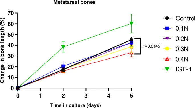
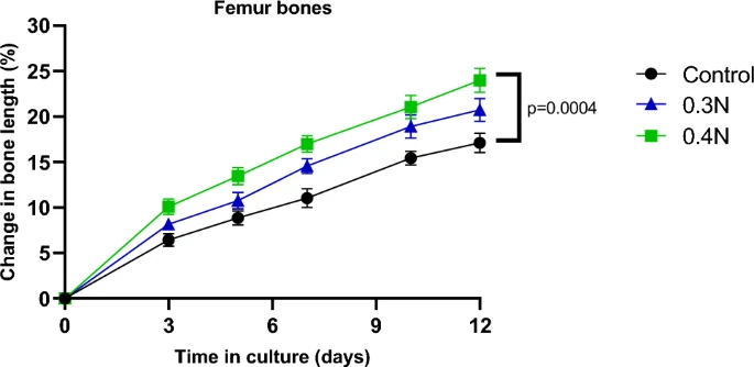
Micromechanical Loading Studies in Ex Vivo Cultured Embryonic Rat Bones Enabled by a Newly Developed Portable Loading Device

Mechanical loading has been described as having the potential to affect bone growth{we want it to affect bone growth post skeletal maturity of couse}. In order to experimentally study the potential clinical applications of mechanical loading as a novel treatment to locally modulate bone growth, there is a need to develop a portable mechanical loading device enabling studies in small bones. Existing devices are bulky and challenging to transfer within and between laboratories and animal facilities, and they do not offer user-friendly mechanical testing across both ex vivo cultured small bones and in vivo animal models. To address this, we developed a portable loading device comprised of a linear actuator fixed within a stainless-steel frame equipped with suitable structures and interfaces. The actuator, along with the supplied control system, can achieve high-precision force control within the desired force and frequency range, allowing various load application scenarios{the potential would be to use the device to induce longitudinal bone growth on skeletally mature individuals}. To validate the functionality of this new device, proof-of-concept studies were performed in ex vivo cultured rat bones of varying sizes. First, very small fetal metatarsal bones were microdissected and exposed to 0.4 N loading applied at 0.77 Hz for 30 s. When bone lengths were measured after 5 days in culture, loaded bones had grown less than unloaded controls (p < 0.05). Next, fetal rat femur bones were periodically exposed to 0.4 N loading at 0.77 Hz while being cultured ex vivo for 12 days. Interestingly, this loading regimen had the opposite effect on bone growth, i.e., loaded femur bones grew significantly more than unloaded controls{femurs have different proportions than metarsal bones, one possibility is that femur bones shape makes it more susceptible to fluid based forces and pressure gradients, due to the femurs longer shape it is possibly more susceptible to deforming forces than metatarsals}. These findings suggest that complex relationships between longitudinal bone growth and mechanical loading can be determined using this device. We conclude that our new portable mechanical loading device allows experimental studies in small bones of varying sizes, which may facilitate further preclinical studies exploring the potential clinical applications of mechanical loading.”

“two use-cases were devised: repetitive impact loading of small force (0.05–0.5 N) at a range of  30–180 repetitions and continuous sinusoidal loading of medium force (0.5–5 N) at a frequency range of 5–20 Hz.”<-something like a massage gun does something similar of repetitive impact loading.

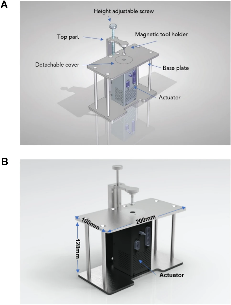
Above is the device used.

So unfortunately for metarsals it does look like yes the device suppressed growth. However, initially the 0.1N load enhanced growth. More studies would have to be done.
In contrast femur bones grew pretty uniformly and the difference is pretty significant. Again, I think it is possible that this could be due to the different shape of the femur bones or maybe the load was too strong for the smaller metatarsal bones.

“Its square shape securely covers the entire cartilage area on each side of the embryonic femur bone, allowing the indenter to apply mechanical loading specifically to the growth plate in a stable manner.”<-just because the growth plate was the thing that was loaded does not necessarily mean that the growth plate was solely responsible for the growth there could be effects elsewhere as well including that which induces longitudinal bone growth.

“we identified that the same load applied to bones of different dimensions has opposing effects on bone growth, suggesting that the effects of mechanical loading on growth are dependent on the magnitudes and relative dimensions of the bones.”<-so they too think that the shape of the bone has an impact on whether growth is induced or not. The fact that the shape of the bones matter also suggests that yes it is possible that not all of the growth is due to the growth plate.

“Embryonic metatarsal bones from Day 19.5 of gestation are approximately 1 mm in length, whereas femur bones are approximately 4 mm, indicating that the level of mechanical loading that stimulates or inhibits bone growth is likely dependent on bone size.”

This is the sentence where the cite Yokota/Zhang’s joint loading study: ” The device’s controller enables sinusoidal loading, which has been demonstrated to have a bone growth-promoting effect in mice “

Study finds that vibration can affect longitudinal bone growth

I am studying vibration due to some anecdotal success using the massage gun on fingers. Looking at vibration results I am seeing mixed results. Sometimes it results in degenerative changes but it is likely that vibration needs to be applied correctly to see results. I believe that like Lateral Synovial joint Loading, vibration would be more effective when applied laterally as that would be more effective in driving fluid flow within the bone. I have written about vibration in the past and in one study I saw that when vibration was applied growth plate parameters increased in the vibrated growth but bone length was about the same so it is possible that bone length at skeletal maturity could be longer in the vibrated group.

Attempt to stimulate longitudinal growth in the dog by mechanical vibration

“In animals, right- and left-handedness leads to larger bones on the dominant side.”

“six miniature partly threaded and self-tapping stainless-steel pins 3 mm in diameter (similar to Denham pins used in orthopaedic surgery) were inserted and allowed to penetrate the skin of the medial sides of the forelegs. The incision was loosely sutured between the pins. Stainless-steel bars were fixed to the ends of the protruding pins. These were grooved and cut to match the yokes on the vibrating rig described below.”<it’s possible these surgical incisions are a confounding factor to longitudinal bone growth.

“The amplitude of vibration was not markedly reduced; this was achieved by incorporating a stiff spring web in the system”

Here’s some images of the vibration mechanism:

“The vibrator enabled the foreleg to be vibrated longitudinally with respect to the radius and ulna”<-so there’s some lateral and torsional loading here.

“In all, seven animals were experimented upon. The first (Dog 1) was experimented upon alone to see if
the study was humane and feasible. Subsequently, and after an encouraging result, four dogs were used
(Dogs 2A-D). In all of these the vibration was sinusoidal, the frequency 20 Hz and the peak-topeak amplitude 4 mm. Finally, in two more dogs (Dogs 3A and B) the same frequency was used but
the amplitude increased to 6 mm”

“As stated above, Dog 1 was used to test the feasibility of the experimental protocol. The result from this single test was an obvious lengthening of the vibrated side{!!!!!} and it indicated to us that
the study should be expanded. Therefore in Group 2, four animals were used, the experimental procedure
being similar except that the daily average vibration period was increased from 1-7h to 2.1h{so it’s possible that these animals did not increase in bone length due to “overtraining”}. The results of Group 2 were disappointing in that, in two dogs, there was actually a small reduction in growth in length on the vibrated side. Group 3 was a more vigorous test in which the amplitude of vibration was increased to 6-8 mm (peak-to-peak). The dogs tolerated this well but, again, no increase occurred. (In one other animal, pins were inserted on one side only and here there was a slight increase in length of both radius and ulna compared with the undisturbed side{so yes this means that the pins are a confounding variable}.)”

Here’s the exciting increase in bone length:

That is frigging massive. The bone looks a lot different in the length increased bone.

“The arteriograms obtained from Dog 1 before sacrifice showed a slight real increase in calibre of the digital vessels on the vibrated side. The difference did not warrant further arteriograms.”<-So vascularity could contribute to part of the increase?

“Histological examination of the growth plates and of the cortical bone at the site of the pins was unremarkable.”<-this is huge as it means that the increase in size could be unrelated to the growth plate i.e. meaning that it could work post skeletal maturity!!!!!

I think there is an overtraining aspect at play here that is possible. As 2 and 3 had stronger stimulus applied and that is why they did not get results.

“The forces were transferred from the radius through the elbow and wrist joints to the arm and paw
respectively. In order to reach these joints, the forces must have passed through the upper and lower
growth plate respectively of the radius, and these will have been subjected to alternating compressive
and distracting forces.”<-one possibility is that the vibration may have induced longitudinal bone growth on the bone but damaged the growth plates. In skeletally mature individuals this would not be an issue as they do not have growth plates. Thus, it would be worthwhile to repeat the experiment on skeletally mature animals.

Here’s the study that was cited in the paper as being about vibration:

The effects of mechanical vibration on bone development in the rat

The study found bone decreased calcium content. This could play a role as the hardness of bone impedes intersititial(longitudinal) bone growth.

And this study too finds slight increase in longitudinal growth!:

Here’s the vibration applied:

“In one series of experiments. lasting for 5 weeks. 20 and 25 Hz was applied for 2.5 hr twice daily with 6
hr of rest intervening. In another series, which lasted for 4 months. the animals received 25 Hz for 12 hr each day without interruption”

So another result that indicates that perhaps by optimizing the vibration stimulus longitudinal bone growth is possible. Also interesting to note that in this study the non vibrated groups grow faster at first. I don’t know what conclusion to draw from that but it is something to explore.

So overall there is not a smoking gun to vibration and longitudinal bone growth but there is some smoke. So it is worth it to keep testing the massage gun method to see if the anecdotal reports continue to have promise. And it is entirely possible that once the method is optimized the results will be stronger than what is reported here.

Yokota/Zhang LSJL related study finds stem cell migration

If migration of stem cells can occur it means that potentially stem cells could migrate to form new growth plates and that cartilage could regenerate at articular cartilage sites potentially leading to height increase via articular cartilage endochondral ossification.

Mechanical Loading Promotes the Migration of Endogenous Stem Cells and Chondrogenic Differentiation in a Mouse Model of Osteoarthritis

This study evaluated whether mechanical loading can enhance anabolic activities by facilitating the recruitment of stem cells for chondrogenesis{anabolic effects could potentially be used for non-osteoarthritic ie normal cartilage}. We evaluated cartilage degradation in a mouse model of OA through histology with H&E and safranin O staining. We also evaluated the migration and chondrogenic ability of stem cells using in vitro assays, including immunohistochemistry, immunofuorescence, and Western blot analysis. The result showed that the OA mice that received mechanical loading exhibited resilience to cartilage damage. Compared to the OA group, mechanical loading promoted the expression of Piezo1 and the migration of stem cells was promoted via the SDF-1/CXCR4 axis. Also, the chondrogenic diferentiation was enhanced by the upregulation of SOX9, a transcription factor important for chondrogenesis. Collectively, the results revealed that mechanical loading facilitated cartilage repair by promoting the migration and chondrogenic diferentiation of endogenous stem cells. This study provided new insights into the loading-driven engagement of endogenous stem cells and the enhancement of anabolic responses for the treatment of OA.”

A vacuole is a space within a cell that is empty of cytoplasm, lined with a membrane, and filled with fluid. Reduction in vacuoles could potentially that new bone is being formed.

“Mechanical stimulation affects the migration and differentiation of MSCs”

“We developed joint loading modalities in which dynamic lateral loads are applied to synovial joints”

“mechanical loading caused cyclic alteration of the intramedullary pressure, driving oscillatory
fluid flow and molecular transport in the lacunocanalicular network in the bone matrix and the medullary cavity”

“female C57BL/6 mice (~ 14 weeks of age, Animal Center of Academy of Military Medical Sciences, China) were used.”

Piezo1 is greater in loading groups even than non ostearthritic controls meaning LSJL(lateral synovial joint loading) meaning that joint loading should enhance stem cell migration in healthy controls.

joint loading promotes adhesion and migration compounds even above controls. These compounds could potentially help induce new cartilage and bone formation.

“Dynamic loads were sinusoidal at 1 N (peak-to-peak) and the frequency was 5 Hz for 6 min/day (each knee, 3 min).”

joint loading increases sdf1 over non-osteoarthritic controls a compound which is again involved in cellular migration.

“After 2 weeks of mechanical loading, the level of Piezo1 in OA mice was significantly increased”

Again strong increase in stem cell migration over controls.

“knee loading contributed to repairing damaged cartilage by recruiting endogenous stem cells and stimulating chondrogenic differentiation.”

Here’s a diagram which explains how loading could be anabolic to cartilage and bone and potentially non osteoarthritic cartilage and bone as well.