Monthly Archives: October 2023

Review paper mentions several papers that show length increase in bones past skeletal maturity

Continuing Periosteal Apposition I: Documentation, Hypotheses, and Interpretation

“This paper reviews 42 studies published since 1964 that have found both significant and nonsignificant
age-related change in various skeletal size dimensions, e.g., length, diameter, width, and area”

“Continuing periosteal apposition (CPA) of lamellar bone in adulthood leads to greater skeletal dimensions in older individuals.”

This is lamellar bone. Periosteal deposition does not usually occur in such a way to increase height but it is possible none the less if only at the top of the skull and the soles of the feet.

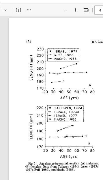
The paper mentions several citations that show changes in skull shape with age including length of skeletal elements. If skull bones can increase in length it is possible that other bones could increase in length too.

Some papers show dramatic length changes while others show smaller or none. Most do seem to trend upward with a decrease between 20 and 30 years of age.

This review paper mentions a Harry Isreal paper as showing that the vertebral body can increase in height past skeletal maturity:

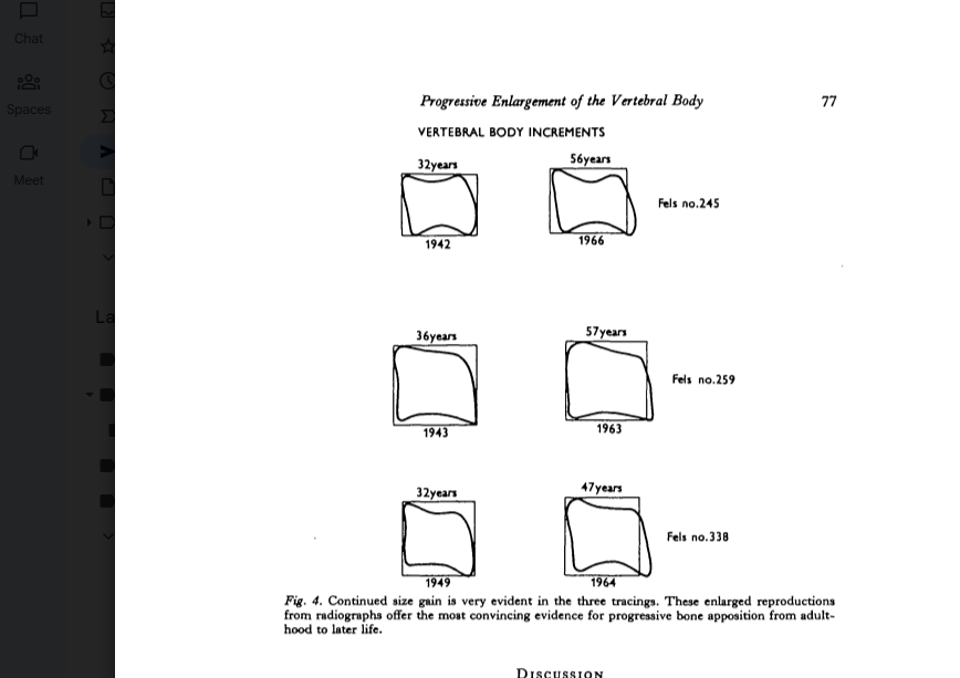
PROGRESSIVE ENLARGEMENT OF THE VERTEBRAL BODY AS PART OF THE PROCESS OF HUMAN SKELETAL AGEING

Both longitudinal and cross-sectional evaluation of the body of the third cervical vertebra reveal
that age-associated continuing enlargement occurs in women through adulthood and into the later
years.
“<-this is exciting stuff as it means it is possible to gain torso height and it could explain the height gain in pregnancy.

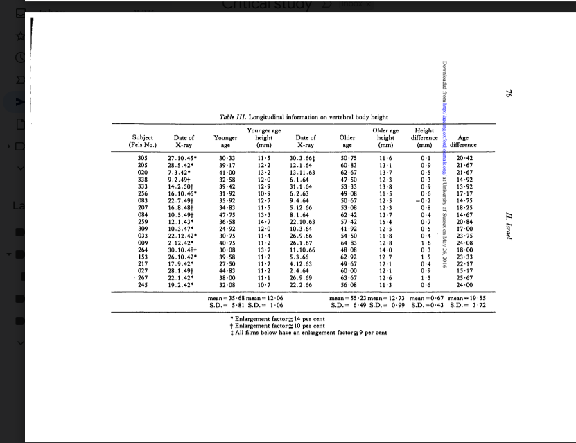
So only one person did not have a age related vertebral body height increase. And amazingly this was a longitudinal study with some people well past their twenties.

Above is an illustration in the change in the cervical vertebrae. A change in height can be seen.

This is the third cervical vertebrae

This study leaves little doubt that continuing growth in the vertebral body, at least the
third cervical vertebra, occurs on an ageing basis among adults.
“!!!!<-very exciting.

The paper Continuing growth in sella turcica with age also looks promising as Sell Turcica is a bone but I could not get the paper.

Here’s another one of the papers mentioned that measured length:

Sex Differences in Age-Related Remodeling of the Femur and Tibia

“In a previous study of a preindustrial sample with high activity levels, both men and women exhibited bone subperiosteal expansion and increase in second moments of area with aging.”

“In preliminary analyses of the data, we found that there was a significant negative trend in femoral and
tibial length with age, particularly among men, where bone length decreased 34 mm per decade on
average”<-if bone length can decrease it is possible that it can increase.

Paper finds that appositional growth can continue post fusion

I emailed Susan Pfeiffer regarding her paper “Age Changes in the External Dimensions of Adult Bone” which showed bioarcheological data that stated that persistent physical activity could stimulate bone growth in length. This paper showed archeological evidence that limb assymetry continued to widen post puberty. The issue with that paper is that it is not a longitudinal study(not on the same subjects over time) so there could be methodological issues. The paper is quite old so I sent her an email about it and she replied ” It looks to me that my paper joins a number of others. Its core conclusions appear to remain valid.”

I asked her to expand on the papers that provided evidence to contribute to the theory that lengthening post epiphyseal fusion is possible and this is one of the ones she suggested. Unfortunately, she stated that her research did continue to go down the direction. I of course was not satisfied with this but I’m lucky I got a response at all. Her is the paper she mentioned: Now it does not show proof of longitudinal growth but it show evidence that appositional growth increase is possible post maturity.

Here’s where the page is hosted if the image above is not sufficient.

IF you look at figure 3 you can see that in some individuals cortical bone continued to increase for the 3rd metacarpal between the ages 20 and 25.

Cortical index is a radiogrammetric parameter that assesses cortical bone stock using a bone X-ray. It is calculated using the ratio between the thickness of the cortical bone and the diameter of the bone shaft.

The exciting statement of course is that appositional continues post fusion at least but not necessarily limited to the ages of 20 and 25.

It looks though if anything and in measuring on gimp that the second metacarpal is longer in the second photo in figure 3. The third metacarpal is too close to call. This is despite the increase in appositional growth being more in the third metacarpal. It is extremely difficult to do an accurate measurement via gimp.