
In a recent post “Increase Height And Grow Taller Using Microfracture Surgery, Part III” I had begun to propose a new height increase minimal invasive technique using the most general idea from microfracture surgery.
Here is how I interpret the entire idea behind microfracture surgery. You cut a section of the cartilage to get to the underlayer of bone. You use a type of thin drill or pick to poke very small holes, the microfractures, into the bone so that the device reaches deep enough to hit the inner of the epiphysis of the long bone. This means that the inner bone marrow, blood, and stem cells can seep out, filling up the hole made, and the clot eventually turns into into fibrocartilage.
I had stated at the end of the previous post…”What could work is that if a series of microfractures in a specific distribution design is created from drills on the side of the epiphysis to completely go around the bone in a closed path. This means that after a few days, the path of drilled microfractures would fill up with stem cells which will eventually turn into fibrocartilage. The fibrocartilage will not be that strong, but before they calcify into bone from vascularization, it would be possible to drill another set of microfractures around the same path to fill up the remaining bone bridges.”
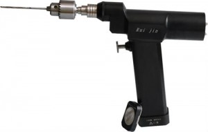 I wanted to explain a little further into this idea so that the reader can understand from a 3 dimensional perspective what I am graphically proposing. The microfracture surgery used an awl (surgical device that punctures) to get the holes in the bones. What I would proposed for the height increase method is to use a similar device, the surgical drill, which would still have the same thickness as the awl.
The drill would go through the outer tissues like the skin and the muscle, just like what would be seen when the traditional ilizarov external method does when the drill is used to add the spokes and wires which are supposed to hold the two bone sides apart and in place when they are being distracted slowly by the long screws.
 The path from looking from a downward z direction would show that the drilled micro holes goes completely around the leg bone. Where the original external fixator method used only 3 drills to put metal spokes or wires into the bones to hole them into place, the drilling for this method would be in the dozens, but will be done under anesthesia. The drilling will go through the bone multiple times to make a path. After the drilling is done, the entire bone will leak out the marrow and stem cells. Something to hold the leaked marrow will keep the clot. After a few days, the outer surface of the bones would be clotted with initial fibrocartilage formation. When the cartilage is formed after 1 week, The rest of the undrilled areas of the bone is done. This would eventually cause the entire cortical area of an entire strip of length in the long bone to be drilled through. The other drilled fractures will also be filled with clots, which turns into cartilage. After the cartilage is formed around the entire bone, covering where the cortical bone is supposed to be, the tensile strength of the long bone will be reduced dramatically. Any type of method like LSJL or weight pulling would lead to cartilage width expansion, thus longitudinal growth.