
I recently found a nice diagram of the signaling pathways of the various proteins in a study (the 1st link below cited). I felt that it would be appropriate right now to look at the diagram since I feel confident now that I am sufficiently prepared to be able to look at the diagram and be reasonable well versed in knowledge to be able to dissect and really do some real analysis on the pathways.
From PubMed study “Mechanisms of Growth Plate Maturation and Epiphyseal Fusion”
Authors: Joyce Emonsa Andrei S. Chaginc Lars Sävendahlc Marcel Karperienb Jan M. Wita
2nd resource : “LOCAL REGULATION OF GROWTH PLATE CHONDROCYTES: Molecular PLATE CHONDROCYTES: Molecular Mechanisms and Implications for Longitudinal Bone Growth” -Author: Anenisia Coelho de Andrade – Thesis for Doctoral Degree (Ph D) 2010
Analysis of the Chart to the right:
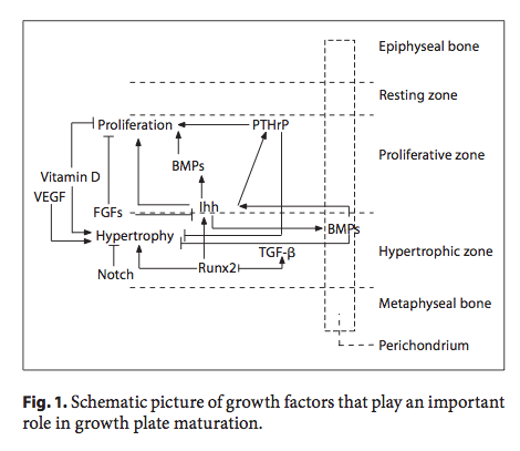 What we are seeing right here is that the growth plates are controlled in each section by certain growth factors we have already seen before many times. However the fact is that we never fully tried to at least summarize all the growth factors together to make a better, more general understanding of the local regulation patterns and pathways that are involved.
Here is what we already know from our studies:
There is basically 3 main areas in the growth plate, the resting zone, the proliferative zone, and the hypertrophic zone. Some authors and medical professionals might even put a zone between the proliferative and hypertrophic zone called the pre-hypertrophic zone, as well as put the layer of ossification and calcification in, but the main idea can be summarized in three zones. The number of chondrocytes we have as the raw material is from the resting zone. From what I have gathered, that area seems to have a finite number of chondrocytes there which does not proliferate. The multiplication in number through cell division seems to happen when it gets to the proliferative zone.
The Legend or Map: All arrow heads indicate that the protein signal is promoting or up-regulating the protein is is pointed to.
The main pathway for the growth factor regulations is one which we have looked at a few times before, on posts like “The Connection Between Regenerating Deer Antlers and The PTHrP, PTH And IHH pathway for Cartilage Regulation, PTHrP Seems To Be The Answer (Big Breakthrough!)”
This is what I would call the IHH-PTHrP negative feedback loop pathway. It shows that the PThrP does two main things. The PTHrP is found mainly in the proliferative zone, as indicated by the diagram of how pathways work in the growth plate above.
- Help stimulate, cause, or increase chondrocyte proliferation
- Help inhibit, down-regulate, or decrease chondrocyte hypertrophy
As for the IHH, it has multiple functions and connections with other growth factors. It seems to be found in both the proliferative and hypertrophic zone, although the diagram suggest that is might be more in the proliferative zone.
It stimulates, up-regulates, and/or increases BMPs in both the proliferative and hypertrophic zones as well as PTHrP
- Runx2 seems to stimulate the production of IHH
- The FGFs however seems to inhibit or down-regulate the protein IHH
One compound in the diagram that has sort of caught my attention is the Runx2 protein/gene. It seems to modulate and maybe help increase the IHH, TGF-Betas, and the chondrocyte hypertrophy.
As for the BMPs, they are multiple types of BMPs, found in both the hypertrophic zone and the proliferative zone. It seems that BMPs in general help increase chondrocyte proliferation, decrease hypertrophy, and has some type of controlling role on the IHH.
In the end, we could try to describe all of the diagram in two major segments by asking the question “Which growth factors stimulate or inihibit the proliferation and hypertrophic zones?
So let’s do a listing of the two main zones.
Proliferation Zone
Stimulating/Increases:
- IHH
- BMPs
- PTHrP
Inhibiting/Decreases:
- Vitamin D
- FGFs
Hypertrophic Zone
Stimulating/Increases:
- Vitamin D
- VEGF
- Runx2
Inhibiting/Decreases:
- Notch
- BMPs
- PThrP
Conclusions:
It is interesting to note that one of the posts which I felt was pivotal in the evolution of this website was the one where I initially suggested that PTHrP was probably the key growth factor we should be trying to get into our bone material to stimulate cartilage regeneration. I came to that conclusion after reading about the fact that abnormal overgrowth after study was shown to be from a high level of PTHrP expression, which happens in cancers.
What I have always believed in my personal opinion was that chondrocyte proliferation was always more important that chondrocyte hypertrophy towards longitudinal growth. Maybe Tyler would believe the opposite, since technically you grow taller in the bones only through hypertrophy of the chondrocytes, where they expand in volume multiple times. It was shown in one study that the rate of the longitudinal increase was proportional to the rate of volume increase in the chondrocytes in hypertrophy.
However I have found enough evidence to show that the real process limiting step in this multiple step process was the fact that you eventually run out of chondrocytes. From the Thesis “LOCAL REGULATION OF GROWTH PLATE CHONDROCYTES: Molecular PLATE CHONDROCYTES: Molecular Mechanisms and Implications for Longitudinal Bone Growth” The Ph. D candidate seems to reference a study “Growth Plate Senescence Is Not a Function of Time per se but of Growth” which I would google and come up with the study “Growth-inhibiting conditions slow growth plate senescence.”
The abstract is pasted below…
Developmental Endocrinology Branch, Program in Developmental Endocrinology and Genetics, Eunice Kennedy Shriver National Institute of Child Health and Human Development, National Institutes of Health, CRC, Room 1-3330, 10 Center Drive, MSC-1103, Bethesda, Maryland 20892-1103, USA.
Abstract
The mammalian growth plate undergoes programmed senescence during juvenile life, causing skeletal growth to slow with age. We previously found that hypothyroidism in rats slowed both growth plate chondrocyte proliferation and growth plate senescence, suggesting that senescence is not dependent on age per se but rather on chondrocyte proliferation. However, one alternative explanation is that the observed slowing of growth plate senescence is a specific consequence of hypothyroidism. We reasoned that, if delayed senescence is a general consequence of growth inhibition, rather than a specific result of hypothyroidism, then senescence would also be slowed by other growth-inhibiting conditions. In this study, we therefore used tryptophan deficiency to temporarily inhibit growth in newborn rats for 4 weeks. We then allowed the animals to recover and studied the effects on growth plate senescence. We found that structural, functional, and molecular markers of growth plate senescence were delayed by prior tryptophan deficiency, indicating that the developmental program of senescence had occurred more slowly during the period of growth inhibition. Taken together with previous studies in hypothyroid rats, our findings support the hypothesis that delayed senescence is a general consequence of growth inhibition and hence that growth plate senescence is not simply a function of time per se but rather depends on growth.
This abstract thus suggest that the reason growth plates develop senescence,or aging is because they run out of chondrocytes. So this means that the step of chondrocyte proliferation or the number of chondrocytes in the resting zone are the limiting steps in the multi-step process. The reason why chondrocyte numbers in the resting zone is another rate limiting step is because of the study “Depletion of resting zone chondrocytes during growth plate senescence.”
And that is why I have felt that PTHrP is more important than IHH, since IHH promotes hypertrophy and PTHrP promotes proliferation.
Indirectly, the IHH stimulates BMPs in both zones which increase proliferation and decrease hypertrophy so maybe after more research, I may have to reverse my conclusion in this (and previous posts) but it is well known that IHH can negatively regulate PTHrP while PTHrP have always been about stimulating cell proliferation and there are no studies which show that PTHrP has any function like inhibiting anything.
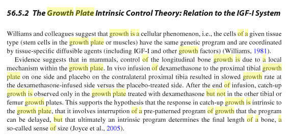
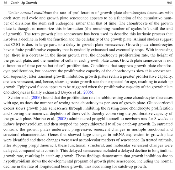
As a final parting message, I would like to paste two clippings from the Medical Reference Book “Handbook of Growth and Growth Monitoring in Health and Disease” by Victor R. Preed, pge 940-941.
Excellent post , i asked you a question in an earlier post on how worlds tallest man Sultan Kosen was gaining adult height 1 inch per year well into his twenties and you reply something about spinal curvature i think you missed the main points 1) his height was officially measured by guiness world records so no chance of measurement error 2).1inch per year is a huge significant increase in adult height .
guinness would measure it and it does seem to show that kosen grew by 2 inches. but let’s remember that a person has the ability to decompress their vertebrate until they are 3% taller than where they are. 8’1″ is 97 inches. 97*.03 = 2.91 inches. So Kosen had the ability to increase his height by quite a bit. Then you add in the fact that almost all 8 footers have severe back curvature and you can say that if Kosen wanted to be taller, he might have started stretching and exercising to realign his back pushing his height even taller.
I dont think Kosen has any need desire or motivation to do exercises to increase his height and even if he did adult height increase is rarely achieved the more likely is that he has grown via sternum spinal disks and possibly articular cartilage from 24cm-251 cm (5cm ) over this 2-3 year timeframe his natural curvature decompression should remain fairly consistent throughout timeframe of official measurements.
I wrote a post about this exact issue. It seems that Kosen has grown and that makes no sense. Maybe we can grow after our growth plates close. but we don’t know how the bones are lengthening without cartilage and chondrocyte stacking into columns.
google reference = worlds tallest man finally stops growing at 8ft 3 in