
I want to go over an article that provides analysis on the structure of the long bones and what it mean for the effectiveness of the LSJL method.
Unfortunately, the paper provides only information about the diaphysis of the long bone rather than the epiphysis which is where we try to induce growth plates with LSJL but it should still provide some insight.
Structure of long bones in mammals.
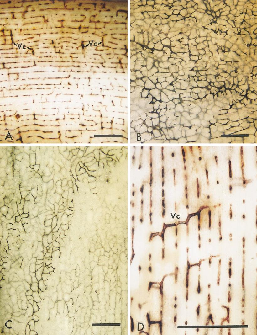
“Bone is a two-compartment system with capillaries and some kinds of connective tissue in one compartment separated from fibers of bone collagen, often forming lamellae, in the other.{cartilage is a form of connective tissue, it is these compartments that would be good candidates for forming new growth plates}. Laminar bone consists of stacks of lamellae separated by vascular spaces containing capillary network sheets. It is deposited at the periosteal and endosteal surfaces. Osteonic bone consists of cylinders of lamellae with central vascular spaces. The primary structure of the shafts of mammalian long bones is laminar and laminae often remain as the main component. Secondary osteons are a replacement within laminae. As laminar bones mature, some of the irregular longitudinal capillary spaces in the network sheets enlarge and become less crooked to form secondary osteons{since the connective tissue compartments are within the laminae perhaps degrading the bone from osteons to more laminar bone would help create a better pro-growth plate micro-environment}. Parts of the random networks become ordered longitudinal ones, resulting in collapse of those network spaces not converted to osteons. The residual capillaries become bloodless, making the surviving network spaces difficult to resolve. For example, laminar bone occurs with osteonic bone in the human femur, although it is rarely figured. Nearly mature bones switch the kind of primary bone deposited at the peripheral (periosteal) surface from laminar to primary osteonic.”
“In the young growing animals examined (beef, sheep, pigs), the shaft is completely made from circumferentially oriented laminae without osteons. In older or even mature animals much of the shaft is still laminar (human, buffalo, deer, horse, oxen, as well as beef, sheep, and pigs).”<-So the presence of osteons in more mature bone could be responsible for the anti-growth plate microenvironment in mature individuals.
Laminar bone is more sheet like whereas osteon bone is more cylindrical. Thus orientation could affect mesenchymal condensation which is the crucial first step for growth plate formation.
Images of young animal laminal bone:
“Mainly laminar bone, with its characteristic vasculature, survives into the adult in a layer below the periosteum. Within this is a layer of mainly osteonic bone. Bone on the endosteal surface is cancellous. The core consists of marrow.”<-maybe this interruption by osteonic bone interferes with growth plate formation.
“The vascular compartment is at first much wider than the compartment destined to contain bone. It declines in width as the lamellae are deposited in the bone layer, eventually shrinking to contain little more than the capillary network”
Figure I shows more osteonic bone:
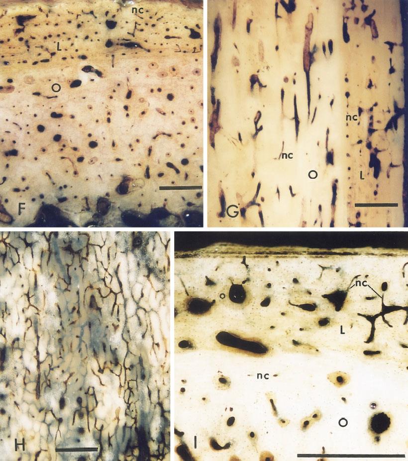
Osteons within laminae bone(mature bone):

So these osteons within laminae in adult bone could also potentially disrupt growth plate formation.
The paper mentions that one of the reasons that bones are white rather than pink is that bones do not have that many blood vessels. This could mean a lack of supply within bone for new growth plate formation.
“The idea that not all vascular compartments contain blood-filled capillaries agrees with the observations that osteons lose their connections with the capillary sheets from which they have been derived”<-restoring this connection might be a key to grow taller.
This study did not address more epiphyseal bone but it does present two problems to neo-growth plate formation and how LSJL might solve them.
1. Osteons might impede new growth plate formation. LSJL degrades bone via fluid flow and shear strain and may disrupt osteonal impedance.
2. Bone loses the connection with the capillary sheets that form stem cells over time. LSJL increases fluid flow which could help restore this connection.
I’m not sure i believe this, i mean why do people emphasis’s get bigger?