
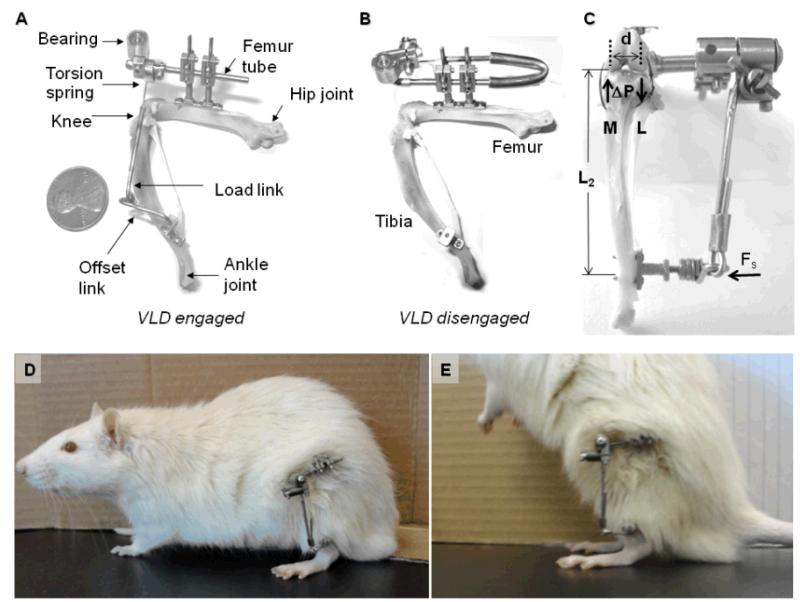
“This study describes the first application of a varus loading device (VLD) to the rat hind limb to study the role of sustained altered compressive loading and its relationship to the initiation of degenerative changes to the tibio-femoral joint. The VLD applies decreased compressive load to the lateral compartment and increased compressive load to the medial compartment of the tibio-femoral joint in a controlled manner.
Mature rats were randomized into one of three groups: unoperated control, 0% (sham) or 80% body weight (BW). Devices were attached to animal’s leg to deliver altered loads of 0% and 80% BW to the experimental knee for 12 weeks. Compartment-specific material properties of the tibial cartilage and subchondral bone were determined using indentation tests. Articular cartilage, calcified cartilage and subchondral bone thicknesses, articular cartilage cellularity, and degeneration score were determined histologically.
Joint tissues were sensitive to 12 weeks of decreased compressive loading in the lateral compartment with articular cartilage thickness decreased in the peripheral region, subchondral bone thickness increased, and cellularity of the midline region decreased in the 80% BW group as compared to the 0% BW group. The medial compartment revealed trends for diminished cellularity and aggregate modulus with increased loading.
The rat-VLD model provides a new system to evaluate altered quantified levels of chronic in vivo loading without disruption of the joint capsule while maintaining full use of the knee. These results reveal a greater sensitivity of tissue parameters to decreased loading versus increased loading of 80% BW for 12 weeks in the rat. This model will allow future mechanistic studies that focus on the initiation and progression of degenerative changes with increased exposure in both magnitude and time to altered compressive loads.”
” The VLD applies altered loads in addition to the normal loads across the joint without disruption of the joint capsule while maintaining full use and range of motion of the joint.”
The mice were 9-month old sprague dawley. The rats had the plates attached surgically so it’s not really an exact approximation of LSJL. And the 0% BW results were drastically different from control so the apparatus definitely had an effect.
“Gross observation of the tibial plateau revealed minimal erosion or fibrillation of the articular cartilage in all experimental groups.”
“The thickness of the articular cartilage in the peripheral region of the lateral compartment decreased 19% in the 80% BW group as compared to the 0% BW group and 26% as compared to the control group. In contrst, the thickness of the subchondral bone increased 38% in the 80% BW group as compared to the 0% BW group”<-So loading increases subchondral bone “thickness” but does this thickness add to height?
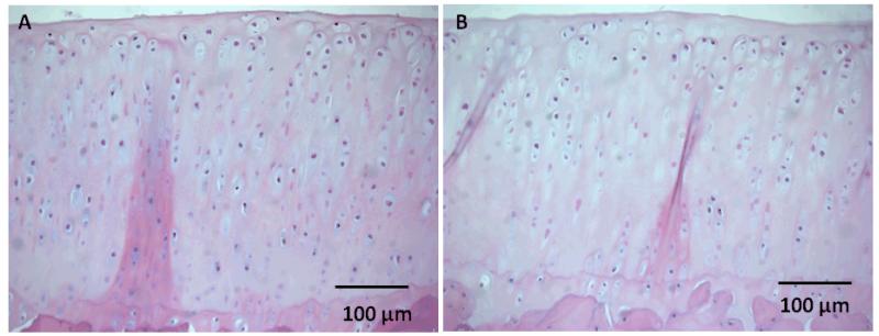
Here’s a comparison between sham control(0% BW) and 80% BW. Loading definitely decreased chondrocyte number but it didn’t seem to decrease articular cartilage height and subchondral bone height seemed to be higher.
Subchondral bone thickness is considered height.