Noninvasive Bone Distraction Using A Shape Memory Implant (Breakthrough!)

This is one of those posts where an idea is talked about which helps height increase researchers turn their head into a completely new direction of research and study.

Analysis & Interpretation

This new way of developing slow distraction in the long bones of the leg is something I have not seen before. It seems to use principles of material science engineering to expand the long bones. What might be difficult for height increase seekers to accept is that idea that two metal rods might first have to be implanted into the long bones for the distraction to happen non-invasively later.

There is indeed a stage where surgery is need to get the rods in initially. However, the later process would be noninvasive.

What is the most interesting about this idea is that it is beyond anything the traditional medical professional like surgeons or genetic researchers can ever come up with. The smart material that is being suggested is a very new technology and the idea of first implanting a precompressed rod and then letting it decompress is very impressive and something I would not have thought of if the idea was not suggested.

In theory the idea could work. The Shape Memory Implants are something which I would have to do much more research on since the field this idea comes from, Material Science Engineering, and maybe even Nanotechnology is very new.

Implications For Height Increase

This idea of using surgery to implant memory shape rods which will eventually extend and decompress to push the length of bones up is reasonable in theory. However the actual implementation of this idea could take decades of research and testing before it would be reasonable for humans to use for limb lengthening. In addition, the idea of getting very invasive surgery to have form changing rods implanted into the bones would not be appealing to most people, not even the most hard core of height increase seekers.

From the PedMed website entitled “Pediatric Medical Devices – Interactive Idea Campaign

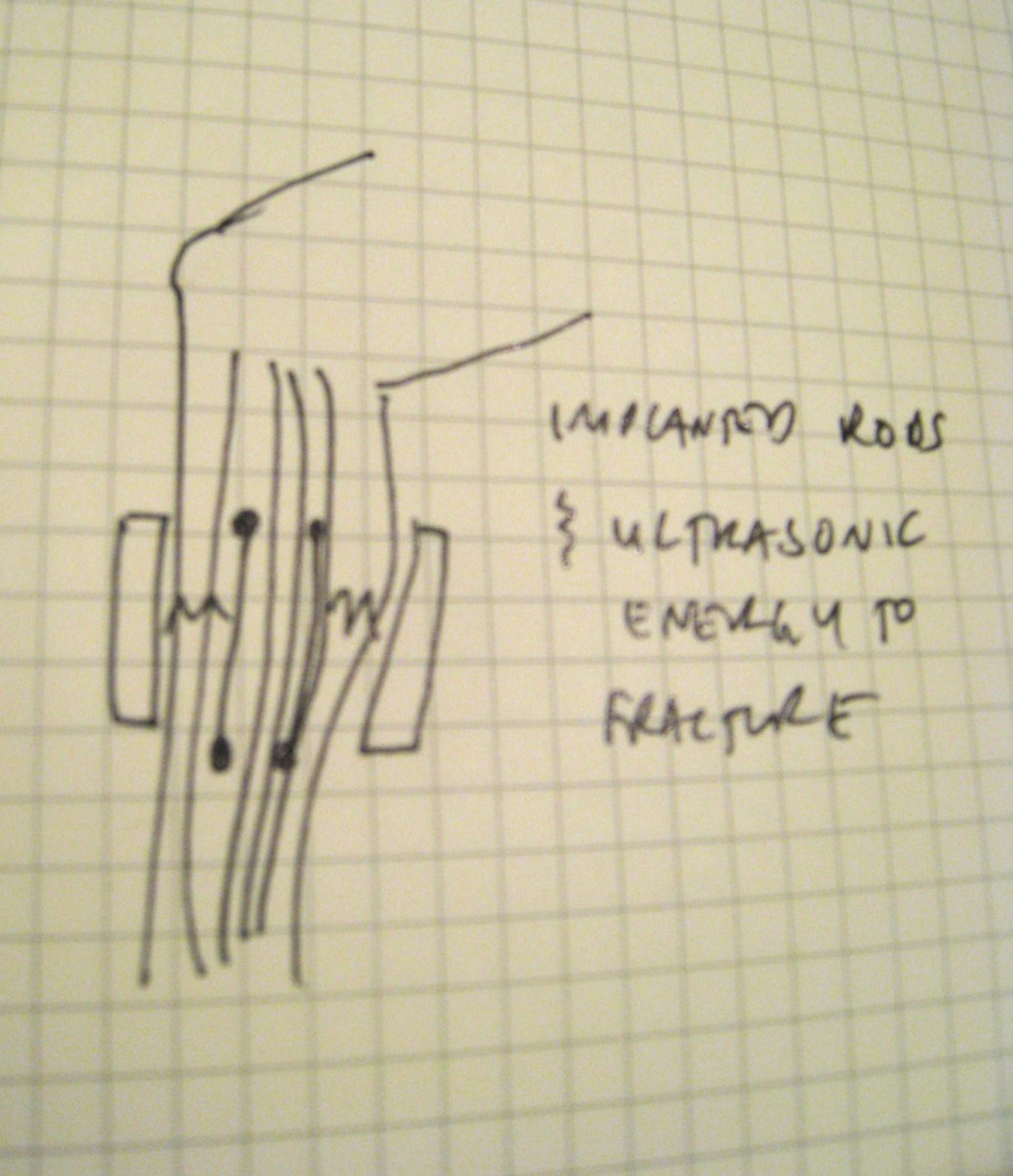
getfileNoninvasive bone distraction using a shape memory implant
D112  Category: Deformity  Submitted by  Avi Feb 24 2010  Status: New idea
Tags:  braceslimbinequality

This idea solves an effective, minimally invasive way to treat lower limb leg length inequality

It involves the implantation of shape memory rods on either bone in the leg.  These rods are placed in a pre-compressed state and are intended to provide axial pressure on the bones.  An external cuff is worn to create mild stress fractures in the bone that are no more painful than a shin splint.  The pre-stressed rods cause distraction osteogenesis.

Analysis Of A Patent For Nutritional Supplements For Stimulating Bone Growth

Recently I found a patent which seems to propose the idea that there might something even simpler and easier we can do to stimulate bone growth. Bone growth implies increasing bone density and possibly also accelerated rates of bone fracture healing. The simpler idea is to create food types with certain chemical inside which can stimulate bone density increase.

This inventor seems to have a patent for certain compounds they have either created or hypothesizes will stimulate bone growth from oral ingestion.

Analysis & Interpretation

The idea of the invention is to reformulate statin and other bone growth enhancing chemicals compounds into edible supplements a person can put in their food already to gain a very easy natural way of bone growth.

The thing is that the inventor is very comprehensive in their research. They admit that the BMP group has osteogenic properties but because they are peptides, they can’t be taken orally but need to be injected subcutenously (under the skin) into the blood stream. Plus, the BMP seems to be produced by different parts of the body and has effects on different parts of the body. If the BMP was taken orally, it would have an overal systemic affect on the entire body, not just the specific local areas the inventor is hoping to target.

For The Researcher, What Is The Science Behind This Claim?

The thing about BMPs are that they are produced when the precursor cells start to differentiate into osteoblasts, which are the cells which produce bone. The cells also seem to produce Type-1 collagen, osteocalcin, osteopontin and alkaline phosphatase. The inventor seems to have found that molecules or chemicals which have bone growth enhancing properties are made of this type of chemical structure…

“…Ar1—L—Arwherein Arand Arare aromatic moieties and L is a linker that separates them by a specified distance.”

It seems however the real group of type of molecules the inventor wants to get a patent for are the “statins”. He states…

“Another class of compounds comprises inhibitors of β-hydroxy-β-methyl glutaric acid CoA (HMG-CoA) reductase. These compounds also enhance bone formation; they are generically known as “statins.” HMG-CoA reductase is the principal rate limiting enzyme involved in cellular cholesterol biosynthesis.”

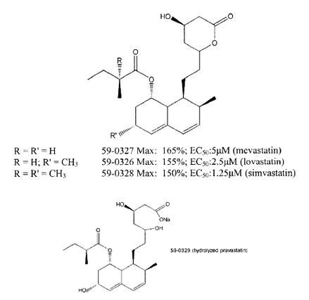
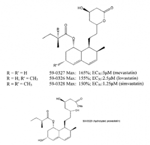
There have been a few other patens which came out, specifically ones by Zymogenetics of Seattle who in the early 2000s were also developing bone growth pills and the primary compound they looked at was “statin”. What I didn’t know was that the statin was an entire group of chemical molecules. These go by the much longer term inhibitors of β-hydroxy-β-methyl glutaric acid CoA (HMG-CoA) reductase. He notes that in a few studies it was shown that lovastatin did seem to decrease osteoporosis but there was no study which showed that lovastatin directly increased bone formation. The invention would be in some type of liquid form which can be added into foods as a supplement to enhance bone growth.
statintypesNutritional supplements for stimulating bone growth – US 6410521 B1

Gregory R. Mundy et al.

A food or food supplement which comprises a compound that enhances bone growth in vertebrates is described wherein the food or foodstuff is formulated so as to provide the desired bone growth enhancing effect.

TECHNICAL FIELD

The invention relates to foodstuffs and nutritional supplements which contain active ingredients that stimulate bone growth in humans and other vertebrates. More specifically, the invention concerns methods to enhance bone growth by supplementing the diet of humans and domesticated animals with edible materials that contain statins or other bone growth enhancers and by formulating such enhancers in edible form.

BACKGROUND ART

Bone is subject to constant breakdown and resynthesis in a complex process mediated by osteoblasts, which produce new bone, and osteoclasts, which destroy bone. The activities of these cells are regulated by a large number of cytokines and growth factors, many of which have now been identified and cloned.

There is a plethora of conditions which are characterized by the need to enhance bone formation. Perhaps the most obvious is the case of bone fractures, where it would be desirable to stimulate bone growth and to hasten and complete bone repair. Agents that enhance bone formation would also be useful in facial reconstruction procedures. Other bone deficit conditions include bone segmental defects, periodontal disease, metastatic bone disease, osteolytic bone disease and conditions where connective tissue repair would be beneficial, such as healing or regeneration of cartilage defects or injury. Also of great significance is the chronic condition of osteoporosis, including age-related osteoporosis and osteoporosis associated with post-menopausal hormone status. Other conditions characterized by the need for bone growth include primary and secondary hyperparathyroidism, disuse osteoporosis, diabetes-related osteoporosis, and glucocorticoid-related osteoporosis.

One group of compounds suggested for enhancing bone formation comprises bone morphogenic proteins (BMPs). The BMPs are novel factors in the extended transforming growth factor β superfamily. Recombinant BMP-2 and BMP-4 can induce new bone formation when they are injected locally into the subcutaneous tissues of rats (Wozney J., Molec Reprod Dev (1992) 32:160-67). These factors are expressed by normal osteoblasts as they differentiate, and have been shown to stimulate osteoblast differentiation and bone nodule formation in vitro as well as bone formation in vivo (Harris S., et al., J. Bone Miner Res (1994) 9:855-63). This latter property suggests potential usefulness as therapeutic agents in diseases which result in bone loss.

The cells which are responsible for forming bone are osteoblasts. As osteoblasts differentiate from precursors to mature bone-forming cells, they express and secrete a number of enzymes and structural proteins of the bone matrix, including Type-1 collagen, osteocalcin, osteopontin and alkaline phosphatase (Stein G., et al., Curr Opin Cell Biol (1990) 2:1018-27; Harris S., et al. (1994), supra). They also synthesize a number of growth regulatory peptides which are stored in the bone matrix, and are presumably responsible for normal bone formation. These growth regulatory peptides include the BMPs (Harris S., et al. (1994), supra). In studies of primary cultures of fetal rat calvarial osteoblasts, BMPs 1, 2, 3, 4, and 6 are expressed by cultured cells prior to the formation of mineralized bone nodules (Harris S., et al. (1994), supra). Like alkaline phosphatase, osteocalcin and osteopontin, the BMPs are expressed by cultured osteoblasts as they proliferate and differentiate.

Although the BMPs are potent stimulators of bone formation in vitro and in vivo, there are disadvantages to their use as therapeutic agents to enhance bone healing. Receptors for the bone morphogenetic proteins have been identified in many tissues, and the BMPs themselves are expressed in a large variety of tissues in specific temporal and spatial patterns. This suggests that BMPs may have effects on many tissues in addition to bone, potentially limiting their usefulness as therapeutic agents when administered systemically. Moreover, since they are peptides, they would have to be administered by injection. These disadvantages impose severe limitations to the development of BMPs as therapeutic agents.

Small molecules that are useful in treating bone disorders in vertebrates are of the general formula Ar1—L—Arwherein Arand Arare aromatic moieties and L is a linker that separates them by a specified distance. These are disclosed in PCT application WO98/17267 published Apr. 30, 1998. These compounds were assessed for usefulness in treating bone disorders by their ability to enhance the production of a reporter protein when the nucleotide sequence encoding the reporter protein is operably linked to the promoter for BMP-2. Similar compounds are disclosed for this purpose in earlier filed PCT applications WO97/15308 published May 1, 1997 and WO97/48694 published Dec. 24, 1997.

Another class of compounds comprises inhibitors of β-hydroxy-β-methyl glutaric acid CoA (HMG-CoA) reductase. These compounds also enhance bone formation; they are generically known as “statins.” HMG-CoA reductase is the principal rate limiting enzyme involved in cellular cholesterol biosynthesis. The pathway is also responsible for the production of dolichol, ubiquinones, isopentenyl adenine and farnesol. HMG-CoA reductase converts 3-hydroxy-3-methyl-glutaryl CoA (HMG-CoA) to mevalonate. Addition of mevalonate at concentrations between 25-800 μM inhibits the activity of mevastatin (100, 25, or 6.25 μM) in the ABA assay described in Example 1 herein. Mevalonic acid has no effect on primary screen activities of bone growth-active compounds outside of the statin family (compounds 59-0008 (see Example 1)). These data indicate that the effect of mevastatin in the ABA assay is mediated by its effect on HMG-CoA reductase. Knowledge of inhibitors of the cholesterol biosynthetic pathway (including SAR or pharmacophore analyses) may be useful in determining appropriate modifications or analogs of the statins that maintain bone growth activity.

U.S. Pat. No. 5,280,040 discloses compounds described as useful in the treatment of osteoporosis. These compounds putatively achieve this result by preventing bone resorption. Related to these compounds are the bisphosphonates—the methylene bisphosphonic acids. These compounds are comprised of two phosphonic acid residues coupled through a methylene linkage. Typical representatives include the clodronates which are simple compounds wherein the phosphonic acid residues are coupled through dichloromethylene. Other representative bisphosphonates include ibandronates, the risedronates, alandronates and pamidronates. These compounds have been shown to inhibit the resorption of bone, presumably by effecting apoptosis of osteoclasts. Luckman, S. P., et al., J Bone Min Res (1998) 13:581-589.

Wang, G.-J., et al., J Formos Med Assoc (1995) 94:589-592 report that certain lipid clearing agents, exemplified by lovastatin and bezafibrate, were able to inhibit the bone resorption resulting from steroid administration in rabbits. However, there is no suggestion in Wang, et al., that lovastatin directly enhances bone formation. An abstract entitled “Lovastatin Prevents Steroid-Induced Adipogenesis and Osteoporosis” by Cui, Q., et al., appeared in the Reports of the ASBMR 18th Annual Meeting (September 1996) J. Bone Mineral Res. (1996) 11(S1):S510. The abstract reports that lovastatin diminished triglyceride vesicles that accumulated when osteoprogenitor cells cloned from bone marrow stroma of chickens were treated in culture with dexamethasone. Lovastatin was reported to diminish the expression of certain mRNAs and to allow the cells to maintain the osteogenic phenotype after dexamethasone treatment. Further, chickens that had undergone bone loss in the femoral head as a result of dexamethasone treatment were improved by treatment with lovastatin. Again, there is no suggestion that lovastatin directly enhances bone formation in the absence of steroid treatment. In addition, PCT publication WO99/45923 describes methods of inhibiting bone resorption by administering an HMG-CoA reductase inhibitor. Again, there is no suggestion that statins or other HMG reductase inhibitors enhance the formation of bone.

The distinction between enhancing bone formation and inhibiting resorption is further elucidated by Ducy, P., et al., Nature(1996) 382:448-52 who have recently reported that osteocalcin deficient mice exhibit a phenotype marked by increased bone formation and bones of improved functional quality, without impairment of bone resorption. Ducy, et al., state that their data suggest that osteocalcin antagonists may be of therapeutic use in conjunction with estrogen replacement therapy (for prevention or treatment of osteoporosis).

The present invention describes means for administering statins and other classes of compounds that enhance bone formation by including them in nutritional supplements, or by administering edible materials that naturally contain these compounds. In addition, the invention is directed to an optimum protocol for administering compounds that enhance bone formation.

What is claimed is:

1. A method to enhance bone formation in a human characterized by a condition selected from the group consisting of osteoporosis, bone fracture or deficiency, primary or secondary hyperparathyroidism, periodontal disease or defect, metastatic bone disease, osteolytic bone disease, post-plastic surgery, post-prosthetic joint surgery, and post-dental implantation, which method comprises administering to said human, as a dietary supplement, an effective amount of Red Yeast Rice.

2. The method of claim 1, wherein said administering is on an intermittent protocol wherein the time period occupied by dosage administration is less than 50% of the time frame over which treatment is administered, as measured by the time between initial administration and assessment of results.

3. A method to enhance bone formation in a human characterized by a condition selected from the group consisting of osteoporosis, bone fracture or deficiency, primary or secondary hyperparathyroidism, periodontal disease or defect, metastatic bone disease, osteolytic bone disease, post-plastic surgery, post-prosthetic joint surgery, and post-dental implantation, which method comprises administering to said human an effective amount of a statin in hydrolyzed or unhydrolyzed form wherein said statin prepared as a pharmaceutical has been added to a liquid foodstuff.

4. The method of claim 3, wherein said statin is in hydrolyzed form.

5. The method of claim 3, wherein said statin is cerivastatin, lovastatin, mevastatin, simvastatin, fluvastatin, pravastatin, or NK-104.

6. The method of claim 3, wherein the liquid foodstuff is grapefruit juice.

7. The method of claim 3, wherein said liquid foodstuff is salad dressing or salad oil.

8. The method of claim 3, wherein said statin is emulsified in said foodstuff.

9. The method of claim 3, wherein said administering is on an intermittent protocol wherein the time period occupied by dosage administration is less than 50% of the time frame over which treatment is administered, as measured by the time between initial administration and assessment of results.

DISCLOSURE OF THE INVENTION

In one aspect, the invention is directed to a method to enhance bone growth in humans or other vertebrate animals by including in their diets foodstuffs or food supplements that contain effective amounts of bone growth enhancing compounds. These compounds can be the small molecule bone enhancing compounds of the formula Ar1—L—Ar2described hereinabove, bisphosphonates, or other bone growth enhancing agents such as BMPs. A particularly preferred group of compounds which can be administered in this was comprises the statins. Typical statins are of the formula:

Figure US06410521-20020625-C00001


wherein X in each of formulas (1), (2) and (3) represents a substituted or unsubstituted alkylene, alkenylene, or alkynylene linker of 2-6C optionally containing one heteroatom which is O, N or S;

Y represents one or more carbocyclic or heterocyclic rings; when two or more rings are present in Y, they may optionally be fused; and

R′ represents a cation, H or a substituted or unsubstituted alkyl group of 1-6C.

Thus, the invention is directed to methods to treat bone disorders by directly stimulating bone formation stimulating bone formation by including in the diet of a subject in need of such stimulation, a food or food supplement which contains a bone enhancing compound, especially a statin compound. Oral administration of the compounds of the invention, in particular the statins, enhances plasma concentration by avoiding hepatic first pass effects. Thus, their effects on bone and non-hepatic tissues are enhanced. In another aspect, the invention relates to administering the compounds that stimulate bone growth on an intermittent schedule in contrast to continuous administration.

 

A Technique To Increase Bone Growth Using Hyaluronic Acid And Growth Factor basic FGF

What I have found recently is a patent which shows how we even as amateur experimentalist can probably get the raw materials needed to do this experiment to show that using bFGF and Hyaluronic Acid can get bones to growth, at least in terms of thickness.

The patent is rather long so I will not be attempting to copy and paste on this post everything that was written in the patent. I only copied the abstract, patent description, and the claims made by the inventor.

Analysis & Interpretation

From a first glance it looks like the patent for the invention involves a two part composition mixture that is supposed to be applied to the area of bone one would like to thicken. The composition that is formed from getting the hyaluronic acid and basic FGF growth factor would have the viscosity level and and biodegradability level to stay in the area of bone long enough to have some type of osteogenic effect.

For the actual growth factor, there might be more than just FGF involved, but also the TGF-Betas 1-3, IGF-1, PDGF, EGF and other elements in the TGF-Beta superfamily. The inventor is better at describing the invention with….

“A method of treating diseased, injured or abnormal bone at an orthotopic or intraosseous site of desired bone growth…”

To get into specifics, if we are choosing the basic version of the FGF growth factor, we would use concentrations at 10.sup.-6 to 100 mg/ml and the Hyaluronic Acid would need to be around 0.1-4.0% by weight percentage.

Implications For Height Increase

This type of invention is interesting to  consider since it might be the type of method or technique which for the height increase seeker means that they might be able to take a type of syringe and inject the bone growth material on the edges of long bones to stimulate layer upon layers of appositional bone growth. The process of using this idea for height gain will be very slow.

However this type of patent is useful to note for later research if the case where we need to form bone growth quickly can be done using this mixture of growth factor and hyaluronic acid.

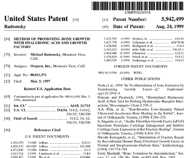
methodMethod of promoting bone growth with hyaluronic acid and growth factors

Michael Radomsky

A bone growth-promoting composition is provided comprising hyaluronic acid and a growth factor. The composition has a viscosity and biodegradability sufficient to persist at the site of desired bone growth for a period of time sufficient to promote the bone growth. Preferably hyaluronic acid is used in a composition range of 0.1-4% by weight and preferred growth factor is bFGF, present in a concentration range of about 10.sup.-6 to 100 mg/ml.

SUMMARY OF THE INVENTION

The present invention provides a bone growth-promoting composition comprising hyaluronic acid and a growth factor such that the composition has a viscosity and biodegradability sufficient to persist at the site of desired bone growth for a period of time sufficient to promote bone growth.

Compositions comprising hyaluronic acid and a growth factor are provided which have the requisite viscosity and biodegradability.

As used herein, the term hyaluronic acid, abbreviated as HA, means hyaluronic acid and its salts such as the sodium, potassium, magnesium, calcium, and the like, salts.

By growth factors, it is meant those factors, proteinaceous or otherwise, which are found to play a role in the induction or conduction of growth of bone, ligaments, cartilage or other tissues associated with bone or joints.

In particular these growth factors include bFGF, aFGF, EGF (epidermal growth factor), PDGF (platelet-derived growth factor), IGF (insulin-like growth factor), TGF-β I through III, including the TGF-β superfamily (BMP-1 through 12, GDF 1 through 12, dpp, 60A, BIP, OF).

What is claimed is:

1. A composition for treatment of diseased, injured or abnormal bone comprising an effective amount of a mixture of a growth factor and hyaluronic acid sufficient to enhance bone growth rate and magnitude and having a sufficient viscosity and biodegradability to persist upon application at an orthotopic or intraosseous site of desired bone growth for a period of time sufficient to enhance said bone growth rate and magnitude.

2. A composition according to claim 1 wherein said hyaluronic acid is uncrosslinked.

3. A composition according to claim 1 wherein said composition comprises 0.1 to 4% by weight of hyaluronic acid in solution.

4. A composition according to claim 1 wherein said growth factor comprises bFGF.

5. A composition according to claim 4 wherein said bFGF is present in said composition in a range of about 10.sup.-6 to 100 mg/ml of said composition.

6. A method of treating diseased, injured or abnormal bone at an orthotopic or intraosseous site of desired bone growth comprising the step of applying to said site a composition comprising an effective amount of a mixture of hyaluronic acid and a growth factor sufficient to enhance bone growth rate and magnitude and having a sufficient viscosity and biodegradability to persist at said site for a period of time sufficient to enhance said bone growth rate and magnitude.

7. A method according to claim 6 wherein said hyaluronic acid is uncrosslinked.

8. A method according to claim 6 wherein said hyaluronic acid in said composition comprises about 0.1-4% by weight of said composition.

9. A method according to claim 6 wherein said growth factor comprises bFGF.

10. A method according to claim 9 wherein said bFGF is present in a range of about 10.sup.-6 to 100 mg/ml in said composition.

EBI Bone Healing System, Another Non-Invasive Bone Growth Stimulation Device

biomet ebi bone healing systemThis device which I found from the BIOMET company’s website is another device that has bone regeneration and bone growth abilities.

EBI Bone Healing System®

Clinically Effective

  • The most studied non-invasive bone growth stimulation device on the market1
  • Heal rates as high as 92%2
  • 2 ½ months earlier healing3
  • 75% of Biomet customers are repeat prescribers4

Scientifically Proven

  • Pre-clinical studies state PEMF has a reproducible osteogenic effect in vitro and simultaneously induces naturally occuring BMP-2 and BMP-45
  • In vivo and in vitro pre-clinical studies demonstrated PEMF exposure more than doubled the rate of angiogenesis6

Cost Efficient

  • According to a published, peer-reviewed study, electrical stimulation was shown to be more cost efficient when compared to no stimulation or LIPUS for the treatment of nonunions7

Analysis & Interpretation:

What a person needs to understand is that if one really thinks that all it takes to lengthen bone is to have a non-invasive bone growth stimulation device, this would be it. The device seems to be something a patient with a broken leg aka fracture on their long bones would wear like a brace. The way the electrical field would be generated and interact with the broken bone would result in accelerated bone healing time.

If a person thinks they just need a bone growth stimulation device, they can try this and put it on their leg and wear it for a few months and check to see whether the one leg they have been wearing it on increased slightly more in length compared to the contralateral leg used as control.

For more information or to see the scientific studies and articles written about the idea that PEMF technology does indeed work, I copy and pasted all of the studies that was referenced below for any researcher’s convenience.

1. Data on file at Biomet Spine & Bone Healing Technologies, most published studies as of 9/7/2012

2. Frykman, G.K., Taleisnik, J., Peters, G., Kaufman, R., Helal, B., Wood, V.E., and Unsell, R.S. Treatment of nonunited scaphoid fractures by pulsed electromagnetic field and cast. J Hand Surg Am, 1986. 11(3): p. 344-9.*

3. Murray, H and Pethica, B. Pulsed Electromagnetic Field (PEMF) Therapy and Fracture Management: An Analysis of the Time to Heal Data. 2012. Data on file at Biomet Spine and Bone Healing Technologies. Disclaimer: Funding for this study was provided by EBI, LLC, d/b/a Biomet Spine and Bone Healing Technologies

4. Data on file at Biomet Spine & Bone Healing Technologies

5. Bodamyali T, Bhatt B, Hughes FJ,Winrow VR, Kanczler JM, Simon B, Abbott J, Blake DR and Stevens CR. Pulsing electromagnetic fields simultaneously induce osteogenesis and upregulate transcription of bonemorphogenetic proteins 2 and 4 in rat osteoblasts in vitro. BiochemBiophys Res Commun 250:458-461, 1998. Disclaimer: Following non invasive electrical stimulation, increases in multiple growth factors have been observed in various pre-clinical in vitro cellular and in vivo animal studies. Although not indicative of human clinical results, these factors have been implicated in various models of bone repair.*

6. Tepper OM, Chang EI, Baharestani S, Galiano RD, Bhatt KA, Hopper RA, Heitman DE, Simon BJ, Gan JC, Levine JP and Gurtner GC. Electromagnetic fields increase in vitro and in vivo angiogenesis through a FGF-mediated VEGF independent mechanism. Submitted to FASEB. Disclaimer: Following non invasive electrical stimulation, increases in multiple growth factors have been observed in various pre-clinical in vitro cellular and in vivo animal studies. Although not indicative of human clinical results, these factors have been implicated in various models of bone repair.*

7. Burden of Illness among Patients Experiencing Bone Fracture Nonunion report, Prepared by Ning Wu, PhD, Yuan-Chi Daisy Lee, MS, Alan Wang, BS, Luke Boulanger, MA, MBA, United BioSource Corporation Copy on file at BS&BHT. Disclaimer: Funding for this study was provided by EBI, LLC, d/b/a Biomet Spine and Bone Healing Technologies

Endogenous Retinoic Acid Negatively Regulates Growth Plate Chondrogenesis And Longitudinal Growth

One compound that I had read about from the very beginning of the research for the website was the fact that there was a type of acid known as retinoic acid which might have certain properties and functions which involved limb regeneration in amphibians, fish, and reptiles. I wanted to go back to look slightly deeper on the function of retinoic acid to see whether it had any potential to be a chondrogenic stimulant and could induce something similar to what we see in deer antler regeneration and the antler longitudinal growth. My hope is to find studies which suggest that the retinoic acid can stimulate chondrocyte proliferation and/or chondrocyte hypertrophy.

These are the PubMed studies I have found which shows the connection between the function of retinoic acid and chondrogenesis.

Study #1: Retinoic acid is a potent regulator of growth plate chondrogenesis.

  • Endocrinology. 2000 Jan;141(1):346-53.
  • De Luca F, Uyeda JA, Mericq V, Mancilla EE, Yanovski JA, Barnes KM, Zile MH, Baron J.
  • Source: Developmental Endocrinology Branch, National Institute of Child Health and Human Development, National Institutes of Health, Bethesda, Maryland 20892, USA.
  • fdeluca@peds.umaryland.edu
  • PMID: 10614657

Abstract

Vitamin A deficiency and excess both cause abnormalities in mammalian longitudinal bone growth. Because all-trans retinoic acid (RA) is synthesized from vitamin A, we hypothesized that RA regulates growth plate chondrogenesis. Consistent with this hypothesis, a single oral dose of RA reduced the height of the rat proximal tibial growth plate. To determine whether RA acts directly on growth plate, fetal rat metatarsal bones were cultured in the presence of RA. In this system, RA inhibited longitudinal bone growth by three mechanisms: 1) decreased chondrocyte proliferation, (assessed by 3H-thymidine incorporation), particularly in the proliferative zone of the growth plate; 2) decreased matrix synthesis (assessed by 35SO4 incorporation into glycosaminoglycans); and 3) decreased cell hypertrophy (determined histologically). The growth-inhibiting effects of RA were completely reversed by a retinoic acid receptor (RAR) antagonist. In the absence of exogenous RA, this antagonist accelerated bone growth, as did an RA-specific neutralizing antibody, suggesting that endogenous RA negatively regulates growth plate chondrogenesis. We conclude that RA, acting through RARs, negatively regulates longitudinal bone growth by inhibiting growth plate chondrocyte proliferation, chondrocyte hypertrophy, and matrix synthesis.

Study #2: Retinoic acid induces rapid mineralization and expression of mineralization-related genes in chondrocytes.

  • Exp Cell Res. 1993 Aug;207(2):413-20.
  • Iwamoto M, Shapiro IM, Yagami K, Boskey AL, Leboy PS, Adams SL, Pacifici M.
  • Source
  • Department of Anatomy-Histology, School of Dental Medicine, University of Pennsylvania, Philadelphia 19104-6003.
  • PMID: 8344389

Abstract

Numerous studies of experimental hypo- and hypervitaminosis A have long suggested that retinoic acid (RA) is involved in chondrocyte maturation during endochondral ossification and skeletogenesis. However, the specific and direct roles of RA in these complex processes remain unclear. Based on recent studies from our laboratories, we tested the hypothesis that RA induces the expression of genes associated with the terminal mineralization phase of chondrocyte maturation and promotes apatite deposition in the extracellular matrix. Cell populations containing chondrocytes at advanced stages of maturation were isolated from the upper portion of Day 18 chick embryo sterna and grown for 2 weeks in monolayer until confluent. The cells were then treated with low doses (10-100 nM) of RA for up to 6 days in the presence of a phosphate donor (beta-glycerophosphate) but in the absence of ascorbic acid. Within 4 days of treatment, RA dramatically induced expression of the alkaline phosphatase (APase), osteonectin, and osteopontin genes, caused a several-fold increase in APase activity, and provoked massive mineral formation while it left type X collagen gene expression largely unchanged. The mineral had a mean Ca/Pi molar ratio of 1.5; Fourier transform infrared spectra confirmed that it represented hydroxyapatite. Mineralization was completely abolished by treatment with parathyroid hormone; this profound effect confirmed that RA induced cell-mediated mineralization and not nonspecific precipitation. When cultures were treated with both RA and ascorbic acid, there was a slight further increase in APase activity and increased calcium accumulation. The effects of RA were also studied in cultures of immature chondrocytes isolated from the caudal portion of sternum; however, RA only had minimal effects on mineralization and gene expression in these cells. Thus, RA appears to be a rapid, potent, maturation-dependent, ascorbate-independent promoter of terminal maturation and matrix calcification in chondrocytes.


Analysis & Interpretation:

From the 1st study, the first thing that really stands out is that the researchers note that both too much and too little vitamin A results in mammalian bone longitudinal growth abnormalities. It seems that Vitamin A is the precursor for retinoic acids, in all of its many variations. The researchers hypothesized that retinoic acid would inhibit long bone longitudinal growth. It seems that just 1 oral dose of the RA by a lab rate resulted in thinner growth plates. It seems that the RA decreased chondrocyte proliferation, hypertrophy, and cartilage extracellular matrix formation. What is the big discovery to us as height increase researchers is what the abstract says next…

“The growth-inhibiting effects of RA were completely reversed by a retinoic acid receptor (RAR) antagonist. In the absence of exogenous RA, this antagonist accelerated bone growth, as did an RA-specific neutralizing antibody, suggesting that endogenous RA negatively regulates growth plate chondrogenesis”

This is a major discovery. It seems that if we can get exogenous RAR close to the growth plates, the longitudinal growth of the long bones can be accelerated. It seems that the RA actually acts through the RAR though. So any endogenous RA will have this effect. This means that since the balance of RA and RAR in the human body is important to keep the bones from being completely inhibited or growth uncontrollably, the concentrations of RA and RAR is kept at a sort of metastatic equilibirum. I would then suggest that we get an exogenous source of RAR which we apply close to the growth plates through injection, or even taken orally can possibly lead to the growth plates having accelerated lengthening and growth.

As for study #2, the researchers state the same initial hypothesis as made by the researchers of study #1. The Retinoic acid causes endochondral ossification to mature aka stop. For their specific study, …

“…we tested the hypothesis that RA induces the expression of genes associated with the terminal mineralization phase of chondrocyte maturation and promotes apatite deposition in the extracellular matrix…”

Chondrocytes were extracted from the chest cartilage of days old chicken. They were grown in a monolayer until they became confluent. The cells were then treated with low doses of RA for 6 days. After 4 days it seems that the chondrocytes showed a very high increase in the expression of alkaline phosphatase,osteonectin, and osteopontin genes. There was a huge accumulation of mineral formation. When the mineral that was formed was testing, it was concluded that the mineral was hydroxyapatite. What is even more interesting is how the researchers made the mineralization go away….

“Mineralization was completely abolished by treatment with parathyroid hormone;…”

It seems that by using the PTH, the researchers made the mineralized hard material of calcium deposits aka hydroxyapatite dissolve.

So the researchers concluded that…

“…this profound effect confirmed that RA induced cell-mediated mineralization and not nonspecific precipitation…”

It seems that if you take the RA with ascorbic acid, there is an even higher level of alkaline phosphatase release or expression and the mineralization and calcification increased too.

What is really strange twist is that if we then take non-mature chondrocytes, the RA injections seem to not have as dramatic of an effect on them as the mature chondrocytes. It would seem that “fresh” chondrocytes don’t get mineralized as easily as mature chondrocytes.

The researchers conclude with this statement which puts the nail on the coffin on the debate on how retinoic acid would effect chondrocytes and growth plates.

“…Thus, RA appears to be a rapid, potent, maturation-dependent, ascorbate-independent promoter of terminal maturation and matrix calcification in chondrocytes.”

Turning This Project Into A Legacy, Beyond Profit And Currency

This website is so big now with almost 900 posts and I have so many things to do that I feel very overwhelmed a lot in recent months. This project has become too large for me to do everything. That is why I don’t plan to put too much energy on finishing the website layout or design just yet. I am looking at this website/project for the long term.

In the last few months I have had to travel extensively for personal reasons and for my businesses and professional projects. However I have gotten back into the pace of things since coming back to the country, I will be producing posts much more freqeuntly now since a lot of my old obligations are done.

There may come a point in my research where everything will be completely researched, from the academic and theoretical perspective. I am sure that at this rate of research everything will be finished in 3-4  years. After that it will come down to the practical application of the information I have been reading and researching.

There will be a day when no more research will be possible. When that day comes, I will go back to old, previous post, rewrite them to polish up the content, grammar, stream of logic, to make it easier to read for future readers. I want to make every single section of the website easy to understand, navigate, and reach. All the information a person would ever need can be found on this one single website.

I plan to get many things done and write many posts which I have always wanted to write about. I still need to get the website speed for uploading increased, create multiple saved files for the website, develop a forum or message board, increase the library, edit other sections, delete spam, answer emails, and add more podcast episodes.

I had said that I would never monetize the website but the amount of work I have put in has now definitely crossed the 2000 hour mark. I want to fund the website in many ways and turn it into a passive form of cash flow so that it can at least pay for itself and the hosting and registering fees. There are fees for the podcasting hosting, the website hosting, the registration, and the email listing program monthly. Yes, I am breaking many of the sacred oaths I had talked about long ago but the website is becoming so much bigger than I have ever hoped it would be. I can not fund everything alone to operate the website for the long term. I need outside help.

I think I have said this many times before but I will say this one more time. This website is something that I will be leaving as my own legacy. When people search the internet even 10 years from now, they will still find this website which they can use as a useful guide to answer any questions they might have about trying to increase their height and grow taller.

This endeavor is what I plan to be working on part time for many years to come. This website will be around, and I will not take it down until I have written up a real complete book on all of my discoveries and research for later visitors to buy and use as a reference for any type of research they would want to do themselves.