Unilateral Activity and Bone and Muscle Development in the Forearm
“Tennis players exercise one arm almost exclusively and mere inspection shows that they develop one arm more than the other. A comparison was made of the arms of tennis players to evaluate alterations in muscular development and bony structure associated with extensive unilateral activity. In order to assess whether differences between arms of tennis players were larger than for normal young males (non-tennis players), a group of soldiers was also studied.”
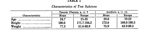
“seven nationally ranked tennis players from the Minnesota-Wisconsin-Iowa area and 11 soldiers from Fort Lee, Virginia, were used as test subjects”
“All of the tennis players had played tennis regularly winter and summer for at least the last seven years. None of the soldiers had engaged in any type of extensive unilateral activity.” “Hand area, third-finger length, wrist width and the forearm circumference measurements differed between the arms of the tennis players but not between the arms of the soldiers.”
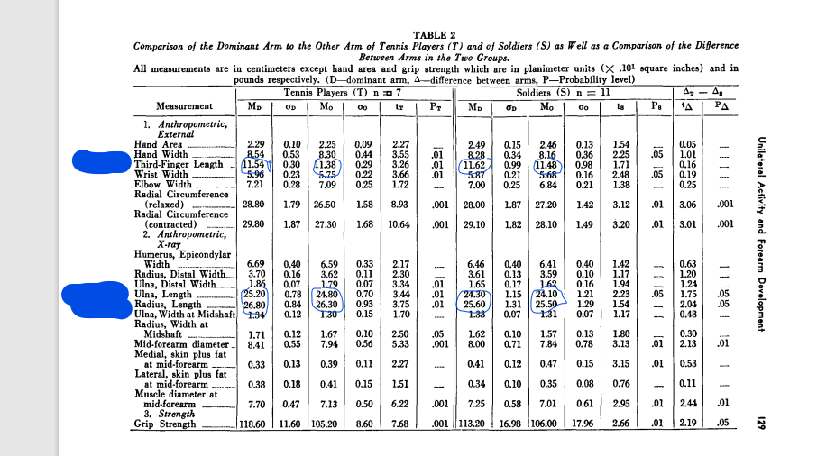
“Ulna length proved to be significantly different in both groups, but a slightly more significant difference was noted between arms of tennis players than soldiers. Radius length differed only in tennis players”
You can see that the tennis players had greater length asymmetry than the soldiers. The third finger length asymmetry was pretty much the same which is interesting.
“Contralateral arm movement is necessary to elevate the ball for the service as well as to steady the racket prior to hitting the ball during the volley and when hitting ground strokes.”<-So rapid inversion/eversion of the arm. Torsional loading to grip the racket. And vibration when the ball hits the racket.
“Bony lengths were increased in the dominant forearm of tennis players, indicating an alteration in the osseous “growth apparatus””
“Hand area, hand width, third-finger length, wrist width, and forearm circumference (relaxed and contracted) differed significantly between the dominant and other arm of tennis players, but only hand width, wrist width and forearm circumference (contracted) differed between arms of soldiers. Radius and ulna length and distal ulna width were different between the arms of tennis players but only ulna length differed between arms of soldiers.”
“small changes in radius and ulna length could be associated with participation in this vigorous unilateral activity.”
It should be noted that soldiers also engage in unilateral activities with their dominant arm. So it is likely that the types of loads that occur in tennis are the kinds of loads that induce growth in length and we should attempt to apply those loads to the legs.
The gut microbiome is definitely something that has a lot of effort put into it by the scientific community so there is potential to optimize the gut microbiome to make people taller.
Island biogeography theory provides a plausible explanation for why larger vertebrates and taller humans have more diverse gut microbiome
“Prior work has shown a positive scaling relationship between vertebrate body size, human height, and gut microbiome alpha diversity.{this means that the more diverse your gut microbiome the taller you were. This could be a correlation rather than causal if there is a factor that increases human height and gut microbiome diversity}. This observation mirrors commonly observed species area relationships (SARs) in many other ecosystems. Here, we expand these observations to several large datasets, showing that this size–diversity scaling relationship is independent of relevant covariates, like diet, body mass index, age, sex, bowel movement frequency, antibiotic usage, and cardiometabolic health markers. Island biogeography theory (IBT), which predicts that larger islands tend to harbor greater species diversity through neutral demographic processes, provides a simple mechanism for positive SARs. Using a gut-adapted IBT model, we demonstrated that increasing the length of a flow through ecosystem led to increased species diversity, closely matching our empirical observations. We delve into the possible clinical implications of these SARs in the American Gut cohort. Consistent with prior observations that lower alpha diversity is a risk factor for Clostridioides difficile infection (CDI), we found that individuals who reported a history of CDI were shorter than those who did not and that this relationship was mediated by alpha diversity.{so increased microbiome diversity reduces the risk of infection}.
We observed that vegetable consumption had a much stronger association with CDI history, which was also partially mediated by alpha diversity. In summary, we find that the positive scaling observed between body size and gut alpha diversity can be plausibly explained by a gut-adapted IBT model, may be related to CDI risk, and vegetable intake appears to independently mitigate this risk, although additional work is needed to validate the potential disease risk implications.”
So this indicates that vegetable intake is a good way to increase microbiome diversity.
“The human gut microbiota has an enormous impact on our phenotype, with almost half of the metabolites circulating in blood significantly associated with cross-sectional variation in the ecological composition of the gut microbiome. One of the key ecosystem functions that the gut microbiota provides to its host is resistance to enteric bacterial pathogens. Niche saturation or nutrient competition are commonly invoked mechanisms for how the microbiota excludes invaders.”
“species-diverse commensal communities are more apt to saturate available metabolic niches so that an invasive pathogen is less likely to colonize, outcompete commensals, and cause disease”<- so one of the ways that the gut microbiome increases height is by competing with invading pathogens and thereby preventing them from stunting growth. However if it is due to preventing infection that height is increased it could mean that for the average person the microbiome has less impact on height if they would not get an infection otherwise. On the other hand, the microbiome has been shown to have other effects that influence height such as via IGF-1.
“Vertebrate body size, which varies over six orders of magnitude, has been shown to be positively associated with gut microbiome alpha diversity, indicating that larger animals with larger guts harbor more species.” <-so the correlation could be possibly caused by larger people having larger guts which enable more microbiome diversity.
“we demonstrate a consistent scaling between body size and gut microbiome alpha diversity across vertebrates and human populations. We find that this association is independent of many potential confounders, like diet, bowel movement frequency (BMF), body mass index (BMI), age, and sex”
What the next steps would be to test the influence of the microbiome would be to compare the growth rates of different animals versus microbiome diversity who have never had an infection.
But with the frequency that kids get sick even if microbiome only reduces infection risk then it is worth it.
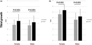
“Our goal was to study the role of mechanical loading (one of the components of ambulation) on endochondral ossification and longitudinal bone growth. Thus, we applied cyclical, biologically relevant strains for a prolonged time period (4 weeks) to one tibia of juvenile mice, while using the contralateral one as an internal control. By the end of the 4-week loading period, the mean tibial growth of the loaded tibiae was significantly greater than that of the unloaded tibiae. The mean height and the mean area of the loaded tibial growth plates were greater than those of the unloaded tibiae. In addition, in female mice we found a greater expression of PTHrP in the loaded tibial growth plates than in the unloaded ones.”
This suggests that possibly boosting PTHrP directly could induce additional longitudinal bone growth.
“Mechanical forces related to gravitational changes, ambulation, and exercise may contribute to modulate bone growth.”<-this suggests that exercises that invert and every may modulate longitudinal bone growth.
“daily physical activities transmit complex mechanical loads including tension, compression, torsion, and shear to the skeleton”<-there are many ways to load bones that are under explored.
“Seventy-five 4-week old TOPGAL mice were exposed to mechanical loading using a Bose ElectroForce 3220 dynamic loading system. Before each loading session, mice were anesthetized with 3.5% isoflurane. Each loading session included 100 compressive loading cycles of 5 Newton (N) force to the right tibia at the frequency of 2 Hz per cycle”<-the reason that cyclic loading is more effective than static loading is likely related to fluid flow.
“mean tibial growth of the loaded tibiae was significantly greater than that of the unloaded tibiae, in the whole sample of mice “
This is a pretty significant different in length.
“Gravity and physical activity generate mechanical forces on the long bones and on the growth plates that may be involved in the regulation of bone growth.”<-one of the reasons why lateral loads are so effective is that it applies loads in a different mannerism against gravity.
“Of note, the stimulatory effect of mechanical loading on tibial growth persisted in the 4 weeks following the cessation of loading.
In addition, at the end of the 4-week loading period the whole growth plate and the epiphyseal zone heights, as well as the overall area of the loaded tibial growth plates were significantly greater than those of the unloaded growth plates. Such finding suggests a loading-mediated enhanced growth plate chondrocyte formation”
The study mentions that too high load can suppress growth. So maybe lighter weights are better and applying it the right way is more important but that may be for growth plates and not bones. Higher loads may be needed to stimulate growth in adult bones as bones are tougher tissue than catilage.
“similar studies in rodents indicate that elevated loading forces tend to inhibit longitudinal growth, while strains of lower intensity (like ours) tend to stimulate it. “
“After 4 weeks of loading, we found a greater Pthrp gene expression in the growth plate chondrocytes of the female mouse loaded tibiae, while no difference was found in male mice” however male mice also had greater length. So this suggests that mechanical loading also influences length by mechanisms not related to PTHrP.
So I stalled out in the old method at 75 1/4” for a long time(several months), it wasn’t until I tried this method that I started increasing the measurement again. I also increased the duration and am moving the vibration device around the hand more. That could play a role.
The old method took me from about 75 to 75 1/4”. I don’t know why I stalled out. Could be over time the body adapts to this stimulus and with this method I too will need to add other modalities to make it more effective.
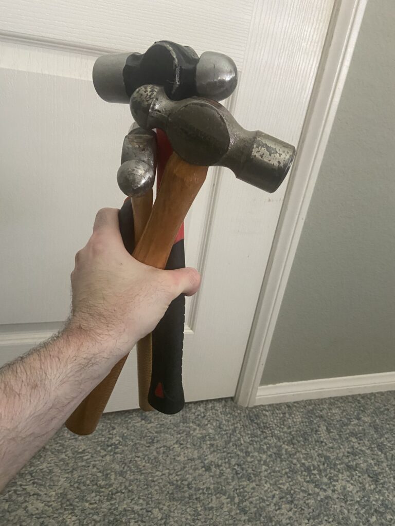
Here’s the video demo:
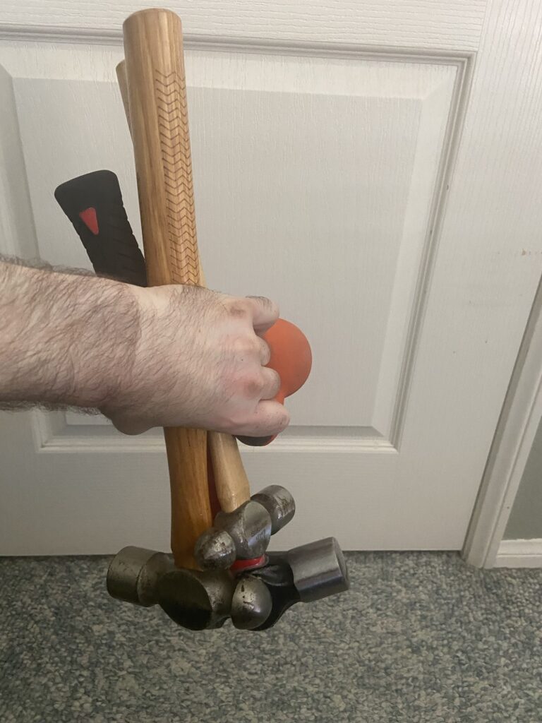
Essentially what I do is I grip some oddly shaped objects for torsional loading. The more oddly shaped and weird the objects the better. You could also use bands, etc. ideally you’d the want the bone to have as many areas of compression and tension as possible. Fluid flows from areas of compression to areas of tension so the more areas of compression and tension there are there. The more fluid is going to flow.
Vibration is another stimulus to enhance fluid flow. The closer the vibration is the target bone/cartilage the better.
As seen in the video I kind of stir the hammers as a way to get more torsional loading and activity in the muscles. Muscles pull on the bone via the tendons creating more elastic bone deformation
The reason for changing the bones axis in relation to gravity I explain below in a an email to Hiroki Yokota the pioneer of the Joint Loading Modality and an expert in fluid flow to stimulate anabolic responses in the bone:
“You mention that the reason why lateral loads in bone is so effective is because of the water bottle analogy in pressing to the side is more effective in moving fluid.
But inversion/eversion is even more effective in moving fluid in the water bottle analogy and the arms undergo rapid eversion/inversion much more frequently than the legs.”
Hiroki Yokota’s response : “
I think your idea may work but we need to think about a basic fluid motion. A Navier-Stokes equation has three major forces to alter the flow. They are:
Pressure change
viscosity
gravity
Since viscosity is to prevent the flow, two driving forces to generate flow are pressure change and gravity. Lateral loading induces pressure change, while inversion activates gravity. We need to evaluate quantitatively the effects of loading-driven pressure change and inversion-induced gravity on fluid low in a bone matrix.”
Below are some studies that show that gravity can alter fluid flow in the bone and can stimulate cellular activity:
According to Fifteen days of microgravity causes growth in calvaria of mice. , microgravity alters interstitial fluid flow. Inversion and eversion would mimic this.
The Effect of the Microgravity Rotating Culture System on the Chondrogenic Differentiation of Bone Marrow Mesenchymal Stem Cells., microgravity rotating culture increased the chondrogenic differentistion of mesenchymal stem cells. Inversion and eversion would mimic this.
That interstitial fluid flow can stimulate bone response is not controversial. That this response includes making the bones longer is controversial. However, baseball pitching, tennis, and arm wrestling all of which have anecdotal reports of increase in bone length all have changing the bones axis in relation to gravity. Diving is the closest thing for legs that I could find that increases bone length and it also has constant rotation inversion/eversion to change the bone’s axis in relation to gravity.
I believe that interstitial fluid flow can make bones longer if this stimulus is sufficient. The reason that arm bones are easier to grow than legs is because the hands can grip things so the arm bones get more direct loading.
Think of an hourglass:
You tip it over the sand moves from one to another but not all right away(the rate at which it flows is affected by vicosity). You could affect the rate by which the sand flows by compressing the sides of the hourglass or vibrating the hourglass to make the sands move faster.
Interstitial fluid flow has the ability to affect osteoblasts, osteoclasts, and stem cells all of which could potentially combine to make a bone longer. The exact mechanism of how this could happen is not yet known. But if interstitial fluid flow can affect all these cells and can affect gene expression then it suggest that there is potentially a method by which interstitial fluid flow can increase bone length is possible even if the exact mechanism is unknown.
The arms are subjected to much better loads than the legs are. Legs are not typically inverted. They femur is kind of inverted in a squat or deadlift but the weight is not close to the femur it’s on the back or in the hands. Standing hamstring curls are typically done on machines which are not as an effective a loading as actually gripping the weight and it’s hard to grip weight with the toes. Iron boots perhaps? Kicks are typically not loaded. I am trying leg swings with ankle weights but the ankle weights are only 20lbs more weight may be needed. reverse crunches also involve inversion of the legs but weight/torsion needs to be on the leg itself
Torso is typically only inverted in good mornings and decline sit-ups and cartilage is easier to stimulate than bone since it is a softer more easily deformable tissue but the issue is it has a poor blood supply so it grows slower.
Finding ways to apply this method if it works can be done for torso and the legs but it will be more challenging.
One other person has reported growth with this method but he was under 25 but over 18 so it may have been natural growth since arms grow longer for longer.
I was originally going to shoot for x-rays around 75 1/4” but that was before I stalled out originally. I want to see a rate of consistent and steady growth. I do have before x-rays. I want some experimenters to try and validate the method. I also think 1/4” is not strong enough above measurement error.
Most people want to move on to the legs already but the legs are harder it needs to be validated on arms first. Wingspan was chosen because it’s easy to see when wingspan begins and ends and I do get some variance in measurement but I go for the peak measurement.
So next phase is:
Try to gain more in wingspan at a steady rate and validate with X-rays
Try to get experimenters to validate. I have gained wrist thickness and muscle mass with this so it has other benefits too. Other experimenters will reduce personal bias.
Gut microbiome is the microorganisms like bacteria that live in your digestive tract. The gut microbiome is influenced by your diet. There is a lot of research into the gut microbiome so if it does affect child’s height significantly improvements in that area could lead to dramatic increases in height for a population. Parent microbiome is passed on to a child so there is the possibility to increase height of a child by manipulating parent’s diet. Microbiome could impact height via IGF-1. Antiobiotic dosing which affects the gut microbiome has been shown to influence height. Changing the gut microbiome is not easy however. ” the composition of the gut microbiota is partially heritable and, once established, does not change substantially without a large or prolonged stimulus.“
“Longitudinal bone growth in children is governed by different genetic, nutritional and other environmental factors acting systemically on the endocrine system and locally at the growth plate. Recent studies have shown that this intricate interplay between nutritional and hormonal regulation of the growth plate could involve the gut microbiota, highlighting the importance of a holistic approach in tackling childhood undernutrition. In this review, I focus on the mechanistic insights provided by these recent advances in gut microbiota research and discuss ongoing development of microbiota-based therapeutics in humans, which could be the missing link in solving undernutrition and childhood stunting.”
“Growth hormone stimulates production of IGF-I in the liver, which then acts as an endocrine factor to stimulate bone growth at the growth plate. Growth hormone also stimulates local IGF-I production in target tissues, such as the growth plate and the intestine, which acts as a paracrine/autocrine growth factor. Nutritional status positively regulates bone growth and maturation of the gut microbiota, which reciprocally promote nutritional intake. The gut microbiota also promotes bone growth, perhaps directly, by stimulating IGF-I production. Possible mechanisms for such stimulation might involve SCFAs and NOD2-mediated bacterial sensing pathways in the intestinal epithelial cells.”
“growth deceleration is associated with the gradual decline in growth plate function, also known as growth plate senescence. Importantly, growth plate senescence is characterized by a gradual depletion of chondrogenic stem cells, decreasing chondrocyte proliferation and hypertrophy in the growth plate. Although growth plate senescence is generally associated with age, it appears not to be driven by age per se but instead depends on how much growth potential has been ‘used up’. In other words, chondrocytes in the growth plate appear to have a finite amount of growth potential, which is depleted gradually as more bone growth occurs, leading to the gradual decline in growth rate and the associated changes of senescence. This is supported by the fact that growth-inhibiting conditions, such as undernutrition, can slow down growth plate senescence, allowing bone growth not only to resume but temporarily to accelerate faster than normal for chronological age once nutritional status improves, a clinical phenomenon known as catch-up growth”<-so height seekers should have the goal to reverse senescence.
“The signal by which the gut microbiota stimulates IGF-I might not even be a metabolite. bacterial cell walls isolated from L. plantarum were sufficient to stimulate IGF-I and bone growth in mice”
“NOD2-activating ligands, such as muramyl dipeptide or the synthetic NOD2-activating adjuvant mifamurtide, alone were sufficient to induce IGF-I and bone growth, suggesting that NOD2 agonists could be a new class of therapeutic agents for improving childhood stunting.”
“another major cause of growth inhibition comes from a local effect of cytokines, which are often elevated in inflammatory diseases. At a systemic level, pro-inflammatory cytokines can inhibit bone growth by suppressing IGF-I. For example, in mice overexpressing interleukin-6 (IL-6), body growth is significantly suppressed, with decreased IGF-I and IGFBP3 but with normal levels of GH”
“The gut microbiota has been shown to influence circulating levels of pro-inflammatory cytokines. Serum IL-1β and IL-6 levels were correlated with the presence of certain bacterial strains in the gut microbiome. Mechanistically, butyrate, one of the SCFAs produced by the gut microbiota, has been shown to inhibit the inflammatory response elicit by lipopolysaccharides, TNFα and interleukins via GRP41 and GRP43, both in endothelial cells and in chondrocytes, suggesting that the gut microbiota could stimulate bone growth by reducing inflammation”
So you can use prebiotics and gut transplants potentially.
“In addition to the bare minimum of improving nutritional status, mitigation of gut microbiota dysbiosis, either by introducing growth-stimulating bacterial strains or by promoting gut microbiota maturation, should be considered as coupling therapeutic strategies.”
According to Fasting challenges human gut microbiome resilience and reduces Fusobacterium, fasting can alter the gut microbiome. Perhaps this could be how fasting affects bone length. “Water-only fasting could have a profound and long-lasting effect on gut microbiome.”
“Microbiome changes due to water-only fasting remained in five subjects even after returning to their normal diet, indicating the resilience of gut microbiome was successfully challenged. “
“skeletal stem cells were shown to be present in the epiphyseal growth plate (epiphyseal skeletal stem cells, epSSCs). Here, we explore the possibility that modulating the number of epSSCs can correct differences in leg length. First, we examined regulation of the number and activity of epSSCs by Hedgehog (Hh) signaling. Both systemic activation of Hh pathway with Smoothened agonist (SAG) and genetic activation of Hh pathway by Patched1 (Ptch1) ablation in Pthrp-creERPtch1fl/fl tdTomato mice promoted proliferation of epSSCs and clonal enlargement. Transient intra-articular administration of SAG also elevated the number of epSSCs. When SAG-containing beads were implanted into the femoral secondary ossification center of 1 leg of rats, this leg was significantly longer 1 month later than the contralateral leg implanted with vehicle-containing beads, an effect that was even more pronounced 2 and 6 months after implantation. We conclude that Hh signaling activates growth plate epSSCs, which effectively leads to increased longitudinal growth of bones. This opens therapeutic possibilities for the treatment of differences in leg length.” According to this paper Mechanotransduction pathways in the regulation of cartilage chondrocyte homoeostasis, “Mechanical stress up‐regulates Indian hedgehog expression (IHH) and activates hedgehog (Hh) signaling” “cyclic tensile strain activates Hh signaling and promotes the expression of ADAMTS‐5 in a primary cilia‐dependent manner, but in a high strain environment, histone deacetylase 6 (HDAC6) causes cilial disassembly and blocks this response”<-So we could potential mimic some of the effects of this study with mechanical loading because it also activates the hedgehog pathway but not too much activation as it blocks the mechanical loading benefits to hedgehog. So potentially with the right mechanical loading we could mimic the benefits of this study.
From the above picture, any method of increasing hedgehog signaling will work including potentially mechanical loading.
“Morphologically and functionally, the growth plate can be divided into the resting, proliferative, and hypertrophic zones. The resting zone contains slowly cycling cells, which, upon recruitment into the underlying proliferative zone, begin proliferating rapidly and arrange themselves into longitudinal columns of flat chondrocytes. Thereafter, these flat chondrocytes undergo further differentiation along with hypertrophy, forming the hypertrophic zone. Thereafter, the hypertrophic chondrocytes die or transdifferentiate, leaving a cartilaginous template on which spongy bone is built.”
“The resting zone contains a unique population of stem cells that express the parathyroid hormone–related protein (PTHrP). Furthermore, these stem cells reside in a niche that governs their abilities of renewal and generation of transit-amplifying proliferative chondrocytes. Interestingly, this niche arises postnatally, probably in association with maturation of the SOC, and only then can the stem cells obtain self-maintaining capacity and the ability to produce stable, long-lasting clones”
“
The importance of the Hedgehog (Hh) signaling pathway in development of the skeleton is demonstrated by the observation that deletion of Indian hedgehog (Ihh) (normally expressed by prehypertrophic and hypertrophic chondrocytes), either globally or specifically in cartilage, virtually eliminates formation of the growth plate. Together with PTHrP, Ihh is involved in a negative-feedback loop that controls the rate of chondrocyte differentiation. More specifically, Ihh produced by prehypertrophic and hypertrophic chondrocytes diffuses to the resting zone, where it stimulates expression of PTHrP, which in turn inhibits the hypertrophic differentiation of chondrocytes.
In addition, if Hh signaling is inhibited either genetically within the growth plate or pharmacologically during postnatal growth, the growth plate fuses abruptly“<-can we manipulate hedgehog signaling to keep the growth plate open for longer?
” Hh signaling stimulates the proliferation and clonal activity of epSSCs independent of age.”<-this is very promising for being able to potentially get the skeletal stem cells and then stimulate Hh signaling in adults.
“Local temporal stimulation of Hh signaling lengthens the legs.”
You can see from F how dramatically Hedgehog signaling increased the growth plate(SAG).
“The bony epiphysis (which develops from the Secondary Ossification Center) appears to be an appropriate location for such intervention, allowing placement of the Hh pathway agonist in close proximity to the epSSCs.”
” In the experiments presented here, some compound is likely released into the bloodstream, since an increase not only in femoral length, where the SAG-containing beads were placed, but also in the tibia of the same leg was observed. The arterial blood flow in extremities goes in the proximo-distal direction, and it is plausible that some levels of SAG diffuse in the same direction as the blood flow.”<-therefore the compound could be put into the blood stream without the beads for overall height!
“Noriaki said he was surprised that the cells in the resting zone “weren’t just lazy and doing nothing, they’re very hardworking cells, they can occasionally wake up and keep making chondrocytes.””<-that is amazing that means that potentially they can form new growth plates.
“It’s been hypothesized for many years that chondrocytes at the bottom of the growth plate die, but these findings show definite evidence that they survive and continue to make bone, he said.”
“The hallmark of endochondral bone development is the presence of cartilaginous templates, in which osteoblasts and stromal cells are generated to form mineralized matrix and support bone marrow hematopoiesis. However, the ultimate source of these mesenchymal cells and the relationship between bone progenitors in fetal life and those in later life are unknown. Fate-mapping studies revealed that cells expressing cre-recombinases driven by the collagen II (Col2) promoter/enhancer and their descendants contributed to, in addition to chondrocytes, early perichondrial precursors prior to Runx2 expression and, subsequently, to a majority of osteoblasts, Cxcl12 (chemokine (C-X-C motif) ligand 12)-abundant stromal cells and bone marrow stromal/mesenchymal progenitor cells in postnatal life. Lineage-tracing experiments using a tamoxifen-inducible creER system further revealed that early postnatal cells marked by Col2-creER, as well as Sox9-creER and aggrecan (Acan)-creER, progressively contributed to multiple mesenchymal lineages and continued to provide descendants for over a year. These cells are distinct from adult mesenchymal progenitors and thus provide opportunities for regulating the explosive growth that occurs uniquely in growing mammals.”
Have to figure out what’s unique about these cells and if there is a way to make these cells reawaken
“Chondrocytes in the growth plate continue to proliferate well into adulthood in mice”
“ In the center of the developing cartilage mold, chondrocytes stop proliferating and become hypertrophic chondrocytes. These cells signal to induce the migration of mesenchymal cells into the marrow space; these cells then differentiate into osteoblasts that then form bone on top of the cartilaginous matrix. Perichondrial precursors expressing osterix (Osx) invade into the cartilage template along with blood vessels and eventually become both osteoblasts and stromal cells in the marrow space”
“growth-related mesenchymal progenitors identified here and adult mesenchymal precursors.” We have to get these back