Increasing Vertebral Bone Growth Using Minimally Invasive Intervertebral Disc Implants

Increasing Vertebral Bone Growth Using Minimally Invasive Intervertebral Disc Implants
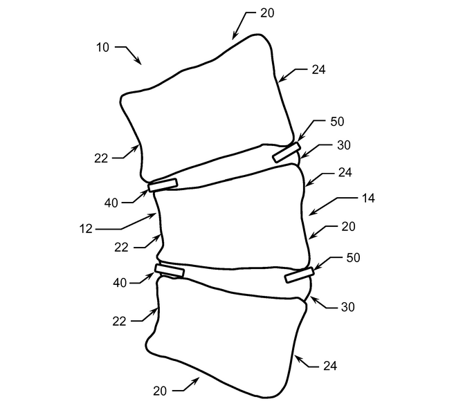
Intervertebral Disc ImplantsThere has always been a lot of talk amount the online grow taller community where people would consider the idea of someone making the overall body bigger by somehow manipulating the vertebrate bones and/or the intervertebral discs. Almost all the talk have been focused on the discs, since the discs are not hard and difficult to remodel unlike the vertebrate bones. Discussions on disc decompression, traction machines, stem cell implants, and growth factor injections have been all been speculated before, but I recently found a new proposed idea which seems to make much more sense than all of them combined.

I agree that stem cell, scaffold, growth factor injections are all quite minimally invasive, so they are all somewhat good. This new idea is probably just as reasonable and attractive as any of the other ones I have suggested before in the past.

I refer the reader to the patent entitled “Method of treating scoliosis using a biological implant” (Patent # US 8123787 B) by Inventor James W. Ogilvie, Kenneth Ward, Lesa M. Nelson.

(The same idea is also available by the patent #s US20090105822,  US20110295369,  WO2009155159A2, WO2009155159A9.)

The method is for treating scoliosis, which is something most American Elementary School nurses check for to make sure children are growing properly and symmetrically but I wonder whether we can use the same type of minimally invasive slow release type of growth factor implants to increase the rate at which maybe adults can increase in their disc height.

From past articles I’ve read, it seems that scoliosis is something that mainly children who are still growing have. When one side of their vertebral growth plates become too active, their vertebrate column starts to become lopsided.
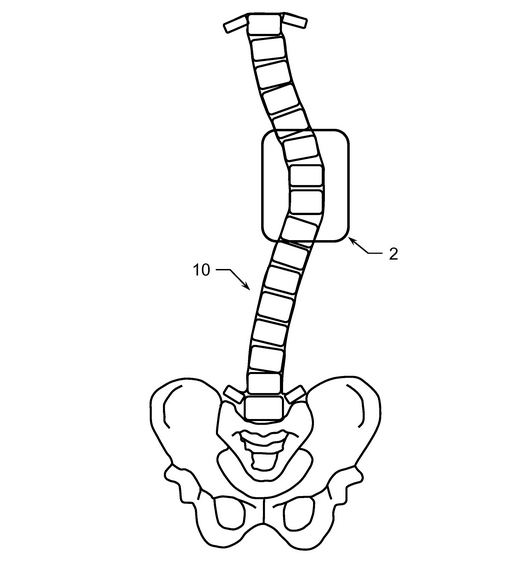
Vertebral Bone GrowthHowever, we do have the medical condition where adults suffer from scoliosis too. The last we checked on WedMD, there is no way to actually easily realign adult scoliosis. The most conservative treatment has been to make sure that the person with adult scoliosis does not suffer from great pain. Treatments has been almost always been for pain management. The other way is the drastic approach, which is to get surgery to correct for the curvature.

From the abstract of the patent we quote below…

The present invention is a bone growth stimulating and promoting cytokine type biological implant preferably comprising PTH coated with a controlled release biodegradable coating that is implanted preferably in the concave side of a scoliotically curved spine in combination with a bone growth inhibiting type biological implant preferably comprising methotrexate or like anti-metabolite coated with a controlled release biodegradable coating that is implanted preferably in the convex side of a scoliotically curved spine. The insertion of the biological implant is highly non-invasion, especially as compared to more conventional spine surgical methods, and the biological implant does not decrease spinal mobility or spinal range of motion”

First, lets forget trying to use any type of bone growth inhibiting implant, but focus exclusively on getting the bone growth increasing cytokine to work. We are aware that Parathyroid Hormone aka PTH has bone growth beneficial effects.

From the other section in the patent….

A method of treating a scoliotically curved spine in a patient being determined to be at risk of scoliosis curve progression comprising placing at least one of a growth stimulant, a medication, and a biological therapy on a concave side of said curve formed in said spine, wherein said biological therapy defines a dissolvable bone growth stimulant biological implant coated with a dissolvable coating, wherein determining said patient is at risk of scoliosis curve progression further defines a determination of genetic predisposition wherein DNA of said patient includes a plurality of genetic markers having an association with adolescent idiopathic scoliosis contained therein and wherein said risk is determined by performing logistic regression on said plurality of adolescent idiopathic scoliosis associated genetic markers

So in the patent, there is more than just the growth stimulant added, but also a type of medication and the patient also should go through a type of biological therapy? Or does it mean that the dissolvable bone growth stimulant do all three parts?

While I am quite sure that this type of method to modulate vertebrate bone growth is going to be quite effective on developing children with functional growth plates, the big question to ask is whether this technique will work on adults.

First, after studying the diagrams and pictures of the structure and setup of the vertebrate bone column, we can say that there is a slight chance that the right type of cytokine might be better for making the discs more robust and tougher to compressive forces.

Now, we are fully aware that even in adults, there is still a very thin layer of hyaline cartilage, between the actual disc and the bone, which is really used as a connective tissue. It is just maybe 2-3 cells thick but it is there after the growth plates are gone. We wonder whether it is possible to get the PTH implants to dissolve slowly to get the few chondrocytes in the hyaline cartilage layer to proliferate and increase the thickness of the layer. If proliferation is possible, then we might be able to slowly, and I am talking very slowly, increase the height of the person with the embedded implants by millimeters over time.

However, like so many of our discoveries, the effects of this idea on young adolescent kids will always be much more effective and noticeable.
Prolotherapy Indicates Increased Disc Height and Reduced Lower Back Pain From Weight Loss

Prolotherapy Indicates Increased Disc Height and Reduced Lower Back Pain From Weight Loss

On the prolotherapy website there was an article that was written back in April of this year (2014) “Obesity, Disc Height Changes and Low Back Pain“. They referenced a study “Intervertebral disc height changes after weight reduction in morbidly obese patients and its effect on quality of life and radicular and low back pain.” (Lidar Z, Behrbalk E, Regev G, Salame K, Keynan O, et al., Spine. 2012;37(23):1947-1952) The study was published in 2012.

It would seem like mostly common sense from many people who are overweight or even obese complain of lower back pain. It is also somewhat around assumed that extra weight would compress the intervertebral discs to a thinner space, making the obese and overweight people slightly shorter than they had to be.

In the study, the test subjects decided to go with a weight reduction surgery known as bariatric surgery. I am not a specialist on the intestinal system but it seems that bariatric surgery means that a large portion of the intestinal system is cut out to restrict the amount of food that the obese person can take in before feeling full.

After the surgery was done, there was on average about a 25-27 kg loss in weight. When the L4-L5 IVD space was measured, the increase in disc height was about 2 mm on average. Maybe the more important thing was that lower back pain was reduced greatly.

This is a very good sign for the people who are readers of the website and are on the heavy side to first try to loss the extra weight on their body before trying to increase in height. That is something which I sort of realized myself, where it is important to first work on weight loss before trying for height gain.

Using Autologous Mesenchymal Stem Cells To Treat Hip and Knee Joint Pain from Osteoarthritis

Using Autologous Mesenchymal Stem Cells To Treat Hip and Knee Joint Pain from Osteoarthritis

I stated about 2 months ago that I wanted to expand the scope of the research that is done on this website, to include other subjects like the biochemistry of cancer, orthopaedic conditions, and similar issues. These next few posts will be dealing with this subjects. Some of the information will be related to what I’ve been researching before.

There was a very recent article (came out on August 3, 2014) that came out in the Australian news website The Age entitled “Stem cell trial hope for osteoarthritis sufferers” which showed that MSCs are being used to treat people who are suffering from knee pain associated with Osteoarthritis. While it may not seem too revolutionary and profoundly new to us who are so used to reading up on Patents and University backed cutting edge research on stem cells, this method would still be very interesting to a large group of the population who are suffering from joint pain from cartilage degeneration.

As the article says, a middle aged female underwent liposuction so that her fat can be sucked and then filtered to get the little bit of stem cells in her fat tissue. Those stem cells will be then be packed together in pure stem cell and high density form, which are injected into her hip joint region to relieve pain.

When looking at the overall procedure from a higher level, I am reminded of a similar type of procedure that is being done by Dr. Peter Wehling at Dusseldorf which is now called the Regenokine/Orthokine method as well as the Platelet Rich Plasma therapy. In all of these methods, the entire thing is minimally invasive.

The hope for the researchers who took the women’s fat out is to get the filtered stem cells to slow down the rate of cartilage degeneration in the woman’s hip. If that is possible, then the need for the complicated and expensive hip prosthetic replacement surgery can be pushed back by more than a full decade. The primary researcher is a Dr Julien Freitag, who would reference a South Korean study. I took the time to track down that exact article and found it. It is suggested in that Korean study that instead of just a single injection of the person’s own adipose derived stem cells, multiple injections will have much better results. 

There will also be a 2nd study looking at how this autologous MSC method would work for cartilage lesions which are the result of trauma which often lead to the onset of osteoarthritis after maybe just 4-10 years after trauma.

The name of the article that is referenced is “Infrapatellar fat pad-derived mesenchymal stem cell therapy for knee osteoarthritis” which is associated with the Department of Orthopedic Surgery, Yonsei Sarang Hospital, Seoul, South Korea. The same group of authors had written also the paper “Mesenchymal stem cell injections improve symptoms of knee osteoarthritis.” which was published in April of 2013.

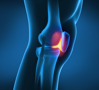
Infrapatellar Fat PadIn the referenced article, the researchers took the stem cells from the infrapatellar fat pad region (aka Hoffa’s Fat Pad). Refer to the picture to the right for reference to where the fat pad is.

The stem cells which are isolated are then injected percutaneously into knees that have been suffering from arthritis.

Note: I might have made a mistake on assumption before. It seems that the method of PRP is used in this technique. Quoted from the article…

After the stem cells were isolated, a mean of 1.89×10⁶ (range, 1.2–2.3 × 10⁶) stem cells were prepared with approximately 3.0 mL of platelet-rich plasma (PRP) and injected in the selected knees of patients in the study group.

I made the mistake of assuming that the stem cells would be reinjected in the body percutaneously and intraarticularly by themselves. I forgot to realize that the platelet-rich plasma would be used as the semi-liquid transport medium since stem cells by themselves with no medium would just shrivel up and dry out. This method is a type of PRP therapy.

After the first injection with stem cells and PRP, 3 mL of PRP was administered every 7 days as the second and third rounds of treatment. The results from the control group and the actual tested group were statistically significant.

Implications

From my search throughout the hundreds of patents for application of stem cells, I’ve already seen at least 2 groups which have put a patent on the technique on using MSCs to treat osteoarthritis or arthritis in some way, whether by the PRP method or some other idea. I quote was is said in the paper “With regard to in vivo studies, the transplantation of MSCs into full thickness articular cartilage defects has been attempted under various conditions.” so there has been many attempts already, and the results have come in have been very fruitful and promising.

Quoted further…

“In 2 reports, experiments on humans [16,17] involving the intraarticular injection of autologous MSCs yielded good results after 6 months. In 2008, Centeno and colleagues reported the use of autologous culture-expanded bone marrow- derived stem cells for knee cartilage regeneration in humans [17]. In their study, the patients’ pain, as determined by the VAS, and range of motion improved, and MRI showed significant articular cartilage growth and meniscus regeneration. “

What I personally got out of it…

I learned that the source of where you get the MSCs is quite important. While it is well known that MSCs can be filtered from bone marrow, bone marrow extractions are often very painful and have a high chance for infections and complications. In addition, as a person ages, the density of MSCs decrease. That seems to be why the researchers here used the infrapatellar fat pad as the source of the stem cells. They were able to get 9.4 g of infrapatellar fat pad which gave out on average about 1.89×10^6 stem cells, which is a nice cell to overall fat density. For future references, instead of asking that we get stem cells from bone marrow biospys, it would be much easier and simpler to use the source of fat from under the patella.

The last important thing that the researchers stated in the study was this ….

the paracrine effects of the cytokines and growth factors released by the grafted cells, which favorably influence the microenvironment by triggering host-associated signaling pathways and lead to increased angiogenesis, decreased apoptosis, and possibly, induction of endogenous generation

I am still trying to understand what this part means and what the implications are.

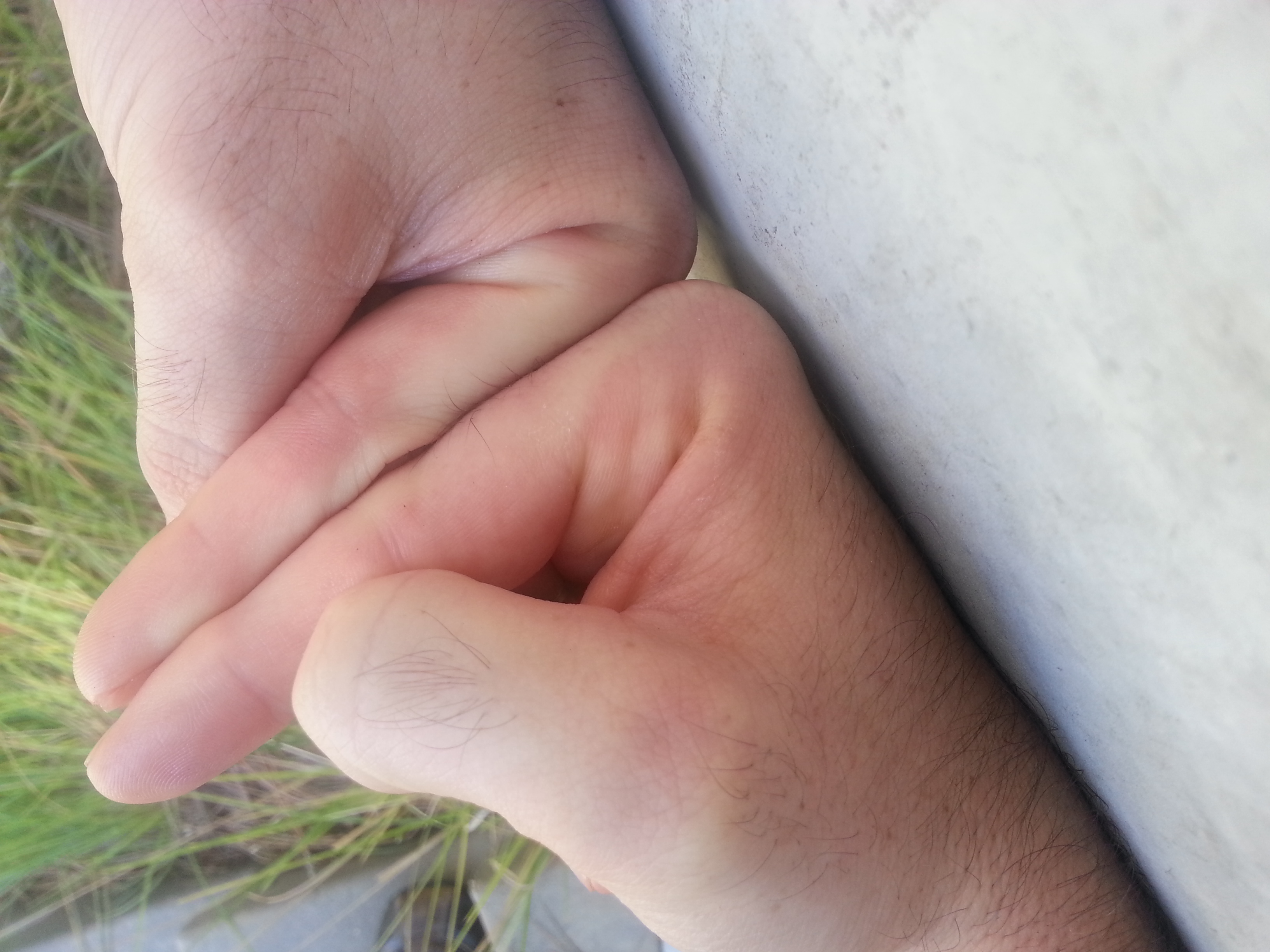
LSJL Progress Update 8-5-14: More finger growth and update on new method

Last time it looked as though my right finger which I loaded via LSJL was about 1/4″ longer.  Now it looks like it’s about .375″ inches longer.  I’ve been loading about every day for about a 100 count on each of the three joints of the finger.  I increase the load as fast as possible, I could do more but I worry about injury because the clamp is so much stronger than the finger.  I might work up to more.  Two joints I load side to side but since the hand is in the way for the knuckle I load from top to bottom.  Here’s a post regarding my previous results and some images about how I perform LSJL. Here’s an image of my fingers now: 20140804_144910 Now it’s an extremely significant increase in finger length that is a result of LSJL.  Now I do have some osteophytes and the finger growth is not the same as normal finger growth.  In some of the other images you can see some finger deformities relative to a normal finger.  But it’s still a strong proof of concept that LSJL works to lengthen long bones. I’d rather prove LSJL sooner rather than later.  Would x-rays help?  I don’t really want to get them if they won’t convince people because it would cost a couple hundred dollar.  A lot of people don’t know exactly what makes you taller.  They can’t connect that long bones make you taller and the finger bones are long bones.  If LSJL can increase the length of finger bones(which are long bones(although they do have some different properties to other long bones)) then LSJL can increase overall height if those long bones are legs. As far as my leg progress though, I find that I can’t get as intense a clamp on my knees as I can on my fingers.  I think part of the reason is that there’s a lot of tissue types you’re clamping when you clamp a synovial joint.  It may take a bit of time before these tissues adapt to the clamping force.  I’ve been clamping for a long time with the C-clamp but there was a lot of slippage so there’s now a lot more force with the Irwin Quick Grip that i’m used to.  So right now I’m clamping with the Irwin Quick Grip to about a count of 130 before the pain in the soft tissues is just too irritating but over time the soft tissues will adapt and I’ll be able to clamp with as much force as I want as I have with bones I’ve been clamping a long time. So I’d recommend not clamping past the point of too much soft tissue pain and just try to increase clamping duration and intensity over time to allow the soft tissues to adapt. Remember, that LSJL is untested so there are guarantees that you won’t get injured or other maladies. Of course, if we could just prove LSJL then more testing can be done.  The question is how can we do it now rather than having to perfect it to increase leg length first?

Michael: The finger seems to be definitely longer, but you said that you clamped in all three joints.

  • Does that mean that the clamping was also at metacarpophalangeal joints?
  • How did you do that, and how can we not make sure that the MSP Joint did not go into inflammation mode aka swelling?
  • There is so much evidence that finger joints can swell up if you hit them on something.

X-Rays seem to be the way to go. We measure the synovial joints of the index finger of the right hand compared to the control of your left hand’s index, which I hoped was never clamped, and see whether the lengthen is from the tissue in the synovial joints thickening as a response. If there is a difference in the distance between the bones in either the PIP and/or MCP joints, then the lengthening was not bone. If the distance in the PIP & MCP joint locations are the same, then we then say that the lengthening was truly bone.

You don’t have to go in for a GP check-up. Look into Urgent Care Centers (Source: Which is Cheaper Out of Pocket: Urgent Care Facility or Hospital ER?). They usually accept Insurance. I’ll even put down $70 for the X-rays if that helps.

An Interview With Dr. Tarek El-Bialy On Using Ultrasound To Regenerate Tooth Dentin

An Interview With Dr. Tarek El-Bialy On Using Ultrasound To Regenerate Tooth Dentin

A few days ago we got a comment by someone calling themselves Pete who linked to an interview given where the Canadian Dr. Tarek El-Bialy was being interviewed on his Ultrasound Device & technology to use to regrow teeth, but stimulating the root of the teeth.

Comment

Originally we had written about the technology in a very old post “Teeth Regrowth Using Low Intensity Pulsed Ultrasound, LIPUS“. The researcher we mentioned in that article was a Dr. Jie Chen, also in Canada who claimed that they could use Ultrasound to not just make bones heal fractures but make people grow taller, which was something that was edited into the Wikipedia article on LIPUS (Low Intensity Pulsed Ultrasound). Tyler really got into the idea of using LIPUS and really deep into LIPUS but I had written in the past year a few times showing multiple articles which disprove the idea that Ultrasound would ever lead to increased longitudinal growth in the long bones. Yes, LIPUS machines are already on sale at rather cheap prices, so if there was real evidence in articles and the scientific literature that shows the really small LIPUS devices does any even the slightest chance of increasing longitudinal growth, I would recommend the portable LIPUS devices on this website, since they are just around $30-$50 each, and may be even free if one has the right type of medical insurance.

In fact, I recently downloaded the PDF of an old article I had eluded to to reread the implications on what would happen if you continuously emit ultrasound in lab test rabbits growth plates. (Effect of continuous therapeutic ultrasound in rabbit growth plates).

Regenerate ToothHowever, the technology of LIPUS on nerve tissue and teeth regeneration is much more promising. As Dr. El-Bialy (Associate Professor of Orthodontics and Biomedical Engineering at the University of Alberta, Canada) seems to suggest, LIPUS can regenerate damaged nerves and root pulp.

You can download the MP3 of that interview wth Dr. El-Bialy Available Here

It may not be our primary focus, but learning about the science of teeth and regeneration of teeth might lead to less money we have to spend at the dentist office in the future.

Salubrinal decreases osteoclastgenesis

This study doesn’t relate directly to height growth but it is by the scientists whose research was the foundation for LSJL.

In vitro and in silico analysis of an inhibitory mechanism of osteoclastogenesis by Salubrinal and Guanabenz

“Synthetic agents such as salubrinal and guanabenz, which attenuate stress to the endoplasmic reticulum, are reported to inhibit development of osteoclasts. However, the mechanism of their inhibitory action on osteoclasts is largely unknown. Using genome-wide expression profiles, we predicted key transcription factors that downregulated nuclear factor of activated T-cells, cytoplasmic 1 (NFATc1), a master transcription factor for osteoclastogenesis. Principal component analysis (PCA) predicted a list of transcription factors that were potentially responsible for reversing receptor activator of nuclear factor kappa-B ligand (RANKL)-driven stimulation of osteoclastogenesis. A partial silencing of NFATc1 allowed a selection of transcription factors that were likely to be located upstream of NFATc1. We validated the predicted transcription factors by focusing on two AP-1 transcription factors (c-Fos and JunB) using RAW264.7 pre-osteoclasts as well as primary bone marrow cells. As predicted, their mRNA and protein levels were elevated by RANKL, and the elevation was suppressed by salubrinal and guanabenz. A partial silencing of c-Fos or JunB by RNA interference decreased salubrinal- and guanabenz-driven reduction of NFATc1 as well as tartrate-resistant acid phosphatase (TRAP) mRNA. Collectively, a systems-biology approach allows the prediction of a RANKL-salubrinal/guanabenz-NFATc1 regulatory axis, and in vitro assays validate an involvement of AP-1 transcription factors in suppression of osteoclastogenesis.”

“Salubrinal and guanabenz are potent chemical agents for the inhibition of protein phosphatase 1 (PP1) that specifically de-phosphorylate eIF2α. Through upregulating the phosphorylated level of eIF2α and reducing translational efficiency of most proteins except for a limited set of proteins, such ATF4, these agents attenuate stress to the endoplasmic reticulum. Gene regulation by salubrinal and guanabenz, however, not only takes place at the level of translation but also at the level of transcription. In osteoclasts, it has been shown that administration of salubrinal and guanabenz suppresses receptor activator of nuclear factor kappa-B ligand (RANKL)-driven activation of nuclear factor of activated T-cells, cytoplasmic 1 (NFATc1) ”

“A partial silencing of c-Fos and JunB decreased the mRNA and protein levels of NFATc1. Furthermore, there was a feedback loop in which a decrease in c-Fos by salubrinal reduced NFATc1 expression, and the reduction in NFATc1 further attenuated the level of c-Fos protein. AP-1 proteins are known to play a critical role in osteoclast differentiation. It is reported that mice lacking c-Fos are osteopetrotic due to abnormal development of osteoclasts”

Unfortunately, no real LSJL insights in this study.