Increase Intervertebral Disk Height Permanently Through Spinal Manipulation

We have known for a long time that if we tried to do some types of physical therapy and chiropractor techniques of decompressing the intervertebral discs, we can gain some temporary height, but I have recently started to believe that some of that height we gain might not be temporary, but much more permanent. When I did research on the words “spinal manipulation height increase” into Google I found some old sources I had used before as well as previous articles that I had written about the subject of decompressing the spine. The article “Can you grow an inch in a week?” Revealed that after a women went through various types of techniques

  1. She gained 1/2 of an inch after 3 sessions using Pilates
  2. She gained 1 full inch after 3 inches using the Alexander Method
  3. She gained 1/4th of an inch after going to the chiropractor.

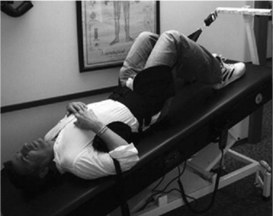
There is a website called the Cox Technic which claims that by using their chiropractor machine called the Cox Technic Flexion Distraction and Decompression you can increase the disk height by 17%.

There was the older post I wrote “Increase Height And Grow Taller Through Non Surgical Spinal Decompression Using Spinal Decompression Therapy” which I showed that the whole process is completely legitimate, at least for temporary effect. However the question was always over how effective was it for long term success.

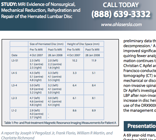
I would find another source Here which revealed that the disk height increase after spinal decompression would be 1-2 mm for each lumbar vertebrate bone.

Spinal Decompression Disc Increase Height

gr3


In the study Radiographic disk height increase after a trial of multimodal spine rehabilitation and vibration traction: a retrospective case series the experimenters used a type of rehabilitation therapy technique known as lumbar vibration traction in the spinal decompression therapy.

There was only 6 patients involved and 1 of them was actually removed to make the data set more smooth. The result was “…achieved improved disk height in 3 of 5 lumbar disks on plain film radiography”. What was shown from the table of disks height was on average an increase of around 1-2 mm for each vertebrate.

The member at the Make Me Taller Forums also made the same theoretical pitch which they called the Intra-Spinal Treatment.

What I am currently proposing is that if we used something like the DRX9000 Non-Surgical Spinal Decompression Machine, (which most medical physicians think is a over-expensive scam product. It might not work at all) to achieve the 1-2 cm of temporary height increase it can definitely do, we might be able to combine it with oral consumption of the compounds…

  • Hyaluronic Acid (aka Hyaluronan) – Dosage: Unknown at this time
  • Glucosamine Sulfate – Dosage: 1000-1500 mg daily (Buy it from Amazon here)
  • Chondroitin Sulfate – Dosage: 1000 daily

This compounds are in the family of the type of organic compound that the GAGS (glycoaminoglycans), Aggrecan, and Proteoglycans are made of which are found in the cartilage and tendon-like tissue found in the intervertebral discs. They are the compounds that make up over 90% of all of the extracellular matrix in cartilage tissue and tendons, forming the Collagen Type 2 and Collagen type 10 (ColX) found in hyaline cartilage.

Glucosamine Sulfate 500 mg dailyA recent commenter did note that after they took glucosamine sulfate (either 1000 mg or 1500 mg) for 3 months, they increased their height by 1/2 cm, which is quite a increase just from oral consumption of a supplement. I spent over half an hour trying to track down that comment made on this website from the hundreds of comments made this recent month but could not find it, clip it, and paste it here showing the actual comment as complete proof. I personally am taking 2 pills of glucosamine sulfate at 500 mg every day myself. What I can show you guys is a picture of my right hand holding the Glucosamine Sulfate 500 mg that I have been taking 2 a day which I bought in Korea –Buy it (at 1000 mg) here from Amazon to help fund the website (I’ll get about $2.50 as an affiliate if you buy the supplement from Amazon)

The combination could lead to upwards of a full 1 cm of permanent height increase which would not go away unless one drastically changes one’s lifestyle and exercise routine. It might sound like much to get just 1 cm but it would a start. This is the real thing.

A Patent For An Apparatus For Stimulating Growth Plate

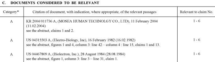
Just something quick to reveal today. After getting back a little to the research, I did manage to find a rather new Patent filed back in 2008 from a Korean researcher, Myung Keun Ji, which wrote up and filed a Patent entitled “Apparatus for Stimulating Growth Plate” which I found from Google Patents. It seems that his idea for using pulsing electrical currents to stimulate growth plate cartilage is very similar to what Brighton talked about in his patent (Method for non-invasive electrical stimulation of epiphyseal plate growth, US 4467809 A), which we have already referenced and reviewed quite extensively. Myung Keun even makes sure to reference Brighton’s patent since his patent seems to be an extension of the older patent. We clipped and linked to the specific patents he references in his Patent. The other patent is Modification of the growth, repair and maintenance behavior of living tissues and cells by a specific and selective change in electrical environment by John P Ryaby

Patent

From the Abstract…

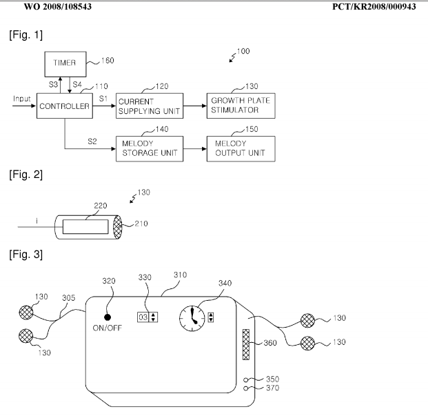
An apparatus for stimulating a growth plate is provided. The apparatus for stimulating a growth plate includes a controller, a current supplying unit, and at least one growth plate stimulator. The controller outputs the driving command signal in response to an external input which determines at least one of an amount of the current and a frequency of the current. The current supplying unit supplies a current corresponding to the driving command signal to the growth plate stimulator. The growth plate stimulator stimulates the growth plate by using the current supplied from the current supplying unit

From a quick Google Search, I also found that it was listed on Patent Scope. From the website…

“…a growth plate stimulating apparatus capable of effectively stimulating a growth plate of the knee even in an asleep time”

So it seems that the device that this Myung Keun Ji has stated might be capable for stimulating the length of the legs closer to the knee area for kids while they are sleeping.

From the Product Description section….

“…The increase in length of the bone is a result of changing a cartilage bone at a distal end of the bone into hard bone cells. If the number of new bone cells is increased, the length of the bone is increased, so that the height of the child is increased”

“…Conventionally, as a growth plate stimulating apparatus, shoes for stimulating the growth plates are mainly used so as to stimulate the ankles or the bottoms of the feet.”

Analysis: What this Korean Medical researcher has revealed is that maybe in traditional Korean medicine, to help young kids become taller, they put on some type of bone lengthening shoes. However he does does in the description that the conventional ways of wearing electrically stimulating shoes to stimulate the growth plates close to the ankle areas was probably not effective.

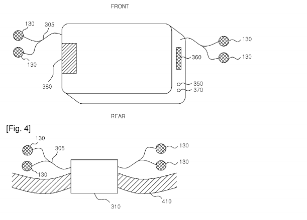
His approach is to stimulate the growth plates in young kids in the knee area.Refer to the pictures below. At some point, we will be breaking down the patent into something much easier to understand for the lay person within the coming months.

Stimulating Growth Plate 1

Stimulating Growth Plate 2

Update #8 – Working On A 2nd Project – November 1, 2013

Update #8 – Working On A 2nd Project – November 1, 2013

Balancing ActThis last month was spent doing three different things, none of which was related to trying to gain height.

  • Traveling – took a short trip to Shanghai, China
  • Building assets which lets me earn what is known as passive income
  • Building a resource on the internet on giving breakthrough ideas and treatments for unique medical disorders and the various chronic pains that result from them

It is absolutely true that my passion is still on trying to go into this endeavor but I also understand that there should be something else in my life.

This website is NOT going anywhere. It is here to stay, and I will be back at some point to focus more on the research. My guess is that based on the other obligations in my life, it would take at least until Jan of 2014 before I start to go back into the research in a consistent fashion.

Changes that have been happening

  • Tyler has become a huge contributor to the website, in revealing multiple new insights and discoveries.
  • I have started to share a percentage of the profits that this website earns with him, so this website is turning more into a side business with business partners.
  • I do wish to produce a book some time either in the coming year, or the year after that.

As for my height, nothing has changed. Maybe writing these monthly reports on updates and changes about the website is pointless at some point if I do not actively get back into increasing my height.

Bmpr1a may be the key to form new growth plates

Bmpr1a is downregulated by aging in bone marrow.  LSJL upregulates Bmpr1b but expression of Bmpr1a is modulated by normal mechanical loadingBMP-2 interacts with Bmpr1a.  LSJL upregulates BMP-2.  Cells in the perichondrial groove express Bmpr1aPersistent upregulation of Hoxa2 which causes short stature downregulates Bmpr1a Bmpr1a is upregulated in a fractured tibia.  These facts are consistent with the observation that bmpr1a could play an on/off-switch in neo-growth plate formation.

BMP Receptor 1A Determines the Cell Fate of the Postnatal Growth Plate.

“Embryos deficient in Bmp receptor (Bmpr)1a or Bmpr1b in cartilage display subtle skeletal defects; however, double mutant embryos develop severe skeletal defects, suggesting a functional redundancy that is essential for early chondrogenesis. In this study, we examined the postnatal role of Bmpr1a in cartilage. In the Bmpr1a conditional knockout (cKO, a cross between Bmpr1a flox and aggrecan-CreER (T2) induced by a one-time-tamoxifen injection at birth and harvested at ages of 2, 4, 8 and 20 weeks), there was essentially no long bone growth with little expression of cartilage markers such as SOX9, IHH and glycoproteins. Unexpectedly, the null growth plate was replaced by bone-like tissues, supporting the notions that the progenitor cells in the growth plate, which normally form cartilage, can form other tissues such as bone and fibrous; and that BMPR1A determines the cell fate.

“During endochondral ossification, the progenitor cells committed act as the stem cells that replenish the pool of proliferative chondrocytes in the resting zone of the growth plate. The resulting daughter cells exit the cycle and undergo maturation via prehypertrophic, hypertrophic and terminal hypertrophic processes, followed by calcified cartilage formation. The vascular invasion then leads to the removal of cartilage, formation of bone marrow, and new bone formation and growth.”

“Is [there] a molecule that initiates and guides the progenitor cell in the resting zone of the growth plate, which solely differentiates chondrogenesis?”<-We want to find this molecule and induce it in adult bone.

” The failure of long bone growth indicates that there is no new progenitor cells added after deletions of Bmpr1a gene by one time injection of tamoxifen at newborn stage.”<-This is bad news that there may be a fixed pool of progenitor cells that is used up.  However, other stem cells could become progenitor cells after the right mechanical stimulus.

An external file that holds a picture, illustration, etc.
Object name is ijbsv09p0895g008.jpg

“The work with a one-time injection of tamoxifen to remove Bmpr1a in cartilage at birth, which leads to a lack of long bone growth postnatally, raises the possibility that the progenitor cell number might already be present in the growth plate at birth with no more new progenitor cells added. This observation may explain why the rodent growth plate remains unfused during the animal’s lifetime but there is essentially no long bone lengthening after 28-30 weeks. In fact, in the 5-month-old control mouse, there is very limited cell proliferation in the growth plate compared to the early stage ”

So the goal in a bone lengthening program is to alter cell morphology of stem cells to be more like the progenitor cells present in the zone of ranvier and then to upregulate bmpr1a expression to encourage a chondrogenic fate.

This fixed pool of progenitor cells is also supported by s1p-ko mice which can form new growth plates from this pool of progenitors.

IGF-2 could play a role in priming MSCs to be more like the progenitor cells that form new growth platesIGF-2 has been used to induce longitudinal bone growth The LSJL scientists have also suggested that IGF-2 injections may be beneficial to LSJL lengtheningCTGF is linked to IGF2 and is another target to increase for supplements as well as HMGA2IGF2 is downregulated almost 1000-fold during senescence which further links IGF2 to the creation of progenitor cells.

I couldn’t find any supplements that increase IGF-2.  There’s a supplement called IGF-2 but that just seems to be the brand name.

Epigenetic consequences of a changing human diet.

“The ultimate methyl donor for epigenetic-methylation reactions is S-adenosylmethionine that is produced by the methylation cycle and it has been reported that periconceptional folic acid use alters the level of methylation within IGF2. A larger study of human pregnancy also observed an effect of folic acid use on IGF2 methylation in the offspring but the effect was restricted to folic acid use after 12 weeks gestation when women are not recommended to take the supplement. Late gestation use of folic acid was also associated with reduced LINE-1 methylation and altered paternally expressed gene 3 (PEG3) methylation. Three of the four significant associations with folic acid use and folate status were negative and one was positive, suggesting that it may be naive to assume that this is a simple substrate limitation effect or that the supply of nutrients involved in the methylation cycle will affect all genes equally.
Imprinting occurs before fertilisation but changes in imprinting methylation in animal models in response to nutritional exposures have been demonstrated into the early post-natal period for IGF2, after which the imprint is apparently fixed

Choline can result in hypermethylation of IGF2 in the liver.  Liver production of IGF2 is not stimulated by GH in adult mammals.  In adult mammals, IGF2 does not seem to respond to physiological status or vary with growth rate.

Expression of bone-related genes in bone marrow MSCs after cyclic mechanical strain: implications for distraction osteogenesis.

“In this study, a single period of cyclic mechanical stretch (0.5 Hz, 2,000 microepsilon) [for 40 minutes] was performed on rat bone marrow MSCs. Cellular proliferation and alkaline phosphatase (ALP) activity was examined. The mRNA expression of six bone-related genes (Ets-1, bFGF, IGF-II, TGF-beta, Cbfa1 and ALP) was detected using real-time quantitative RT-PCR.
The results showed that mechanical strain can promote MSCs proliferation, increase ALP activity, and up-regulate the expression of these genes. A significant increase in Ets-1 expression was detected immediately after mechanical stimulation, but Cbfa1 expression became elevated later. The temporal expression pattern of ALP coincided perfectly with Cbfa1.
The results of this study suggest that mechanical strain may act as a stimulator to induce differentiation of MSCs into osteoblasts{but could be chondrocytes with increase in bmpr1a levels}.”

This was the load applied to the cells during the study it’s reasonable to expect that the load is similar to that which is applied by LSJL.

“The transcription profiles of IGF-Ⅱand TGF-β were similar. Both genes reached maximum transcription immediately after mechanical strain and mRNA levels then decreased with time, except for a slight increase at 6 hours.”

The age of rats is not given and is vital information given that IGF2 is an age related gene.

It’s conceivable that LSJL modulates Bmpr1a expression given the study that shown it can be modified by mechanical loading and that LSJL modulates IGF2 expression given the study above that showed that it was upregulated by mechanical loading.

But still finding supplements that upregulate IGF2 and Bmpr1a would be exceptionally helpful.

General studies on fluid pressure

Biomechanical and biophysical environment of bone from the macroscopic to the pericellular and molecular level.

“Bones with complicated hierarchical configuration and microstructures constitute the load-bearing system. Mechanical loading plays an essential role in maintaining bone health and regulating bone mechanical adaptation (modeling and remodeling). The whole-bone or sub-region (macroscopic) mechanical signals, including locomotion-induced loading and external actuator-generated vibration, ultrasound, oscillatory skeletal muscle stimulation, etc., give rise to sophisticated and distinct biomechanical and biophysical environments at the pericellular (microscopic) and collagen/mineral molecular (nanoscopic) levels, which are the direct stimulations that positively influence bone adaptation. While under microgravity, the stimulations decrease or even disappear, which exerts a negative influence on bone adaptation. A full understanding of the biomechanical and biophysical environment at different levels is necessary for exploring bone biomechanical properties and mechanical adaptation. In this review, the mechanical transferring theories from the macroscopic to the microscopic and nanoscopic levels are elucidated. First, detailed information of the hierarchical structures and biochemical composition of bone, which are the foundations for mechanical signal propagation, are presented. Second, the deformation feature of load-bearing bone during locomotion is clarified as a combination of bending and torsion rather than simplex bending. The bone matrix strains at microscopic and nanoscopic levels directly induced by bone deformation are critically discussed, and the strain concentration mechanism due to the complicated microstructures is highlighted. Third, the biomechanical and biophysical environments at microscopic and nanoscopic levels positively generated during bone matrix deformation or by dynamic mechanical loadings induced by external actuators, as well as those negatively affected under microgravity, are systematically discussed, including the interstitial fluid flow (IFF) within the lacunar-canalicular system and at the endosteum, the piezoelectricity at the deformed bone surface, and the streaming potential accompanying the IFF. Their generation mechanisms and the regulation effect on bone adaptation are presented. The IFF-induced chemotransport effect, shear stress, and fluid drag on the pericellular matrix are meaningful and noteworthy. Furthermore, we firmly believe that bone adaptation is regulated by the combination of bone biomechanical and biophysical environment, not only the commonly considered matrix strain, fluid shear stress, and hydrostatic pressure, but also the piezoelectricity and streaming potential. Especially, it is necessary to incorporate bone matrix piezoelectricity and streaming potential to explain how osteoblasts (bone formation cells) and osteoclasts (bone resorption cells) can differentiate among different types of loads. Specifically, the regulation effects and the related mechanisms of the biomechanical and biophysical environments on bone need further exploration, and the incorporation of experimental research with theoretical simulations is essential.”

“bone  adaptation is regulated by the combination of bone biomechanical and biophysical environment, not only the commonly considered matrix strain, fluid shear stress, and hydrostatic pressure, but also the piezoelectricity and streaming potential”

“At the macroscopic level in long bone such as the femur, cortical bone with compact structures
and low porosity forms the hard shell, and trabecular bone with a three-dimensional interconnected  network of trabecular rods and plates forms the inner surface.  In flat bone, e.g. the calvaria and iliac crest, the cortical bone and trabecular bone form the cortical-trabecular-cortical sandwich structure”

the pericellular spaces between osteocytes and the lacunar-canalicular wall are filled with interstitial fluid and pericellular matrix (PCM), which is a gel-like fiber matrix thought to be composed of proteoglycans and other matrix molecules ”

” Due to  the low permeability of mineralized bone matrix, interstitial fluid flow (IFF) is principally generated during alteration of intramedullary pressurization (ImP) and bone matrix deformation ”

” Uniform pressurization [such as that due to decreased intramedullary cavity resulting from elevated bone marrow lipids induced by high level of corticosteroid administration will generate radial flow from the intramedullary compartment to the endosteal surface and into the LCS due to the pressure gradient from the marrow cavity to the bone matrix”

” Non-uniform pressure gradients within the intramedullary cavity [such as those due to local heterogeneous permeability or fluid displacement changes in the intramedullary compartment from the interaction between mechanical loading/oscillatory muscle stimulation and capillary filtration in bone tissue  will cause tangential fluid flow to the endosteal surface ”

“The matrix deformation generated pressure gradient within LCS stimulates the interstitial fluid to move towards the lower pressure zone”

“the profiles of IFF within the LCS are sophisticated and can be considered as a combination of
oscillating and unidirectional fluid flow ”

“Three stimuli induced by fluid flow, namely chemotransport effects, IFF-induced shear stress, and fluid drag on the PCM, have been shown to regulate osteocyte activity”

“IFF within the LCS serves as the primary transport mechanism between the blood supply and osteocytes. Furthermore, the shear stress induced by IFF provides potent mechanical stimulation for osteocytes ”

” In the inverse piezoelectric effect, subjecting bone to an electric field induces deformation”

“varying degrees of deformations, or even irreversible deformations, will also be evoked in collagen fibrils and mineral crystals oriented in different directions”

Huge Breakthrough: New LSJL study with device

Development of a Portable Knee Rehabilitation Device That Uses Mechanical Loading

development of portable knee device<-Full study there

“Joint loading is a recently developed mechanical modality, which potentially provides a therapeutic regimen to activate bone formation and prevent degradation of joint tissues. Few joint loading devices are available for clinical or point-of-care applications. Using a voice-coil actuator, we developed an electromechanical loading system appropriate for human studies and preclinical trials that should prove both safe and effective. Two specific tasks for this loading system were development of loading conditions (magnitude and frequency) suitable for humans, and provision of a convenient and portable joint loading apparatus. Desktop devices have been previously designed to evaluate the effects of various loading conditions using small and large animals. However, a portable knee loading device is more desirable from a usability point of view. We present a device that is designed to be portable, providing a compact, user-friendly loader. The portable device was employed to evaluate its capabilities using a human knee model. The portable device was characterized for force-pulse width modulation duty cycle and loading frequency properties. The results demonstrate that the device is capable of producing the necessary magnitude of forces at appropriate frequencies to promote the stimulation of bone growth and which can be used in clinical studies for further evaluations.”

Note how the device looks like a table clamp:med_007_04_041007_f011.png

med_007_04_041007_f012.png

Image Not Available

“Dynamic loads applied laterally to the knee joint have been found to stimulate new bone formation not only in the distal femur and proximal tibia epiphyses, but along the entire length of each bone.”

“Knee loading is applied laterally to the epiphysis of the femur and tibia, which consist mostly of trabecular bone modeled to resist axial stresses{if the trabecular bone is modeled to resist axial strain than it is less resistant to lateral strain}. This allows greater deformations than are possible with similar loads in different loading modalities.  Dynamic deformations of the epiphysis cause alterations in fluid pressure in the intramedullary cavity, driving oscillatory fluid flow and molecular transport in the lacunocanalicular network in the bone matrix and in the medullary cavity”<-It is alterations in fluid pressure that could induce chondrogenic differentiation of the MSCs in the epiphysis forming new micro growth plates.

“The device applies cyclic loading of magnitudes up to 30N at frequencies ranging from 1 to 20 Hz and can cause small deformation in the knee.”

“The device proposed in this paper differs [from LIPUS and PEMF] in that it stimulates the bone tissue through direct mechanical loading at low frequencies.”

“A cyclic force applied on [the knee] would force a slight shift of the fluid within the bone towards the opposite end of the bone in a controlled fashion.”

“The device must provide sufficient force in a transverse load to the joint without being bulky or unbalanced.”<-This is a problem we may have too.  We might have to use a larger clamp for the knee.  The standard c-class clamp may work for smaller joints but a larger clamp may perform better for the knee.

“The force magnitudes that the device can produce must be at least 30 N”

“power generation units used in conjunction with a mechanism comprised of linkages was proposed as a means of shifting the power generation component into a position that allowed for better balance.”

“Operating the device at frequencies between 1 and 5 Hz is meant to simulate the therapeutic and rehabilitative effects of walking or running without the need to put the patient’s weight on the leg.”<-But would the effect be more significant than walking or running?  Walking and running have a lot of evidence showing that they don’t make you taller.

“the motor shaft is acting as the sliding block while the linkage connecting to the base plate serves as the crank. The rigid pivot arm that connects the motor shaft and crank before ultimately connecting with the pad at the top is the connecting arm.”<-much like a table clamp.

“Aluminum was chosen for the base plate, side plates, and the adjustable side as it is lightweight, but still provides the necessary strength. The pivot arm and lever arm were made of O1 steel. O1 steel has a high carbon content and a correspondingly high modulus of elasticity. This is crucial as these parts are subjected to higher stresses than are found in any other part of the device; it is important to minimize the deflection in these pieces since deflection of these parts would directly affect the range of displacement provided by the mechanism and may also contribute to damping{a decrease in the amplitude of an oscillation as a result of energy being drained from the system to overcome frictional or other resistive forces.}. High density polyethylene was selected for the pads as it allows a slight amount of deformation to increase comfort to the user while still being sufficiently rigid to transfer most of the force to the knee being loaded.”<-Could be helpful in designing a self-made LSJL device

“For the static loading, a 6.9 kPa pressure is evenly distributed across the 64.5 cm2 surface of both side of the pads directed outwards. A 44.5 N force is applied to the center of the motor shaft pushing away from the motor. This loading simulates the force applied by the motor and the reaction on the pads from the knee being loaded. “<-a lot of studies make it clear though that dynamic load is needed.

According to figure 11, Pressure generated is 6000Pa Or 0.006MPa.  Most figures show 0.1 to 10 MPA is what’s needed for chondroinduction but perhaps the pressure generated on the outside is not reflective of the pressure generated inside the bone which could be higher.

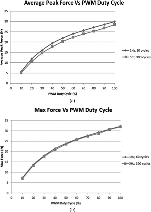
Here’s some force generated measurements:
LSJL device force

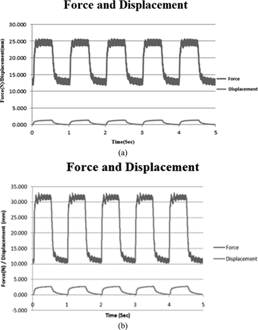
Here’s some displacement measurements:
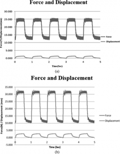
displacement

“the force and corresponding displacement [of the artificial knee] over a period of five cycles at duty cycles of 50% and duty cycles of 100%. Such experiments when performed on an actual knee could be used to assist in the study of the force versus displacement of human tissue, a highly nonlinear reaction, as well as the effects of light mechanical loading to the bone tissue.”

The mechanics definition of displacement is the final position of a point (Rf) relative to its initial position (Ri).  Tissue displacement refers to the change in the form or position of the tissues as a result of pressure.  So the displacement of the artificial knee was about 2.5mm when the force generated was about 30N.  Since the displacement turns to normal it is referred to as an elastic deformation a plastic deformation would be a permanent change.  Thus the only way this device can induce new longitudinal bone growth is if it stimulates neo-endochondral ossification.

Here’s some information about the effects of tissue deformation on tissue deformation from a paper called Biomechanics of Tissues from the Journal of Rheumatology: “In a creep test, an instantaneous step or ramp load is applied to the tissue sample and held constant for an extended period of time. This is considered a load control test, and the resulting tissue displacement is measured. The displacement shows an initial elastic response of the tissue followed by a gradual increase of lengthening of the tissue. As the test proceeds, fluid is exuded from the tissue, and the solid components of the tissue are supporting the applied load. A “solid-like” material will be distracted to a point at which the solid components of the tissue will balance the applied load and will not elongate further. The modulus of the tissue is calculated as the stress of the tissue divided by the end displacement of the tissue. A “fluid-like” material will not be able to balance the applied load and will continue to elongate.“<-I don’t believe this occurred during my LSJL finger lengthening as the fingers increased in width greatly as well.

No mention of using the device for lengthening.  The basis of using LSJL on lengthening is based on studies that show that hydrostatic pressure can induce chondrogenic differentiation and that LSJL upregulates genes that do not only stimulate chondrogenesis of chondrogenitors but also of MSCs such as FGF-2, Gli3, and Cyr61 and of signs of mesenchymal condensation in LSJL images.  Also, in those images is apparent degradation of trabecular bone which would be permissive to neo-growth plate formation.

The condensed stem cells in the images are most consistent with granulocytes which are capable of chondrogenic differentiation.  According to Comparative study of the biological characteristics of mesenchymal stem cells from bone marrow and peripheral blood of rats., peripheral blood MSCs are more chondrogenic than bone marrow MSCs but chondrogenesis of bone marrow MSCs is not impossible.

This study does not provide more evidence that LSJL can increase length but it does support the notion that a table clamp is a perfect home made solution for performing LSJL.

Here’s a study with more about the effects of tissue deformation on the joint region:

The effects of manual therapy on connective tissue<-connective tissue therapy

“a low level of CT[connective tissue] damage must occur in order to produce permanent elongation. The collagen breakage will be followed by a classical cycle of tissue inflammation, repair, and remodeling”<-This paper doesn’t mention cartilage specifically but it mentions other tissues made of collagen fibers.

“Connective tissue that is loaded more quickly will behave more stiffly (will deform less) than
the same tissue that is loaded at a slower rate”

In figure 5, a graph is shown where it appears that microfailure of a tissue(the kind of stimulus that must occur in order to generate permanent lengthening of connective tisues) occurs 4-6mm(with a safer region being between 4-4.5mm) of displacement beyond which is complete failure of the tissue.  The displacement generated by the LSJL device is 2.5mm which is below this level to generate permanent lengthening of connective tissues.