Monthly Archives: September 2015

More on Eden Alsberg’s Biochemical signal gradients

Biochemical and physical signal gradients in hydrogels to control stem cell behavior.

“Cells continuously receive biochemical and biophysical stimuli from their microenvironment. These environmental stimuli drive cellular behavior and function during development and tissue regeneration.”<-We can alter the microenvironment via mechanical means to alter cellular behavior and ideally create neo-growth plates.

“Cell interactions with the extracellular matrix (ECM) and physical signals such as matrix rigidity and mechanical stimuli can also have strong effects on cellular phenotype and tissue formation.”<-The bone ECM likely has a strong negative regulatory effect on neo-growth plate formation.  But we can alter the bone ECM via mechanical means as well.

“[we prepared] HP-ALG hydrogels with incorporated gradients of heparin binding growth factors BMP-2, a potent osteogenic growth factor, and TGF-β1, a chondrogenic growth factor, in opposite directions. When the concentration of growth factors in segments of gradient HP-ALG hydrogels was quantified, linear gradient distributions of BMP-2 and TGF-β1 in opposite directions were observed”

“As the BMP-2 concentration increased, ALP expression significantly increased along the BMP-2 gradient. In contrast, GAG production of encapsulated hMSCs significantly increased as the TGF-β1 concentration increased”

Regulation of Stem Cell Fate in a Three-Dimensional Micropatterned Dual-Crosslinked Hydrogel System.

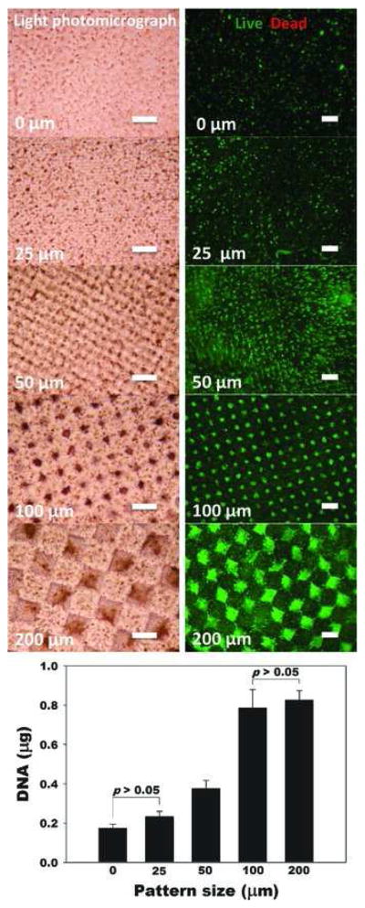
“By manipulating micropattern size while keeping the overall ratio of single- to dual-crosslinked hydrogel volume constant, the physical properties of the micropatterned alginate hydrogels are spatially tunable. When human adipose-derived stem cells (hASCs) are photoencapsulated within micropatterned hydrogels, their proliferation rate is a function of micropattern size. Additionally, micropattern size dictates the extent of osteogenic and chondrogenic differentiation of photoencapsulated hASC. The size of 3D micropatterned physical properties in this new hydrogel system introduces a new design parameter for regulating various cellular behaviors, and this dual-crosslinked hydrogel system provides a new platform for studying proliferation and differentiation of stem cells in a spatially controlled manner for tissue engineering and regenerative medicine applications.”

“cell behaviors such as differentiation and proliferation are known to be affected by cell cluster size”

” micropattern size dictated the extent of osteogenic and chondrogenic differentiation of photoencapsulated hASC.”

“the aggrecan expression of hASCs gradually increased as the micropattern size increased”<-But some markers increased at 200 micrometers so the optimal micropattern size should be around 100-200 micrometers.

HEre’s a visualization of micropatterns:

NIHMS492427.html

Decellularized tissue and cell-derived extracellular matrices as scaffolds for orthopaedic tissue engineering.

“The reconstruction of musculoskeletal defects is a constant challenge for orthopaedic surgeons. Musculoskeletal injuries such as fractures, chondral lesions, infections and tumor debulking can often lead to large tissue voids requiring reconstruction with tissue grafts. Autografts are currently the gold standard in orthopaedic tissue reconstruction; however, there is a limit to the amount of tissue that can be harvested before compromising the donor site. Tissue engineering strategies using allogeneic or xenogeneic decellularized bone, cartilage, skeletal muscle, tendon and ligament have emerged as promising potential alternative treatment. The extracellular matrix provides a natural scaffold for cell attachment, proliferation and differentiation. Decellularization of in vitro cell-derived matrices can also enable the generation of autologous constructs from tissue specific cells or progenitor cells. Although decellularized bone tissue is widely used clinically in orthopaedic applications, the exciting potential of decellularized cartilage, skeletal muscle, tendon and ligament cell-derived matrices has only recently begun to be explored for ultimate translation to the orthopaedic clinic.

“ECM is a product of cells that functions to maintain tissue and organ structure, organization and function. It is a complex network of proteins and polysaccharides forming an intricate meshwork within tissue that interacts with the resident cells to regulate cell behavior, such as migration, proliferation and differentiation. The ECM exists in a state of dynamic equilibrium with its resident cells and is constantly being built, reshaped and degraded in response to changing environmental conditions and to cellular, tissue and organ demands”<-So we should try to alter the bone ECM to be more favorable to cartilaginous tissues.

“Fracture healing requires an intricate and well-organized series of cellular and molecular events. It involves interactions between cortical bone, the periosteum, undifferentiated fascial tissue surrounding the fracture and the bone marrow. Fracture healing is divided into three stages: inflammation, repair and remodeling. After an injury, there is initial bleeding from the damaged bone ends and surrounding tissue resulting in the formation of a hematoma, which provides a source of hematopoietic cells capable of secreting growth factors. The invasion of inflammatory cells, fibroblasts, mesenchymal cells, and osteoprogenitor cells at the fracture site forms granulation tissue around the fracture ends{To induce neo-growth plates we have to allow this invasion}. Fractures that are anatomically aligned with absolute stability, such as those surgically repaired with compression plates, undergo primary bone healing or Haversian remodeling, in which there is direct osteonal healing within the cortex by intramembranous ossification”

” in closed reduced fractures, secondary bone healing occurs with the formation of a bridging soft callus consisting of cartilage tissue connecting the fracture ends. Over time, bone formation occurs under the periosteum and calcification of cartilage results in the formation of hard callus or woven bone by endochondral ossification”

“injuries that penetrate the subchondral bone often result in the formation of fibrocartilage which is biomechanically insufficient compared to hyaline cartilage, resulting in further damage over time”

“the peak force transmitted through the Achilles tendon while running is 9 kN, which is about 12.5 times the body weight”

Sesamoid Bone

A sesamoid is a bone formed within a tendon or muscle while we’re looking for is more interosseous chondroification(the formation of new cartilage tissue within the bone) or probably maybe even first interosseous epithelialification as you might need to transition mesenchymal to epithelial tissues first.

Coincident development of sesamoid bones and clues to their evolution.

“Sesamoid bones form within tendons in regions that wrap around bony prominences. They are common in humans but variable in number. Sesamoid development is mediated epigenetically by local mechanical forces associated with skeletal geometry, posture, and muscular activity. In this article we review the literature on sesamoids and explore the question of genetic control of sesamoid development. Examination of radiographs of 112 people demonstrated that the relatively infrequent appearances of the fabella (in the lateral gastrocnemius tendon of the knee) and os peroneum (in the peroneus longus tendon of the foot) are related within individual. This finding suggests that the tendency to form sesamoids may be linked to intrinsic genetic factors. Evolutionary character analyses suggest that the formation of these sesamoids in humans may be a consequence of phylogeny. These observations indicate that variations of intrinsic factors may interact with extrinsic mechanobiological factors to influence sesamoid development and evolution.”

“As many as 42 sesamoid bones can be found in some individuals. Mechanically, sesamoid bones serve to protect the tendon from damage and, in some cases, increase the efficiency or mechanical advantage of their associated muscle.”

“Most sesamoid bones in humans are 5 to 10 mm in diameter or smaller and are present in 1 to 100% of individuals.”

“Tendon chondrometaplasia[tendon turns into cartilage] and sesamoid bones tend to develop within tendons in areas that experience both tensile strain and hydrostatic compressive mechanical stresses.”<-So tendonous tissue may help form neo growth plates.  Tendon enthesis attach into the bone.  Tendon is capable of undergoing  chondrometaplasia.  So potentially you could form neo growth plates at these tendon entheses.  Originally I thought that ligament enthesis had more potential for neo-growth plate formation but tendon enthesis may have potential too.

” fibrous tendon tissue can form regions of fibrocartilage in areas that wrap around bony prominences (fibrocartilage is a tissue whose phenotype is intermediate between fibrous and cartilaginous tissue, consisting of chondrocytes embedded in aligned bundles of type I collagen)”

“compression loading and treatment with transforming growth factor beta (TGF-β) each resulted in upregulation of aggrecan and biglycan synthesis in fetal bovine tendon, suggesting that one aspect of the response of cells to compressive load is increased TGF-β synthesis which, in turn, stimulates synthesis of extracellular matrix proteoglycans and leads toward fibrocartilage formation. The process of fibrocartilaginous metaplasia in tendons, which is a direct response to an altered mechanical loading environment, appears to represent an intermediate step in the formation of a sesamoid cartilage.”

Sesamoid bones:

sesamoid-bone

Their position won’t help you grow taller but they could if they were in the articular cartilage.  Maybe if tendonous tissue was inserted in the articular cartilage.

“Hox A11, had profound effects on the developing mouse skeleton, including abnormal sesamoid bone development in both the forelimbs and hindlimbs.”

How might we turn tendons(and especially tendenous enthesis) into cartilage tissues.  What if we form a sesamoid bone within the enthesis?

Expression of Bone Morphogenetic Protein-2 in the Chondrogenic and Ossifying Sites of Calcific Tendinopathy and Traumatic Tendon Injury Rat Models

“Ectopic chondrogenesis and ossification were observed in a degenerative collagenase-induced calcific tendinopathy model and to a lesser extent, in a patellar tendon traumatic injury model. We hypothesized that expression of bone morphogenetic protein-2 (BMP-2) contributed to ectopic chondrogenesis and ossification. This study aimed to study the spatial and temporal expression of BMP-2 in our animal models.
Seventy-two rats were used, with 36 rats each subjected to central one-third patellar tendon window injury (C1/3 group) and collagenase-induced tendon injury (CI group), respectively. The contralateral limb served as controls. At week 2, 4 and 12, 12 rats in each group were sacrificed for immunohistochemistry and RT-PCR of BMP-2.
For CI group, weak signal was observed at the tendon matrix at week 2. At week 4, matrix around chondrocyte-like cells was also stained in some samples. In one sample, calcification was observed and the BMP-2 signal was observed both in the calcific matrix and the embedded chondrocyte-like cells. At week 12, the staining was observed mainly in the calcific matrix. Similar result was observed in C1/3 group though the immunopositive staining of BMP-2 was generally weaker. There was significant increase in BMP-2 mRNA compared to that in the contralateral side at week 2 and the level became insignificantly different at week 12 in CI group. No significant increase in BMP-2 mRNA was observed in C1/3 group at all time points.
Ectopic expression of BMP-2 might induce tissue transformation into ectopic bone/cartilage and promoted structural degeneration in calcific tendinopathy.”

“the presence of chondrocyte phenotype and ectopic ossification in a collagenase-induced patellar tendon injury model”

Hiroki Yokota Grant progress on LSJL

What can we discern about the plans from LSJL from the grants?  Yokota is one of the primary scientists behind the study Lengthening of Mouse Hindlimbs with Joint Loading.  There does not seem to be very much on the LSJL length effects since the expiring of Ping Zhang’s 2010 grant.  We either have to study the lengthening effects on our own or help Ping Zhang get more funding.

Yokota doesn’t mainly study the lengthening effects which are primarily studied by Ping Zhang as shown by this grant.

Here’s Hiroki Yokota’s 2015 grant:

“The long-term objective of the proposed studies is to elucidate the mechanism of mechanotransduction in bone. Our present bioengineering-oriented project developed a high-resolution piezoelectric mechanical loader and evaluated the role of mechanical stimulation in bone using cultured osteoblasts. The results reveal that (a) deformation of 3D collagen matrix can induce strain-induced fluid flow;(b) strain-induced fluid flow, and not strain itself, predominantly activates the stress-responsive genes in osteoblasts;and (c) architecture of 3D collagen matrix establishes a pattern of strain-induced fluid flow and molecular transport{We are not interested so much on the effects on osteoblasts but more on the effects of fluid flow on bone degradation and fluid flow on mesenchymal stem cells to create neo growth plates}. Many lines of evidence in animal studies support enhancement of bone remodeling with strain of 1000 – 2000 microstrains. An unclear linkage between our in vitro studies and these animal studies is the role of strain and fluid flow in bone remodeling. In vitro osteoblast cultures including our current studies use 2D substrates or 3D matrices that hardly mimic the strain-induced fluid flow in vivo. This difference between in vitro and in vivo data makes it difficult to evaluate the role of strain and fluid flow in bone remodeling and anti-inflammation. First, microscopic strain in bone might be higher than the macroscopic strain measured with strain gauges. A local microscopic strain higher than 1000 – 2000 microstrains may therefore drive fluid flow in bone. Second, the lacunocanalicular network in bone could amplify strain-induced fluid flow in a loading-frequency dependent fashion{This we should try to modify the frequency of LSJL to amplify strain}. Lastly, interstitial fluid flow in bone might be induced by in situ strain as well as strain in a distant location, such that deformation of relatively soft epiphyses induces fluid flow in cortical bone in diaphyses{We are more interested in deformation of the epiphysis as that’s where growth plates typically occur but deformation of the epiphysis in one end may induce fluid flow in the epiphysis in the other}. This renewal proposal will use mouse ulnae ex vivo as well as mouse in vivo loading to examine the above possible explanations for the data divergence.
Specific aims i nclude: (1) fabricating a piezoelectric mechanical loader for ex vivo and in vivo use;(2) quantifying ex vivo macroscopic and microscopic strains using electronic speckle pattern interferometry as well as molecular transport using fluorescence recovery after photobleaching;(3) conducting bone histomorphometry to evaluate ex vivo data;and (4) examining load-driven adverse effects with gene expression and enzyme activities (e.g., matrix metalloproteinases). Mechanical loads will be given in the ulna-loading (axial loading) and elbow-loading (lateral loading) modes{he’s planning on doing another LSJL loading study!}. These two modes have been shown to enhance bone remodeling in the diaphysis with different patterns of strain distribution. Successful completion of the proposed renewal proposal will provide basic knowledge about induction of fluid flow in bone and establish a research platform for devising therapeutic strategies for strengthening bone and preventing bone loss.”

Here’s the Hiroki Yokota 2014 grant:

Mechanical Loading and Bone

“The long-term objective of this study is to elucidate the mechanisms underlying loading-induced bone remodeling and develop unique loading-based therapies for preventing bone loss. The specific goal of this study, based on our most recent observations, is to determine how mechanical loading to the knee (knee loading – application of mild lateral loads to the knee) may exert global suppression of osteoclast development not only in the loaded (on-site) bone but also in the non-loaded (remote) bone{This is unfortunately not very promising for height growth as osteoclast driven remodeling is pretty significant for growth plate formation}. As a potential regulatory mechanism, we will focus on secretory factors (e.g., Wnt3a, NGF?, TNF?, etc.) and low-density lipoprotein receptor-related protein 5 (Lrp5) mediated signaling. In the parent project, we have shown that knee loading enhances bone formation in the tibia and the femur through the oscillatory modulation of intramedullary pressure. However, its effects on bone resorption have not been well understood. Preliminary studies using a mouse ovariectomized model, which mimics post-menopausal osteoporosis, indicate that knee loading can suppress development of multi-nucleated osteoclasts from bone marrow cells, and the loading effects are observed not only in the loaded femur but also in the non-loaded contralateral femur. In this competitive renewal project, we will test the hypothesis that joint loading (knee/elbow loading) can suppress an OVX-induced osteoclastogenesis in a systemic manner through Lrp5-mediated Wnt signaling with Wnt3a as a secretory factor, as well as interactions with other secretory factors. To examine this hypothesis, we propose two specific aims using a mouse loading model (knee loading, elbow loading, ulna bending, and tibia loading), and assays for bone remodeling and primary bone marrow cells.
Aim 1 : Determine the local and global effects of joint loading on osteoclastogenesis Aim 2: Evaluate the role of load-modulated secretory factors in osteoclastogenesis In response to mechanical loading, we will conduct X-ray imaging and colony forming unit assays{The x-rays will be highly useful in determining whether LSJL can induce neo-growth plate formation although the effects would have to be large to show up on the xray}. We will also examine expression of critical secretory factors such as Wnt3a, NGF?, TNF?, OPG, RANKL, etc. in the serum. Primary bone marrow cells will be cultured, and the mechanisms underlying loading-driven regulation of osteoclastogenesis will be investigated. We will examine expression of regulatory factors, including NFATc1 (master transcription factor for osteoclastogenesis) and osteoclast markers such as OSCAR, cathepsin K, etc. We will employ Lrp5 KO mice (global, and conditionally selective to osteocytes), as well as neutralizing antibodies and RNA interference (loss of a function), and plasmids (gain of a function). We expect that this project will contribute to our basic understanding of load-driven regulation of bone resorption and development of loading regimens useful for global prevention of bone loss. ”

Let’s look at Hiroki Yokota’s 2013 Grant:

“The long-term objective of the proposed studies is to elucidate the mechanism of mechanotransduction in bone. Our present bioengineering-oriented project developed a high-resolution piezoelectric mechanical loader and evaluated the role of mechanical stimulation in bone using cultured osteoblasts. The results reveal that (a) deformation of 3D collagen matrix can induce strain-induced fluid flow{If it is fluid flow that can induce neo-growth plate formation via stem cell simulation then we need to make sure that LSJL deforms the 3D collagen matrix};(b) strain-induced fluid flow, and not strain itself, predominantly activates the stress-responsive genes in osteoblasts;and (c) architecture of 3D collagen matrix establishes a pattern of strain-induced fluid flow and molecular transport. Many lines of evidence in animal studies support enhancement of bone remodeling with strain of 1000 – 2000 microstrains{2000 microstrain is about a 0.2% change in bone length.  LSJL laterally compresses the bone so the compression has to be by at least .1 or .2% to work}. An unclear linkage between our in vitro studies and these animal studies is the role of strain and fluid flow in bone remodeling. In vitro osteoblast cultures including our current studies use 2D substrates or 3D matrices that hardly mimic the strain-induced fluid flow in vivo. This difference between in vitro and in vivo data makes it difficult to evaluate the role of strain and fluid flow in bone remodeling and anti-inflammation. First, microscopic strain in bone might be higher than the macroscopic strain measured with strain gauges. A local microscopic strain higher than 1000 – 2000 microstrains may therefore drive fluid flow in bone. Second, the lacunocanalicular network in bone could amplify strain-induced fluid flow in a loading-frequency dependent fashion. Lastly, interstitial fluid flow in bone might be induced by in situ strain as well as strain in a distant location, such that deformation of relatively soft epiphyses induces fluid flow in cortical bone in diaphyses{of course our goal is to create new growth plates in the epiphysis but the fluid flow from compressing the ends of the epiphysis may flow deeper helping to induce mesenchymal condensation to induce neo growth plates closer to where the epiphysis meets the diaphysis}. This renewal proposal will use mouse ulnae ex vivo as well as mouse in vivo loading to examine the above possible explanations for the data divergence.
Specific aims include: (1) fabricating a piezoelectric mechanical loader for ex vivo and in vivo use;(2) quantifying ex vivo macroscopic and microscopic strains using electronic speckle pattern interferometry as well as molecular transport using fluorescence recovery after photobleaching;(3) conducting bone histomorphometry to evaluate ex vivo data;and (4) examining load-driven adverse effects with gene expression and enzyme activities (e.g., matrix metalloproteinases). Mechanical loads will be given in the ulna-loading (axial loading) and elbow-loading (lateral loading) modes. These two modes have been shown to enhance bone remodeling in the diaphysis with different patterns of strain distribution. Successful completion of the proposed renewal proposal will provide basic knowledge about induction of fluid flow in bone and establish a research platform for devising therapeutic strategies for strengthening bone and preventing bone loss. ”

The grants from 2013-2006 are virtually the same.  It’s only 2014 which is different however it’s unfortunate that it’s not focusing on the lengthening effects.

Unfortunately Ping Zhang’s grant Load-Driven Bone Lengthening only ran from 2008-2010.

How Much Will It Cost To Fund This Biomedical Project?

There were a few people in recent days who wanted to start a KickStarter project to fund this type of project. The project is the idea of using regenerative medicine, stem cell therapy, tissue engineering, and 3D Bioprinting to get an alternative to limb lengthening surgery into market.

What they were discussing was a recent post I wrote entitled – “How Close Are We Towards Growth Plate Regeneration To Grow Taller?”

In response, I have to show these people just how crazy it is to get something new in medicine to the public. Refer to Forbes.com post written back in 2014 entitled – “The Truly Staggering Cost Of Inventing New Drugs”

Eli Lilly, states that it would cost on average about $1.3 Billion USD to get just one drug approved and into the market. $1 Billion Dollars!

If you then take into consideration just how easy it is to have drugs fail trials and testing, then the real cost could be around $4 Billion. The high range is $11 Billion for just one drug to be approved.

I did a quick search on Google to find out what is the project in Kickstarter’s history which got the most funding. It seems that based on this article in Entrepreneur.com it would be the Pebble Time based Watch, with a grand total of $20 Mil. $20 Mil is indeed a lot of money to fund such a project. Of course I have been to trade shows for the latest electronic gear in Hong Kong and China before so I have seen at least 4-5 companies who have also come out with similar type products, aka a smart watch which looks suspiciously similar in design to the Apple Watch.

What we would need to get this project started would be something at least 50X that amount. How many people would even believe us that this idea we have is even possible?

I have emailed someone named Harald from the beginning of this website back in 2012 and he has still not been able to raise even $100 K  to get people to fund what he says is research. He claims he is part of a biomedical team of researchers. It has been 10 years and so far no one has been willing to step up. The honest truth is that no one will step up. The first $50 Mil will be just wasted money, since it will just be used as initial startup cost, which will be sunk cost which would take maybe 20 years to get back, if the project is successful.

This idea I have shown to be quite valid would require a truly herculean amount of will, effort, and money to get it done. There is probably less than 10 people in the entire world who would have the financial ability and incentive to fund us. Of course, for any type of potential investor to invest in us, (since no one wants to loose money on a bad investment), we would have to first show very clearly that the science and the technical details are completely valid and it will work.

Trying to get people to open their wallets is one of the hardest things one learns to do as an adult. Selling is truly the most important skill to learn.

Instead of going on Kickstarter, me and Tyler would have to go on Shark Tank and ask Mark Cuban, who actually has a billion, and the other less rich sharks to give us at least $100 Mil to get started on this project. Of course, then once we reach the first goal, we would have to go back and ask for another $300 Mil. At $300 Mil, not even someone like Kevin O’Leary would have the money to fund our endeavor.

I can see Mark Cuban, who loves basketball and owns a basketball team being very interested in funding a company or project like this, as well as the other sharks. Unlike Cuban, who is 6′ 3″, the others are of short stature. Daymond John, Kevin O’Leary, Robert Herjavek, Barbara Corcoran, they are all on the shorter side. I can see a trip on Shark Tank being most likely to work out. It would at least give our project the big exposure we need, and the short statured “sharks” would definitely be interested in getting something in life which even they can’t buy with their millions. Money can buy limb lengthening surgery for these guys but they probably will never be willing to put up with the pain, loss of time, and helpless feeling.

The other options is to find a rich Arab/Saudi Sheik or Prince who might have a height complex and is willing to give us maybe $50-$100 Mil to get the project started. Before when I used to work in the alternative energy sector in a former life, the CEO at the time said that the company I was at was in talks with a Saudi Family in getting investments (as well as the billionaire T Boone Pickens). My CEO told in passing that apparently the real net worth of the Saudi Family was a total of $1 Trillion!! (True Story).

The third option is to look for a crazy billionaire who was a scientist or biologist in a former life. Off of the top of my head, I am thinking Patrick Soon-Shiong. Patrick is supposed to be the richest person in all of Los Angeles. He is the type of person who is willing to take moon-shots, similar in style to Google. At the current moment Soon-Shiong is willing to put his own billions in looking for a cure for different types of cancers. If he is willing to put billions down to search for a cure for cancer, he just might be willing to also put a billion down to get this stem cell therapy to work out. I honestly believe that if this form of limb lengthening surgery alternative reaches the public market, it will become a multi-billion dollar industry within 10 years.

The last option is just a crazy billionaire who is willing to believe in our idea. Richard Branson. From Necker Island. Is someone like Branson willing to talk to a person like me? Personally, I have met Branson before when I was in Las Vegas in an audience where he was giving a speech and Q&A. He was very inspirational but he is also a very smart, shrewd, calculated business man who is always willing to get a good deal on a business.

If we are going to present our idea to any of these billionaires, I suggest that first we actually really dig deep into the science, learn everything we can, write out a 1000+ page book out to explain all of the technical details, and find someone who knows these types of investors in maybe 5 years.

This is assuming that the researchers at EpiBone don’t come along and get further into the research and development than us.