Tag Archives: plastic deformation

Bone Deformation

Understanding deformation will understand how to help grow taller as bone lengthening is a form of bone deformation.  The problem with tensile(stretching) loading of bone is that it’s plastic so it’ll bounce back to it’s original length.  It’s also hard to generate enough force to stretch bone.

Material heterogeneity in cancellous bone promotes deformation recovery after mechanical failure.

“Many natural structures use a foam core and solid outer shell to achieve high strength and stiffness with relatively small amounts of mass. Biological foams, however, must also resist crack growth. The process of crack propagation within the struts of a foam is not well understood and is complicated by the foam microstructure.  in cancellous bone, the foam-like component of whole bones, damage propagation during cyclic loading is dictated not by local tissue stresses but by heterogeneity of material properties associated with increased ductility of strut surfaces. The increase in surface ductility is unexpected because it is the opposite pattern generated by surface treatments to increase fatigue life in man-made materials, which often result in reduced surface ductility. the more ductile surfaces of cancellous bone are a result of reduced accumulation of advanced glycation end products compared with the strut interior. Damage is therefore likely to accumulate in strut centers making cancellous bone more tolerant of stress concentrations at strut surfaces. Hence, the structure is able to recover more deformation after failure and return to a closer approximation of its original shape{we would not want this in terms lf lengthening bone, we don’t bone to return to it’s original shape we want it to be longer}. Increased recovery of deformation is a passive mechanism seen in biology for setting a broken bone that allows for a better approximation of initial shape during healing processes and is likely the most important mechanical function. Our findings suggest a previously unidentified biomimetic design strategy in which tissue level material heterogeneity in foams can be used to improve deformation recovery after failure.”

“Whole bones consist of a dense shell of cortical bone surrounding a foam-like tissue called cancellous bone. Bone tissue itself is a hierarchical composite consisting of a mineral component (primarily impure hydroxyapatite) and an organic polymer component (primarily type I collagen).”

“Completed remodeling sites have highly mineralized boundaries known as cement lines that contribute to crack deflection, thereby increasing tissue toughness”

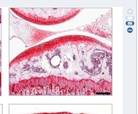
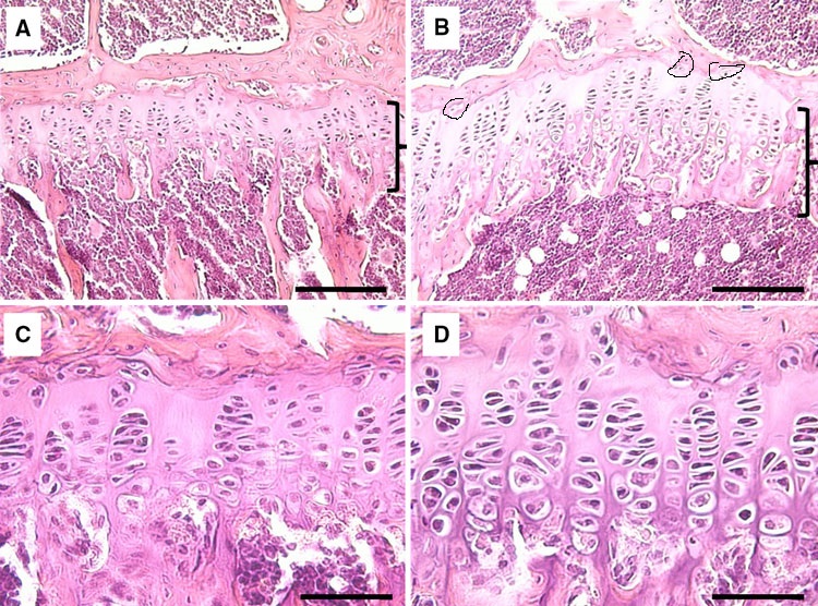
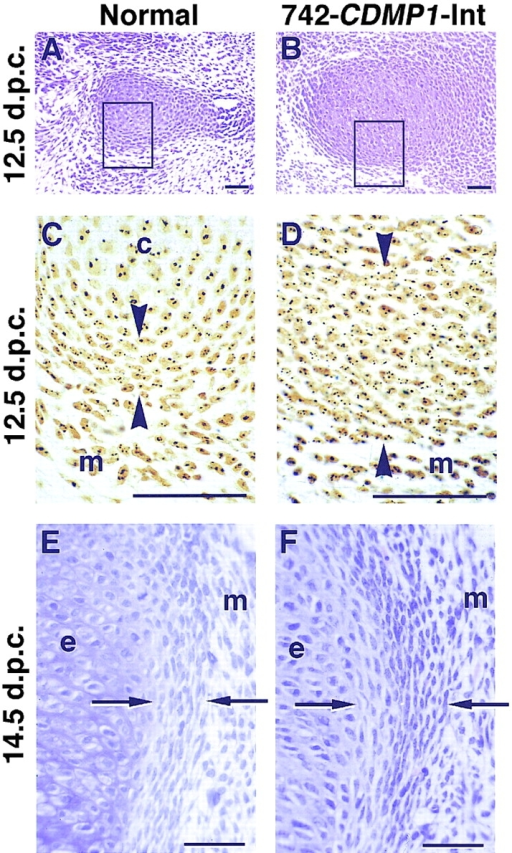
Here’s an image showing what microcracks look like in trabecular bone although to get bone length you’d to cause microcracks in cortical bone:

microcracks

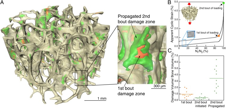
“the presence of a more ductile strut surface forces tissue damage and associated permanent deformations into strut centers.”

“On unloading, struts that accumulate tissue damage in the center (where stresses are lower) will recover more deformation from bending and torsion than struts accumulating damage at surfaces.”

Here’s a paper that shows plastic deformation:

Acute plastic deformation of the ulna in a skeletally mature individual

“Acute plastic deformation of a bone refers to traumatic bending or bowing without a detectable cortical defect{we don’t want bending or bowing we want longitudinal stretching but this shows that plastic deformation is possible}. We present a case that is unusual in that bowing of the ulna occurred in a skeletally mature individual and was associated with injury to the distal radioulnar joint. In this patient, the symptoms were severe enough to warrant an ulnar osteotomy. The patient regained satisfactory function. Acute plastic deformity should be suspected whenerver abnormal curvature of a long bone is noted, even in adults. If the distal radioulnar joint is dislocated, the deformation should be corrected as soon as possible to avoid permanent loss of forearm rotation.”

Couldn’t get the full study but just the abstract is pretty much a breakthrough because it proves that plastic deformation is possible in a skeletally mature individual.  But longitudinal plastic deformation is very difficult.

New study shows LSJL induces Bone Deformation

Bone Deformation is change in the bone shape or structure.  This deformation can be compression of various cavities, stretching of the bone, twisting, and so son.  This is important as bone deformation is one way to increase hydrostatic pressure by decreasing the cavity size.  Hydrostatic pressure is the pressure exerted by a fluid at rest.   Compressing the bone laterally inhibits the fluids ability to move thus increasing hydrostatic pressure.   If there is more fluid within a smaller space than it follows that hydrostatic pressure increases. Hydrostatic pressure has been consistently shown to induce chondrogenic differentiation.  Chondrogenic tissue is the key for longitudinal bone growth as traditionally chondrogenic tissue is capable of interstitial(growth from within) whereas bone is not.  Only interstitial bone growth has been shown traditionally to induce significant longitudinal bone growth but there are potentially other ways to stimulate longitudinal bone growth.

Knee loading inhibits osteoclast lineage in a mouse model of osteoarthritis.

“Osteoarthritis (OA) is a whole joint disorder that involves cartilage degradation and periarticular bone response. Changes of cartilage and subchondral bone are associated with development and activity of osteoclasts from subchondral bone{Since osteoarthritis does affect the subchondral bone that does affect our ability to say that LSJL affects bone deformation in a normal bone but there is no reason why it shouldn’t}. Knee loading promotes bone formation. Knee loading regulates subchondral bone remodeling by suppressing osteoclast development, and prevents degradation of cartilage through crosstalk of bone-cartilage in osteoarthritic mice{This “crosstalk” may stimulate chondral tissue within the bone as well}. Surgery-induced mouse model of OA was used. Two weeks application of daily dynamic knee loading significantly reduced OARSI scores and CC/TAC (calcified cartilage to total articular cartilage), but increased SBP (subchondral bone plate) and B.Ar/T.Ar (trabecular bone area to total tissue area). Bone resorption of osteoclasts from subchondral bone and the differentiation of osteoclasts from bone marrow-derived cells were completely suppressed by knee loading{Knee loading affects the differentiation of bone marrow-derived cells which is the first step in proving that it causes chondrogenic differentiation}. The osteoclast activity was positively correlated with OARSI scores and negatively correlated with SBP and B.Ar/T.Ar. Furthermore, knee loading exerted protective effects by suppressing osteoclastogenesis through Wnt signaling. Overall, osteoclast lineage is the hyper responsiveness of knee loading in osteoarthritic mice. Mechanical stimulation prevents OA-induced cartilage degeneration through crosstalk with subchondral bone. Knee loading might be a new potential therapy for osteoarthritis patients.”

“Daily dynamic knee loading was applied at 1 N, 5 Hz, 5 min/day for 2 weeks”

joint loading on subchondral bone

Compare the OA+ loading to the control bone.  The subchondral bone plate looks much more dynamic.  There are three bone marrow regions rather than two(bone marrow is the blue dots).

You’ll also note that loading+OA increased the ratio of calcified cartilage out of total articular cartilage(but not above statistic significance.  It did not fully restore the thickness of the bone plate.  Alendronate is an anti reobsorption agent.  Given that the ALN and loading group is different we can say that change in subchondral bone shape is likely not related to inhibiting osteoclast activity and is something unique to the loading group.

loading affect on subchondral bone2

Compare the joint capsule region of control and OA+loading group.  The Joint Capsule is the region that’s not inside the bone.  The cells are a lot more spread out.  There’s a dense redness in the control group which is not present in the OA+Loading group

We can see that the loaded group again has distinct characteristics and we can also see the growth plate.  The growth plate of the LSJL group is distinct and there does seem to be signs of cellular migration.  I’ll have to blow it up.

cell migration

Circled is the region of possible cell migration.

LSJL-growth-plates

Similar signs of migration in earlier in LSJL studies(above taken from Lengthening of Mouse Hindlimbs with Joint Loading).

“the expression of Wnt3a was significantly increased by knee loading. However, the protein and mRNA levels of NFATc1, RANKL, TNF-α, and Cathepsin K were significantly suppressed by knee loading”

“Female C57BL/6 mice (~14 weeks of age)”

If you look at this image of bone marrow derived cells extracted from the loaded group and the other groups you can see that the cells are more condensed and condensation is a prerequisite for chondrogenic differentiation,

Loaded cells are more condensed

This is an image of what mesenchymal condensation looks like:

mesenchymal condensation