Author Archives: Tyler

New paper that provides insight on baseball players longer arms

It’s been stated empirically that baseball players have longer throwing arms than non-throwing arms.

The following study suggests that baseball actually hinders growth in the growth plate in adolescents.  Now this could mean that either what the scientist interpret as growth hinderence is actually growth stimulation or that the baseball players having longer arms is not related to the growth plate at all and is longitudinal bone growth by other means which would be very promising for adult longitudinal bone growth.

Cartilage degeneration at symptomatic persistent olecranon physis in adolescent baseball players.

“Elbow overuse injuries are common in adolescent baseball players, but symptomatic persistent olecranon physis is rare, and its pathogenesis remains unclear. Purpose. To examine the histopathological and imaging findings of advanced persistent olecranon physis. Methods. The olecranon physes of 2 baseball pitchers, aged 14 and 15 years,{only two players were analyzed so perhaps the damage to the growth plate was an outlier.  There’s also selection bias where they likely sought out those who had injury.} were examined by preoperative magnetic resonance imaging (MRI), and surgical specimens were examined histologically. Results. T2-weighted MRI revealed alterations in the intrachondral signal intensity possibly related to collagen degeneration and increased free water content. Histological findings of specimens stained with hematoxylin-eosin showed complete disorganization of the cartilage structure, hypocellularity, chondrocyte cluster formation, and moderately reduced staining. All these findings are hallmarks of osteoarthritis and are suggestive of cartilage degeneration. Conclusion. Growth plate degeneration was evident in advanced cases of symptomatic persistent olecranon physis. These findings contribute to understanding the pathogenesis of this disease.”

I wasn’t able to copy and paste the image but if you at figure 2 you can examine the following:

“Figure 2: (a) Growth plate remnant with a sclerotic margin. Moderate reduction in hematoxylin-eosin staining and disorientation of chondron columns in the olecranon physis. (b) Magnified view showing a decreased number of chondrocytes in the lesion. (c) Chondrocyte cluster formation in the lesion. (d) Proliferating cell nuclear antigen-positive cells in a chondrocyte cluster. Bars: 200 μm (a), 100 μm (b), 50 μm (c), and 20 μm (d).”

The key factor is whether these athletes throwing arm had stunted growth.  Unfortunately, I couldn’t find any long term studies on this.  And I couldn’t find any studies that show whether moderate pitching without overuse increases growth plate disorganization.

Does anyone know of anyone with elbow overuse due to pitching and whether it stunted growth in their dominant arm?

Compressive Loading and the Growth Plate

A lot of interesting stuff here:

In vivo dynamic loading reduces bone growth without histomorphometric changes of the growth plate

“This in vivo study aimed at investigating the effects of dynamic compression on the growth plate. Rats (28 days old) were divided into three dynamically loaded groups, compared with two groups (control, sham). A device was implanted on the 6th and 8th caudal vertebrae for 15 days. Controls (n = 4) did not undergo surgery. Shams (n = 4) were operated but not loaded. Dynamic groups had sinusoidal compression with a mean value of 0.2 MPa: 1.0 Hz and ±0.06 MPa (group a, n = 4); 0.1 Hz and ±0.2 MPa (group b, n = 4); 1.0 Hz and ±0.14 MPa (group c, n = 3). Growth rates (µm/day) of dynamic groups (a) and (b) were lower than shams. Growth plate heights, hypertrophic cell heights and proliferative cell counts per column did not change in dynamic (a) and (b) groups compared with shams. Rats from dynamic group (c) had repeated inflammations damaging tissues; consequently, their analysis was unachievable. Increasing magnitude or frequency leads to growth reduction without histomorphometric changes. However, the combined augmentation of magnitude and frequency alter drastically growth plate integrity. Appropriate loading parameters could be leveraged for developing novel growth modulation implants to treat skeletal deformities.”

“Three main factors contributing to bone growth are cellular enlargement in the hypertrophic zone (40–50%), matrix synthesis (35–45%) and cell duplication in the proliferative zone (10%).”

“different frequencies and magnitudes of the dynamic loading applied for 10 min per day in rats forelimbs, and found that growth was reduced proportionally to load magnitude and not the average load.”<-Magnitude is the peak load.  So the peak load you obtain with say LSJL has the largest impact on the stimulus.  Note that the loads advocated here are compressive loads rather than the more tensile load of LSJL.

“among dynamic parameters, we suggest that frequency has more impact on growth compared to magnitude.”<-It’s a lot harder to modulate frequency than magnitude with LSJL.

Dynamic Compressive Loading decreased growth in the Cd7 vertebrae.  Produced a minor growth rate reduction on Cd5.  However, it increased growth rate on CD9 so the various disc growth plates may respond to mechanical stimuli differently.
growthplates  A and B are control group.  C and D are the compressed group.  You can definitely see the compression of cells.

How does a medieval torture rack affect the body?

Since the medieval rack is cited so often as a mechanism to get taller it would be interesting to see how it works.

The torture or stretch arthritis syndrome (A modern counterpart of the medieval ‘manacles’ and ‘rack’)

“A characteristic symmetrical bilateral polyarthritis syndrome is described, affecting the compartments of the wrist joints in a characteristic sequence. The pisiform-triquetral joint gives first, exposing the inferior radioulnar joint so that pisiform pain and tenderness are followed by painful supination{Rotation of the forearm and hand are rotated forward or up}. The proximal radiocarpal joint slackens next, followed by the mid-carpal joints. The lunates subluxate{dislocate} as their posterior attachments stretch. Synovial oedema{swelling} may produce additional median and ulnar carpal tunnel syndromes. The best radiologic sign of this distraction is anterior subluxation of the lunates. Nocturnal arthralgia becomes severe; grip and the ability to write are lost and stretching now shows as a traumatic arthritis in the clavicular joints. The patient is incapacitated and therefore progression halts. A history of excessive straining and lifting is obtained eg with a wheel-barrow, iron pots or strenuous rowing{so if the rack worked to increase height you could just row instead for some of the effects?}. All serologic tests for rheumatoid disease are negative. Serum uric acid levels and blood sedimentation rates remain normal. There are clear analogies with old descriptions of the effects of torture by stretching from manacles or gauntlets or by the rack.”

IF the rack were to increase height it would have to occur before the pisiform-triquetral joint “gives”.

How do growth plates regenerate?

Growth plate regenaration has been covered before here and here.  Ultimately, this provides more evidence that the presence of germ immature cells are the key limiting factor in creating new growth plates.  Thus our primary focus should be on developing mechanical and supplemental methods that alter the structure of cells to be more like these growth plate chondrocyte precursor cells.

Regeneration of the Growth Plate

Pdf-growth plate regeneration

“The occurrence of growth plate regeneration has been doubted. However, in 5 different series of experiments reported between 1950 and 1986 regeneration of injured parts of growth plates in long bones of rabbits and pigs could be demonstrated. The 1st series implied partial X-ray injury of growth plates in rabbits aged 3–6 weeks.The 2nd series implied autotransplantation of the head of the fibula in rabbits aged 10–21 days. The 3rd, 4th and 5th series implied transplantation of autologous fat grafts into provoked defects of growth plates in rabbits and pigs. The findings show that regeneration of a growth plate occurs when a part of it is injured in such a manner that a bone bridge is not formed between the epiphysis and the metaphysis. Regeneration of a plate is much faster in relation to the growth in length of the bone in the rabbit than in the pig. The 1st and 2nd series suggest that regeneration takes place by interstitial proliferation of cells from the germinal layer of the uninjured parts of the plate. Signs of partial regeneration of growth plates have been seen in radiographs after operation for partial closure of growth plates in children. ”

Growth plate regeneration refers to the healing of damaged growth plates but if we learn the mechanisms by which growth plates are repaired we could potentially use those mechanisms to create new growth plates.

“Microscopic examination showed that injured portions of cartilage were pushed aside by the unirradiated carti­lage growing into the irradiated area transversely in rela­tion to the axis of the bone. In several experiments defor­mities and radiolucent foci appeared which showed similarity to those seen in dyschondroplasia{ectopic masses of cartilage}. When one  half of the distal  growth  plate  of the radius  had  been  irradiated then lagging  behind of  ossitication could  be demonstrated after 9 days.”  Part of the injured cartilage was left behind in the metaphysis as the bone was growing.  Another part adhered to the bony plate of epiphysis.  In between, there was what appeared to be normal growing regenerating cartilage.

2nd series. Another set of experiments describes growth plate regeneration via interstitial growth.  This type of growth would be very promising in terms of inducing adult height growth.  No perichondrium and no zone of ranvier ossification groove was present.  So there was no pool of typical growth plate progenitor cells.

“The regeneration seemed to take place from the viable parts of the germinal cell layer.”<-Bone and cartilage tends to come from the mesodermal layer.  But mesodermal cells implies the type of immature cells like embryonic stem cells.  But perhaps the right mechanical forces can still induce more adult mesenchymal stem cells into becoming more immature cells and into pre-chondrocyte growth plate cells.

“lt is generally accepted that interstitial latitudinal growth occurs in the very young animal, when the germinal zone cells of the growth plate are adjacent to a largely cartilaginous epiphysis as the tissues are plastic.”<-Plastic refers to irreversible deformation rather than elastic deformation where the object returns to normal.  You can get plastic deformation in adult bones it’s just much harder.

Later the study states that interstitial growth can still occur even against a more rigid epiphysis as long as the germ cells are present.

Occurding to Figure 16 area C, the germinal cells look much like resting zone cells.  We’ll have to study how much LSJL and other mechanical stimuli can induce mesenchymal stem cells to be more like mesodermal germinal cells.

A Mechanical Jack-like Mechanism Drives Spontaneous Fracture Healing in Neonatal Mice

“To study the mechanism underlying spontaneous regeneration of fractured bones, we left humeral fractures induced in newborn mice unstabilized, and rapid realignment of initially angulated bones was seen. This realignment was surprisingly not mediated by bone remodeling, but instead involved substantial movement of the two fragments prior to callus ossification. Analysis of gene expression profiles, cell proliferation, and bone growth revealed the formation of a functional, bidirectional growth plate at the concave side of the fracture. This growth plate acts like a mechanical jack, generating opposing forces that straighten the two fragments. Finally, we show that muscle force is important in this process, as blocking muscle contraction disrupts growth plate formation, leading to premature callus ossification and failed reduction.”

“The ossification of soft callus bares similarities to the process of endochondral ossification during skeletogenesis”

“another type of growth plate known as synchondrosis{these do fuse though} is located between the bones of the skull base. The synchondroses exhibit a remarkably organized structure, as each consists of two mirror-image growth plates facing opposite directions. These growth plates are fed by a shared resting zone located between them. The formation of double layers of prehypertrophic and hypertrophic chondrocytes drives growth in opposite directions, leading to expansion of the skull volume”

“the growth plate must also be able to generate considerable forces. Indeed, studies in various animal models as well as in humans suggest that the different growth plates can generate forces that range from the equivalent of 40% to 200% of body weight”

“an active bidirectional growth plate is formed at the concave side of the callus to mediate bone growth. This finding strongly supports our hypothesis that bone growth by the bidirectional growth plate generates the force required for the movement of the two fracture fragments during reduction, similar to a “mechanical jack” mechanism.”

“Expression analysis of SRY box containing gene 9 (Sox9), Col2a1, and Col10a1 revealed that the absence of muscle contraction led to symmetric chondrogenesis and loss of the bidirectional growth plate organization”

“in the absence of muscle contraction the callus fails to organize and act as a growth plate and undergoes early ossification”

“all of the characteristics of an active growth plate exist in the callus at the concave side of the fracture site, including gene expression profiles, cell proliferation, and bone growth. We therefore argue that the growth plate in the callus serves not only for intermediate stabilization, but also to actively promote bone reduction. However, unlike the epiphyseal growth plates and similar to the synchondroses that mediate cranial base expansion, the bidirectional growth plate at the fracture site drives growth in opposite directions. This generates force that moves the fragments toward straightening in a mechanical jack-like effect.”

Game Changing Breakthrough OTC Height Supplement-Meclozine

Meclozine is available without a prescription: Meclizine Chewable Tablets – 25mg – Model 85207 – Btl of 100.  Open growth plates only though but it looks like it could be very effective as it is similar to CNP which has a pretty big impact on height.

Even if you don’t understand anything below.  Please spread the word about this study.  It looks like a quite promising OTC height increase supplement.

Unfortunately, there have been no studies on Meclizine and human height and as shown by the study below there is cell toxicity to Meclizine.  Since Meclizine is a well established supplement, anyone who is currently undergoing longitudinal growth and wants to grow taller should take Meclizine at dosages recommended on the bottle and following all other directions about directed use.

Meclozine Facilitates Proliferation and Differentiation of Chondrocytes by Attenuating Abnormally Activated FGFR3 Signaling in Achondroplasia.

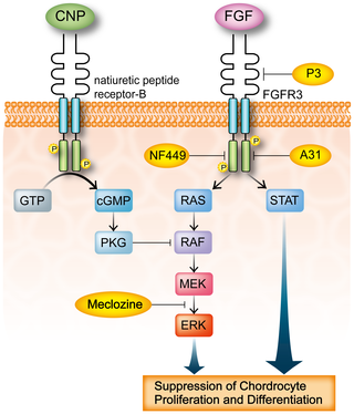
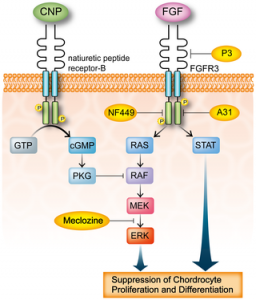
“Achondroplasia (ACH) is one of the most common skeletal dysplasias with short stature caused by gain-of-function mutations in FGFR3 encoding the fibroblast growth factor receptor 3. We used the drug repositioning strategy to identify an FDA-approved drug that suppresses abnormally activated FGFR3 signaling in ACH. We found that meclozine, an anti-histamine drug that has long been used for motion sickness, facilitates chondrocyte proliferation and mitigates loss of extracellular matrix in FGF2-treated rat chondrosarcoma (RCS) cells. Meclozine also ameliorated abnormally suppressed proliferation of human chondrosarcoma (HCS-2/8) cells that were infected with lentivirus expressing constitutively active mutants of FGFR3-K650E causing thanatophoric dysplasia, FGFR3-K650M causing SADDAN, and FGFR3-G380R causing ACH. Similarly, meclozine alleviated abnormally suppressed differentiation of ATDC5 chondrogenic cells expressing FGFR3-K650E and -G380R in micromass culture. We also confirmed that meclozine alleviates FGF2-mediated longitudinal growth inhibition of embryonic tibia in bone explant culture. Interestingly, meclozine enhanced growth of embryonic tibia in explant culture even in the absence of FGF2 treatment!!!!!!!. Analyses of intracellular FGFR3 signaling disclosed that meclozine downregulates phosphorylation of ERK but not of MEK in FGF2-treated RCS cells. Similarly, meclozine enhanced proliferation of RCS cells expressing constitutively active mutants of MEK and RAF but not of ERK, which suggests that meclozine downregulates the FGFR3 signaling by possibly attenuating ERK phosphorylation{Since everything . We used the C-natriuretic peptide (CNP) as a potent inhibitor of the FGFR3 signaling throughout our experiments, and found that meclozine was as efficient as CNP in attenuating the abnormal FGFR3 signaling!!!!!!!!{And CNP is a huge height increase disease}. ”

Loss of function of FGFR3 leads to tall stature and Meclozine decreases FGFR3 function even in normal cells.

“CNP has a short half-life and continuous intravenous infusion is required for in vivo experiments. The CNP analog with an extended half-life, BMN 111, has recently been developed and significant recovery of bone growth was demonstrated in ACH mice by subcutaneous administration of BMN 111″

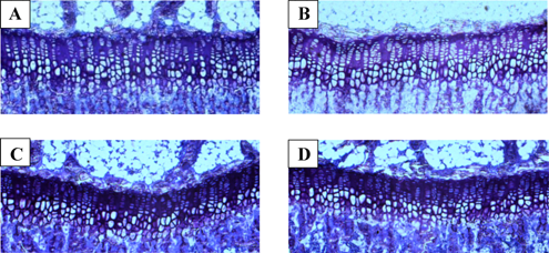
” 0, 1, 2, 5, 10, and 20 µM of meclozine exhibited dose-dependent increases in RCS[Rat Chondrosarcoma cells] proliferation. We did not observe dose-dependency at 50 µM, which was likely due to cell toxicity. We also confirmed that 10 and 20 µM of meclozine increased the number of RCS cells”

“treating RCS cells with FGF2 for four hours induced expressions of matrix metalloproteinase 10 (Mmp10), Mmp13, and a disintegrin-like and metalloproteinase with thrombospondin type 1 motif 1 (Adamts1) transcripts{LSJL upregulates Admts1 and MMP13 but still increases height, LSJL in conjunction with Meclozine could increase height more}. We found that meclozine and CNP significantly suppressed expressions of these matrix metalloproteinases. We also quantified expressions of Col2a1 and Acan transcripts, but FGF2 treatment for 72 hours did not reduce the expression levels of these genes in RCS cells”

meclozine longitudinal bone growth
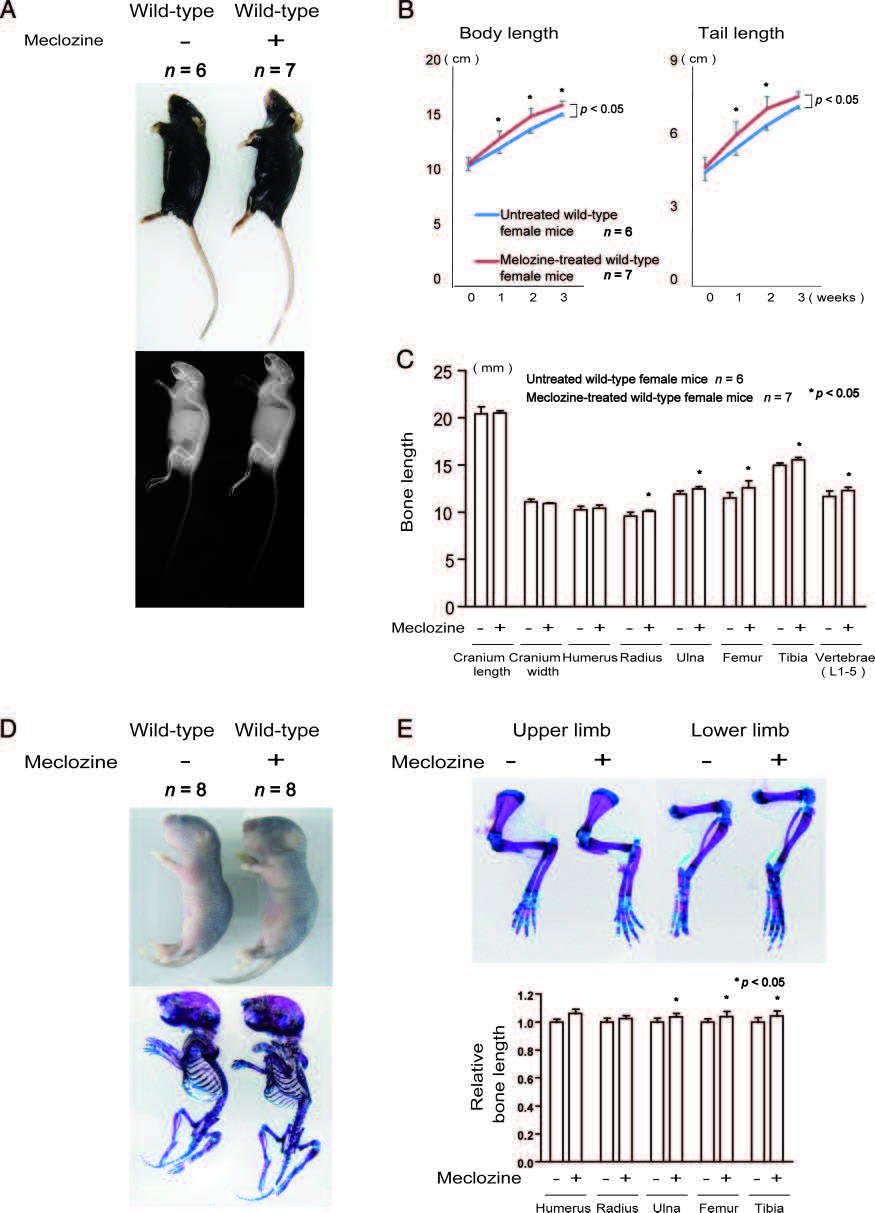
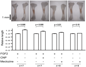
The rightmost figure(the one with n=6) is the Meclozine solo group.  Eyeballing it, it looks like it could be about 5% increase in longitudinal growth.  Note that a 5% increase on 5’9″ is 6’0″.

FGFR3 signalingFGFR3 signaling in chondrocytes.  LSJL does increase ERK phosphorylation.  Maybe that is a side effect of the boost of LSJL on longitudinal growth and not a cause of longitudinal growth.  And that both Meclozine and LSJL would have additive effects.

” a synthetic compound A31 is an inhibitor of the FGFR3 tyrosine kinase by in silico analysis. They demonstrated that A31 suppresses constitutive phosphorylation of FGFR3 and restores the size of embryonic femurs of Fgfr3Y367C/+ mice in organ culture. In addition, A31 potentiates chondrocyte differentiation in the Fgfr3Y367C/+ growth plate.  P3 has a high and specific binding affinity for the extracellular domain of FGFR3. They showed that P3 promotes proliferation and chondrogenic differentiation of cultured ATDC5 cells, alleviates the bone growth retardation in bone rudiments from TD mice (Fgfr3Neo-K644E/+ mice), and finally reversed the neonatal lethality of TD mice”

“These novel FGFR3 tyrosine kinase inhibitors, however, may inhibit tyrosine kinases other than FGFR3 and may exert unexpected toxic effects in humans. Meclozine may also inhibit unpredicted tyrosine kinase pathways, but we can predict that there will be no overt adverse effect, because meclozine has been safely used for more than 50 years.”

Here’s a study that will provide some insight into FGFR3 and why it inhibits growth in some cases but not in others:

The paradox of FGFR3 signaling in skeletal dysplasia: why chondrocytes growth arrest while other cells over proliferate.
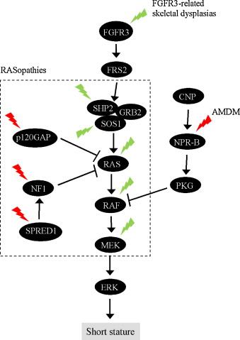
Somatic mutations in receptor tyrosine kinase FGFR3 cause excessive cell proliferation, leading to cancer or skin overgrowth. Remarkably, the same mutations inhibit chondrocyte proliferation and differentiation in developing bones, resulting in skeletal dysplasias, such as hypochondroplasia, achondroplasia, SADDAN and thanatophoric dysplasia{So maybe the longitudinal growth induced by LSJL is not produced by chondrocytes but rather by the stem cells which would be promising evidence for post LSJL-inducable height growth}. A similar phenotype is observed in Noonan syndrome, Leopard syndrome, hereditary gingival fibromatosis, neurofibromatosis type 1, Costello syndrome, Legius syndrome and cardiofaciocutaneous syndrome. Collectively termed RASopathies, the latter syndromes are caused by germline mutations in components of the RAS/ERK MAP kinase signaling pathway. This article considers the evidence suggesting that FGFR3 activation in chondrocytes mimics the activation of major oncogenes signaling via the ERK pathway. Subsequent inhibition of chondrocyte proliferation in FGFR3-related skeletal dysplasias and RASopathies is proposed to result from activation of defense mechanisms that originally evolved to safeguard mammalian organisms against cancer.”

“FGFR3 [inhibits] chondrocyte proliferation. FGFR3/ERK signaling triggers disintegration of the cyclin D3-cdk6 complex in the G1 phase of the cell cycle, followed by increased association of p21WAF1 and p27Kip1 cell cycle inhibitors (CKI) with cyclin-cdk2 and cyclin-cdk4 complexes, leading to inhibition of their kinase activities. Upon FGFR3 activation, CKIs accumulate at the protein level, due to the interaction with transcriptionally induced cyclin D1. cyclin D1 upregulation also mediates the pro-mitogenic effects of FGFR/ERK signaling. Duration, magnitude and timing of cyclin D1 and p21WAF1 induction in the G1 phase of a cell cycle determines the nature of the response to an ERK signal. A strong but transient ERK activation in the early G1 induces intermittent p21WAF1 accumulation and stable cyclin D1 expression, leading to cell proliferation. In contrast, robust and persistent ERK activation leads to stable p21WAF1 accumulation and growth inhibition despite the concomitant induction of cyclin D1.

In chondrocytes, unlike most other cell types, FGFR3 activation elicits highly prolonged ERK activation lasting for up to 24 h. This phenotype is likely to stem from the maintenance of ERK pathway activation within the protein complexes interacting directly with FGFR3. ”

So FGFR3 inhibits growth in chondrocytes but not other cell types is because FGFR3 activates ERK for too long which leads to growth inhibition due to excess levels of p21WAF1.

Another FGFR3 pathway
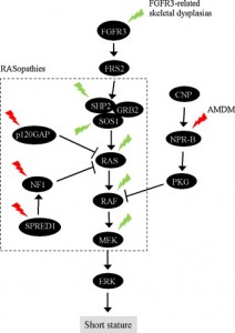
“In chondrocytes, the premature senescence caused by FGFR3 activation does not involve the p53 pathway but appears CKI-dependent as induction of several CKIs (p21WAF1, p27Kip1, p16INK4a, p18INK4c, and p19INK4d) accompanies FGFR3-mediated inhibition of chondrocyte proliferation in vitro and in vivo”

NEW:

Meclozine Promotes Longitudinal Skeletal Growth in Transgenic Mice with Achondroplasia Carrying a Gain-of-Function Mutation in the FGFR3 Gene.

“Achondroplasia (ACH) is one of the most common skeletal dysplasias causing short stature owing to a gain-of-function mutation in the FGFR3 gene, which encodes the fibroblast growth factor receptor 3. We found that meclozine, an over-the-counter drug for motion sickness, inhibited elevated FGFR3 signaling in chondrocytic cells. To examine the feasibility of meclozine administration in clinical settings, we investigated the effects of meclozine on ACH model mice carrying the heterozygous Fgfr3ach transgene. We quantified the effect of meclozine in bone explant cultures employing limb rudiments isolated from developing embryonic tibiae from Fgfr3ach mice. We found that meclozine significantly increased the full-length and cartilaginous primordia of embryonic tibiae isolated from Fgfr3ach mice. We next analyzed the skeletal phenotypes of growing Fgfr3ach mice and wild-type mice with or without meclozine treatment. In Fgfr3ach mice, meclozine significantly increased the body length after two weeks of administration. At skeletal maturity, the bone lengths, including the cranium, radius, ulna, femur, tibia, and vertebrae were significantly longer in meclozine-treated Fgfr3ach mice than in untreated Fgfr3ach mice. Interestingly, meclozine also increased bone growth in wild-type mice. The plasma concentration of meclozine during treatment was within the range that has been used in clinical settings for motion sickness. Increased longitudinal bone growth in Fgfr3ach mice by oral administration of meclozine in a growth period indicates potential clinical feasibility of meclozine for the improvement of short stature in ACH.”

“A CNP analog with an extended half-life, BMN-111, has recently been developed, and significant bone growth recovery was demonstrated in amouse model of ACH by subcutaneous administration of BMN-111”

“Meclozine was administrated to 2-week-old wild-type mice for 3 weeks. As wild-type mice were weaned at 2 weeks after birth, we started meclozine treatment 1 week earlier than that for Fgfr3ach mice.  The body length of meclozinetreated mice was significantly longer than that of untreated mice after 1 week

“the plasma concentrations of meclozine used in the current study (0.2 or 0.4 g of meclozine per kilogram food).”

“Meclozine, an OTC H1 inhibitor, has been safely used for motion sickness for more than 50 years, and its optimal dose and adverse effects have already been established”

“in patients with ACH [given treatment with meclozine], the patients could be expected to increase 6.7 to 7.1 cm in height, based on the average height of adults with ACH.”


melcozone
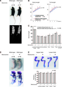
<-Treatment of mice with meclozine without FGFR3 deficiencies.  The meclozone treated mice are noticeably taller and lengthier.

“A-C, Wild-type mice were treated with meclozine 2 weeks after birth for 3 weeks. A, Visual images and soft X-ray images of wild-type female mice with or without meclozine. Meclozine-treated mice were larger than the untreated mice. B, Body and tail lengths of meclozinetreated
wild-type female mice were significantly longer than those of untreated wild-type female mice. Statistical significance analyzed by two-way ANOVA is shown on the right side of each graph. *P  .05 by Fisher’s LSD test for each pair. C, The lengths of the radius, ulna, femur, tibia, and vertebrae on the soft X-ray films were significantly increased by meclozine treatment by unpaired t test. D and E, Pregnant mice were treated with meclozine from embryonic day 14. D, Visual images and skeletons stained with Alizarin red and Alcian blue of wild-type mice at postnatal day 5, with or without meclozine. Meclozine-treated offspring were larger than untreated offspring. E, The lengths of the ulna, femur, and tibia measured using stained skeletons were significantly increased after meclozine treatment, as assessed by unpaired t test.”

Very promising evidence that LSJL induced longitudinal bone growth in my finger

The coloration of regions of an area in an xray reflect their density.  If a region of bone is grayer than normal that bone is less dense.  If a bone is less dense that could mean that it is a region of new growth or that the bone is naturally less dense.  But even though some regions of less density are natural, if these regions of decreased density are expanded it is very possible that this could be indicative of increased growth.  If this expansion occurs in the longitudinal direction then it is indicative of longitudinal bone growth.  If you compare my right and left lateral finger xrays below you can see that my right bone has much greater expansion of these gray regions than the left and the regions extend into the longitudinal direction thus being indicative of longitudinal bone growth.

I need your help to confirm or deny this theory.

I statement I found on white versus black regions in x=rays: “On x-ray, bone and dense materials such as barium are white. Black would be air (lungs or air in the GI tract). Other tissues are in shades of gray. You can see the outline or shadow of the kidneys or certain muscles, for example. But you cannot see these things in detail. X-ray does not show the difference between normal vs. “dead” tissue. A CT scan, or even better, a MRI would be better suited for that type of diagnosis. ”

The darker regions of the bones are more likely to be less dense or possibly new grown bone.  Some bones are naturally less dense than others.  So we have to compare the bones also to standard bones.  If there are more gray areas than normal then it’s likely that there was new bone growth.

Here’s one lateral index finger view:
right lateral index finger

Which my finger looks more like.  Here’s one that my left finger looks more like(this is also a right index finger):
left proximal index finger

Have to compare color intensity of areas of right versus left bone.

Here’s a finger with some gray areas.  There is a large gray region on the tip of the proximal finger and some on the bottom part of the distal region of the medial index finger.  Note this is a fractured finger so we’d expect there to be some anabolic growth.

xray-mallet

So, it’s normal to have those regions of darker bone.

There are other potential reasons why one part of the bone can be much darker than the other but one strong possibility is that it is new LSJL induced bone growth.  Since LSJL involves lateral loading it makes sense that much of the growth can be better observed than a lateral angle(the differences in coloration can not be seen as well from an overhead view).  From the overhead view the right index finger looks to be whiter than the left index finger infact.  Click on the images below to enlarge them.

LSJL-right hand illustrated

LSJL left hand illustratedEach of the circled points A-D represents a region of bone that is much lighter than the other.  On the left side these regions are present but the are much lighter.  At point A it seems to blend better with the whiter region of the bone in contrast to the right side where it is more a bulbous head.  The bone at B looks totally different.  C and D are similar but the right(LSJL loaded side the darker region of the bone is much bigger).

Percentage growth based on darker area versus lighter area:

Proximal Head(Tip of dark region to tip of light): 23.4 pixels

(bottom of proximal index finger bone to top of light region): 170.4 pixels

13.7% growth.

However, I’d guess that my left bones are about 1% longer than my right.  If my right finger did grow longer than 10% my right proximal finger would measure longer than my left which is not the case.  You can see from the above images of other images that the normal finger has these darker regions, those regions are just expanded above normal.  So maybe the the proximal finger bone didn’t grow the entire gray region but the gray region just expanded in response to the stimulus.  In fact a study has shown that the finger bones naturally grow into adulthood.  So these darker regions of bone could be a sign of adult finger growth.  The same study stated that metacarpals tend to decrease in length so the fact that my right metacarpal grew longer is a good sign that this growth could extend to other bones.

What’s a bit unfortunate is that the growth plates are on the proximal(bottom) end of the finger bones and the gray regions are on the distal(top) ends of the finger bones.  However, the distal regions of my right finger bones do seem to be a bit more gray than the left bones so it is possible that the growth plate region was stimulated by LSJL.

Below is some more information about x-rays.

Here’s something I found describing xrays:

” The bone along the joint is usually whiter (called “sclerosis”) and may have little points of bone growing out (called “osteophytes”). There may be holes in the bone ( called “cysts”) and the bones may be starting to slide out of alignment (called “subluxation”).”<-None of these images quite explains the growth in LSJL.

Here’s a comment I found from Yahoo! Answers:

“Bones always appear as white images in regular xrays. However they arent really white because the film is clear, the plastic on the view box is white. But on xray film it is coated with a metalic layer. The xrays make the metalic layer stick to the film. So the black part of the film is where all the xrays reach the film. If the area on the xray is gray, then that means that some of the xrays are getting through implying that the structure is more dense than the black part. Soft tissue appears gray. The bone is a very dense structure allowing minimal to no xrays getting through. The film is then put through the processor and the film runs through chemicals that make the picture “stay” on the film. It is then rinsed and dried. Since the xrays make the metalic layer stick, none stick to the area of the bone because no radiation got through and the layer was washed off during processing. This is why bones appear white, they are the densest structure.”

Here’s another response from Yahoo! Answers regarding why one part of the bone is darker than the other:

“When a bone X-ray is taken, radiation is momentarily irradiated on the area being examined. X-rays pass through the body to produce an image on film. Structures like bone that are dense and have a high atomic number absorb a lot of X-rays, so less X-rays reach the film and the bone appears white on the film. Muscle, fat, tumour, and fluid absorb less X-ray than bone, and appear darker than bone on the film. Air distributed in various pockets within the body (usually due to an abnormality) has a low atomic number and density, and consequently appears black, because most X-rays pass through without being absorbed. You would be advised to consult the doctor or radiologist dealing with this matter and enquire as to the suspected cause of the description that you describe. You may find that further tests/examinations will result from the X-Ray result which will determine or assist the diagnosis.
The information provided here should not be used during any medical emergency or for the diagnosis or treatment of any medical condition. A licensed physician should be consulted for diagnosis and treatment of any and all medical conditions. “<-I don’t think air pockets could explain the larger gray area on the right side bone.

On this page is an image of tissue types in regard to whiteness: http://radiologymasterclass.co.uk/tutorials/physics/x-ray_physics_densities.html#top_first_img.  The coloration is consistent with soft tissue.

Here’s an image of a periosteal chondroma:

periosteal chondroma“In this x-ray of the wrist, the saucer-like hollow in the radius bone is a classic indication of a periosteal chondroma. “<-That’s not really consistant with what’s going on in my bone.

As for my ganglion cysts according to here, they are usually caused by myxoid(mucous) degeneration of collagen and usually lined with flattened mesothelium.

I can’t really find any adequate explanation as to the brighter regions of my finger.

Update by Michael:

From what I remember from being in some medical derivative courses back in my university days, I do remember the idea of X-rays penetrating into human tissue. White light means that the X-rays did not completely go through the tissue, while the black color is an indicator that the X-rays went through less dense tissue, like fat and tendons. 

I don’t have the enlarged X-rays pictures with me anymore but I don’t remember the left or right index fingers having any major differences in color. I think the important thing to remember is that we are looking for bone length differences, not bone density variations.

Now it is theoretically accurate to say that if we can show that LSJl caused bone density changes, causing the X-ray to cause a darker band on a metatarsal bone then LSJL might lead to bone longitudinal growth. However, on a much more practical level, even if the BMD is lowered, that doesn’t seem to translate to bone length increases, even after half a decade of doing joint loading on a consistent basis. I don’t think that it is worth trying to validate the LSJL theory based on a slight difference in color/density from comparing the left and right X-rays of the index fingers. That is too much of a stretch even for me.

As for the fingers growing longer, that actually makes a lot of sense. The human nose gets longer over time, the human ears droop lower and lower, and even the human mandibular joint area grows longitudinally if you have a pituitary problem.

For the old time regular readers, remember my study on why deer antlers can fall off, and regrow back in length every season? It is because of the occurrence of a laceration/osteonomy at the beginning, causing an open wound. A blastema develops in that region. That blastema eventually grows into a pseudo “growth plate”. The top part of the horn is where the mesenchyme is, and it is covered by a layer of perichondrium, NOT periosteum. That means that the top roof surface that is encapsulating the “rest zone’ type cells in the mesenchyme is flexible. It is only being pushed down by air and gravity, not an entire body, like the grow plates in the tibia of an adult human, which can be over 200 lbs of weight. Similarly, the human fingers have the same type of structure, a human anatomical peninsula, like the human tibia-knee area, which is more like an isthmus.

What I am trying to say is that fingers is similar to the horns on the deer. I have already shown a few times that some stem cell powder can regrow fingers. It is not that a big of a stretch to then conclude that human fingers, like noses, and ears, and pregnant female’s feet can grow throughout one’s life.