Author Archives: Tyler

Huge Breakthrough study-hypertophic chondrocytes transdifferentiate into osteoblasts

This is a study that could have massive ramifications on height increase but the the exact mechanism is unknown.  But that cells that have the genetic material of growth plate cells are retained in adult bone is huge news.

Hypertrophic chondrocytes can become osteoblasts and osteocytes in endochondral bone formation.

hypertrophic chondrocytes<-full study

“Chondrocytes and osteoblasts are considered independent lineages derived from a common osteochondroprogenitor. In endochondral bone formation, chondrocytes undergo a series of differentiation steps to form the growth plate, and it generally is accepted that death is the ultimate fate of terminally differentiated hypertrophic chondrocytes (HCs). Osteoblasts, accompanying vascular invasion, lay down endochondral bone to replace cartilage.  [Can] HC become an osteoblast and contribute to the full osteogenic lineage? Here we use a cell-specific tamoxifen-inducible genetic recombination approach to track the fate of murine HCs and show that they can survive the cartilage-to-bone transition and become osteogenic cells in fetal and postnatal endochondral bones and persist into adulthood. This discovery of a chondrocyte-to-osteoblast lineage continuum revises concepts of the ontogeny of osteoblasts, with implications for the control of bone homeostasis and the interpretation of the underlying pathological bases of bone disorders.”

This is huge because it means that the transdifferentiated osteoblasts retain some of the chondrocytic genetic material which means they could possibly dedifferentiate back into chondrocytes!

“The expression of preosteoblastic markers in LHs before the formation of the POC raises the possibility that these cells may transition to an osteoblastic fate”

“HC-Derived Cells Are Present in Fetal, Neonatal, and Adult Bone.”

“The HC-derived cells, morphologically resembling osteoblasts, were found close to the chondro-osseous junction, on the surface of trabeculae, and in the endosteum”<-that means they are in a good position to be involved in neo growth plate formation.

“HC[hypertrophic chondrocytes] Derivatives Transit to the Primary Spongiosa and Become Col1a1-Expressing Cells.”<-the majority of HC derivatives become osteoblasts.

“HC-to-bone transition occurs during postnatal bone growth and that HC-derived cells may be long-lived within the mature bone”

Here’s an image of the HC lineage:
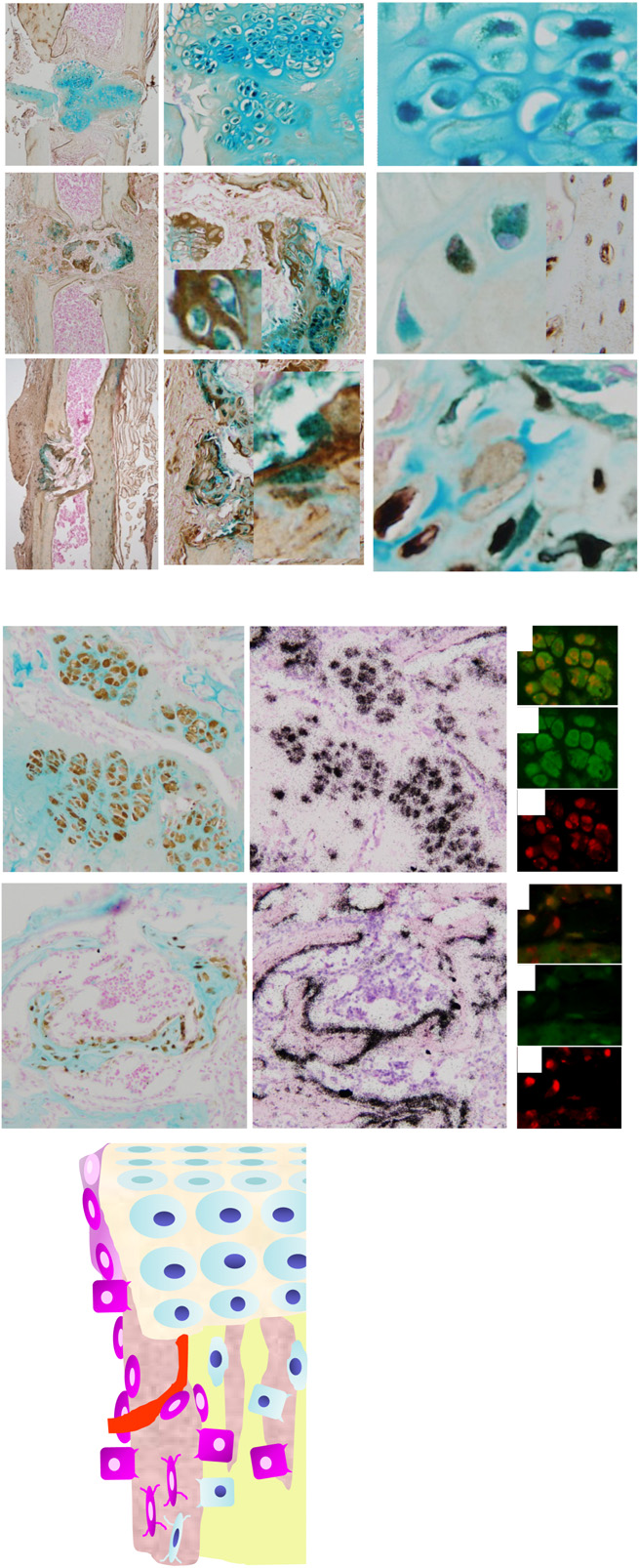
hypertrophic chondrocyte lineage

“The reversion of HCs to a prehypertrophic- like state in response to endoplasmic reticulum (ER) stress suggests that hypertrophy is not an irreversible state in vivo”<-Could we revert HC-derived osteoblasts back to hypertrophic chondrocytes back to pre-hypertrophic cells to reform growth plates.

(Michael: That actually makes a lot of sense if one thought about it. It can’t be all dead inorganic bone ECM matter where the physeal cartilage turns into bones (aka Primary Spongiosa). How would bone cells even be able to travel to that middle-region later on if the chondrocytes completely died out and the lucanae was covered by calcium minerals. For the smooth gradual transition from the hypertrophic layer to the vascularization/calcification layer to the mineralization layers to work out, there would be some minority of chondrocytes which would change identities. If all the chondrocytes died out and the layer of the lucanae was completely mineralized, I would not be sure that osteoblasts would be able to reach the primary spongiosa layer.

Here is what I have found years ago. (Source: Chondrogenesis just ain’t what it used to be) Prehypertrophic chondrocytes secrete IHH while the chondrocytes from the perichondrium secrete PTHrP, which effect the PTH/PTHrP receptors which are on the prehypertrophic chondrocytes. In addition, we have to consider the effects of the VEGF. VEGF is secreted by hypertrophic chondrocytes, and that it acts as a major inducer of vascular invasion.

The big thing is the following…

“…demonstrated that Ihh is not only required for chondrocyte hypertrophy, but also for expression of Cbfa1, a transcription factor required for osteoblast differentiation”

If the molecular biologist researchers have been able to identify all of the  major players which causes the chondrocytes to go into apoptosis, and also transdifferentiation, then we at least know which transcription factors causes the changes. I suspect it is Cbfa1.

Refer also the seminal work “Indian hedgehog couples chondrogenesis to osteogenesis in endochondral bone development

That study proved conclusively that it is IHH that starts everything. IHH stimulates the Cbfa1, which causes the vascularization. I am going to make a guess that somewhere along the way, the two peptides also caused the transdifferentiation.

So how do we actually figure out what type of chemical would lead the osteoblast/osteocytes types to go in reverse and de-differentiate aka some type of reverse transdifferentiation?

We first have to assume that there is some type of external stimuli (mechanical, chemical, electrical) which would be able to even do the de-differentiation.

I propose this idea for Tyler…

Let’s assume that if we change the environment, the cell will start to change its identity. Remember the study which showed that the path which the MSCs will differentiate into can be determined by the shape of space that they are placed into? That suggest that if we change the environment that the osteoblasts/bone cells are in, maybe they will change as well.

Here is my first idea: I propose that we try to remove the calcium crystals/de-mineralize the area. To do that, remember that the PTH and the PTHrP balance from the parathyroid glands controls the level of calcium that is dissolved into the human blood (refer to “Chapter 5 – The parathyroid glands and vitamin D“)

In bone, within 1 or 2 hours, PTH stimulates a process, known as osteolysis, in which calcium in the minute fluid-filled channels (canaliculi/lacunae) is taken up by syncytial processes of osteocytes and transferred to the external surface of the bone and, thence, into the extracellular fluid. Some hours later, it also stimulates resorption of mineralized bone; a process that releases both Ca2+ and Pi into the extracellular fluid. The Pi is rapidly removed from the circulation because the most dramatic effect of PTH on the kidney is to inhibit reabsorption of Pi in the proximal tubule and markedly increase its excretion

Let’s increase the level of PTHrP in the local region, which is what I had proposed in a post I had written more than a year ago (The Connection Between Regenerating Deer Antlers and The PTHrP, PTH And IHH pathway for Cartilage Regulation, PTHrP Seems To Be The Answer (Big Breakthrough!)). Decrease the concentration of CA2+ from the ECM, and see what happens to the osteoblasts. Would they de-differentiate into chondrocytes if their environment changes?

It might be that the formation of osteoblasts and the increased levels of mineralization/vascularization/calcification is a positive feed back loop where each part feeds upon itself, which is initialized by VEGF. If we break the positive cycle at the process of mineralization, would the osteoblasts also go in reverse?
LSJL Update 8-7-14-How to Obtain a Hand X-ray?

Summary:

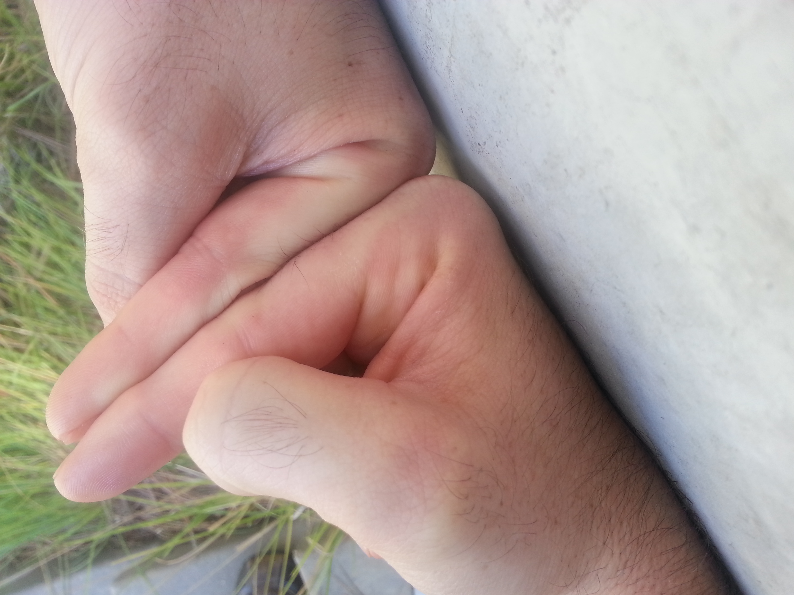
* LSJL increased right finger to be longer than the left.  Need X-ray to see if there are growth plates.  Easiest way to do this?

* LSJL increased left thumb over right thumb.  Thus LSJL can be reproduced.

* There are indications that LSJL is effective at increasing leg height.

* Clamping at the knee may be more effective than clamping at the ankle due to structural differences.

As you know, I’ve proven that due to LSJL my right index finger is now longer than my left.  The issue is that people are “So what?”  They don’t understand that the finger is made up of long bones(albeit with slightly different properties than the leg bones) and if you can induce length increase there it follows that you can increase bone length in the legs as well.

Originally, I ruled out getting an x-ray because people wouldn’t care about an increase in finger length but if I have successfully recreated a growth plate then that would successfully prove LSJL for the masses.  The problem is actually getting an x-ray of ideally both hands without having to get a doctors appointment first (and paying out of pocket for that).  I just want to go in, get an X-ray and then pick it up.  I can afford it for the $200ish I’ve seen especially since it’ll prove LSJL.  There’s a risk that there won’t be a growth plate but I am absolutely positive that my finger has grown longer by some mechanism and that it’s not due to bone thickening.  It is possible that it grew by some mechanism relating to articular cartilage endochondral ossification but people with osteoarthritis have endochondral ossification and I have not found any reports of bone lengthening.

So how can I get a hand X-ray with as little medical red tape as possible?  I want to walk in, get the x-ray, pay, and pick it up.

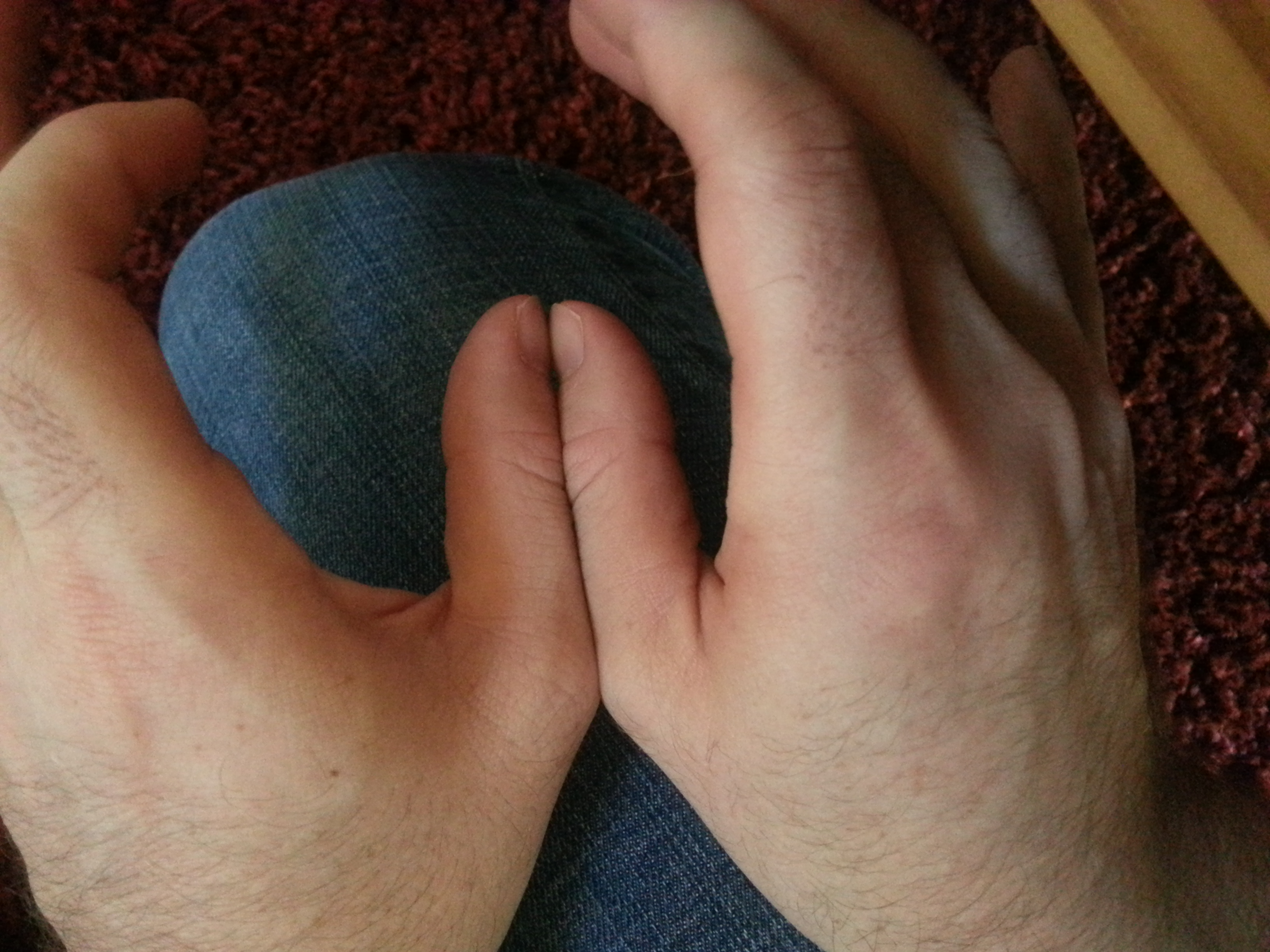
I’ve also been loading my left thumb and there’s a small but significant and noticeable difference between my left and right thumb.  I’m working on finding the best way to photograph it.  It does establish that the finger lengthening is not a fluke and is reproducible.

Another good sign is my legs.  My epiphysis has been changing in shape to become more hammerhead in appearance like my fingers dead.  Even if LSJL induces such “deformities” it would certainly be worth it to some people to grow taller.

And it seems as though my left leg is now longer than my right. The renewed growth can be explained by the following: I used to load my leg with a C-class clamp but now am using an Irwin Quick Grip to reduce slippage.  Now I am focusing much more on intensity of clamping but with shorter duration

(Note:  Since I cannot yet specify an ideal intensity I cannot guarantee against injury!).

Now I always used the Irwin Quick Grip on my ankle but didn’t really get good results there and haven’t seen a lot ankle changes despite clamping harder.  I think the reason for this could be that the knee is different structurally than the ankle and it makes LSJL more effective on that area.

So the reason why this routine could lengthen my left leg more than my right is that I load my left leg first as it is my weaker leg.  If I can get up to say a count of 130 on clamping my left leg(starting over at 0 if the clamp slips) then I clamp to 130 on my right leg.  So my left leg is guaranteed to get maximal clampage whereas the right leg is not.

Now there are other explanations as to why I feel my left leg has grown longer than my right:

1) Placebo effect.  I want my left leg to be longer to prove LSJL.  Although when I extend my legs my left leg is longer than my right and if I stand on my left leg I’m taller then if I stand on my right leg.

2)  Hip Rotation.  Which would explain the symptoms of the left leg extending longer without any actual lengthening.  But what would be the stimulus?

3)  My left leg has always been longer than my left.  I think I would’ve noticed it before.

So this effect isn’t perfect proof of LSJL but I think with the devolpment of more hammerhead-esque bones it is a good sign of LSJL’s effectiveness in increasing leg height.

——————–

Michael: One could go to Urgent Care, which is a type of walk-in facility which would let one get their bodies X-rayed. You’d still have to pay the costs though, but it would be faster. When I was looking at how deer antlers grow, I noted that the antlers where able to grow in length only because there was no physical constraint against the upper horn part from getting longer. Human legs have that constraint since we have to constantly be putting loadings on the feet from walking. if we could put our bodies into some bed for months on end while clamping, maybe there would be much bigger results. The effects on fingers, which are just jutted out and not being pushed down consistently on a flat surface would see much more noticeable effects.

LSJL Progress Update 8-5-14: More finger growth and update on new method

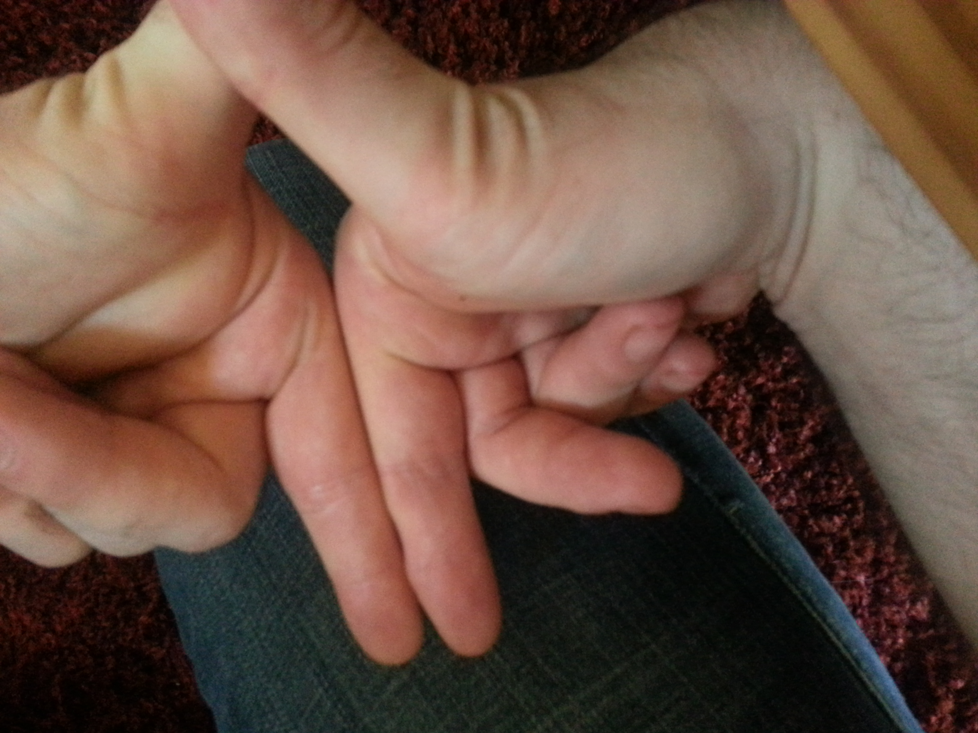
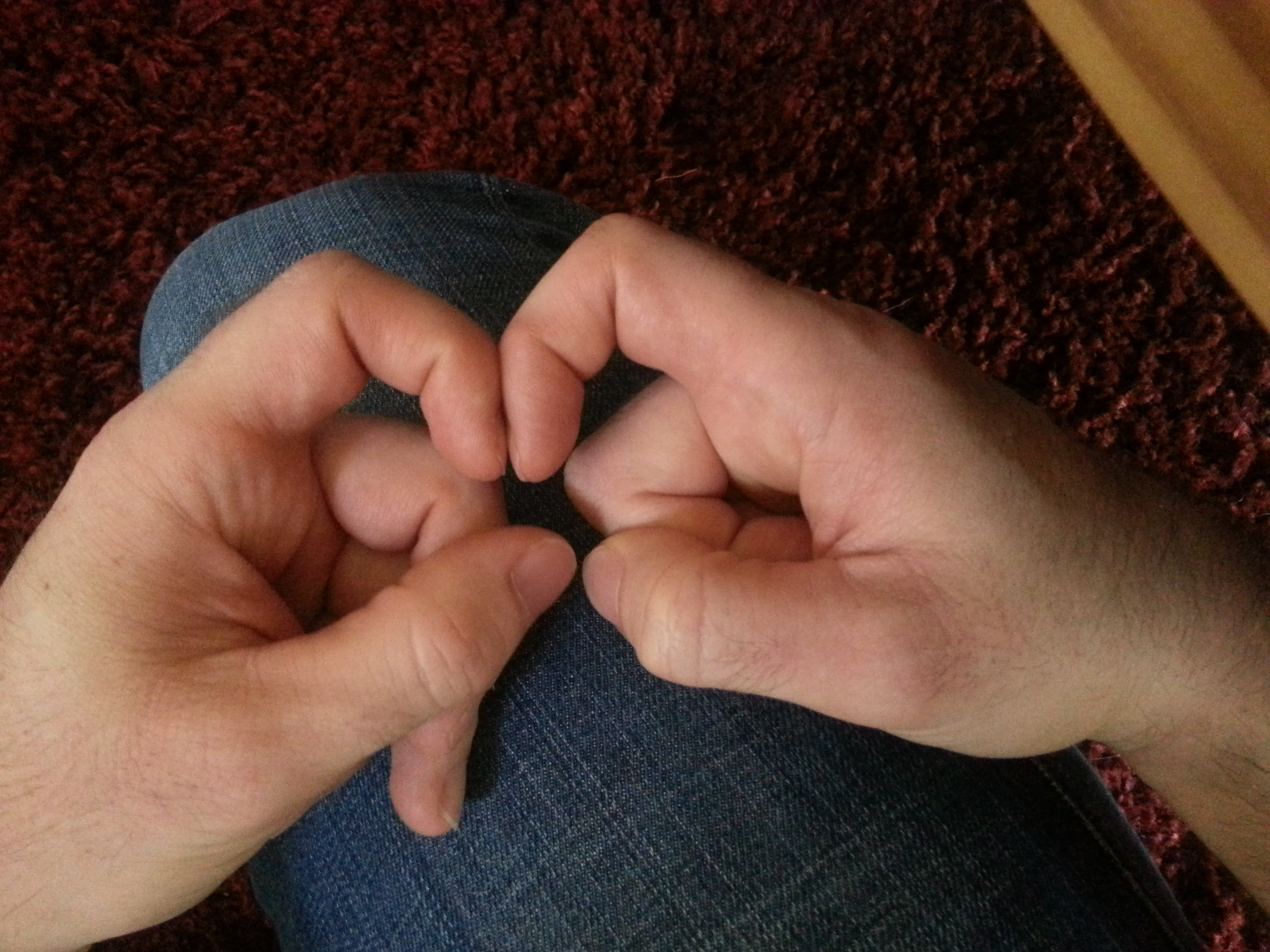
Last time it looked as though my right finger which I loaded via LSJL was about 1/4″ longer.  Now it looks like it’s about .375″ inches longer.  I’ve been loading about every day for about a 100 count on each of the three joints of the finger.  I increase the load as fast as possible, I could do more but I worry about injury because the clamp is so much stronger than the finger.  I might work up to more.  Two joints I load side to side but since the hand is in the way for the knuckle I load from top to bottom.  Here’s a post regarding my previous results and some images about how I perform LSJL. Here’s an image of my fingers now: 20140804_144910 Now it’s an extremely significant increase in finger length that is a result of LSJL.  Now I do have some osteophytes and the finger growth is not the same as normal finger growth.  In some of the other images you can see some finger deformities relative to a normal finger.  But it’s still a strong proof of concept that LSJL works to lengthen long bones. I’d rather prove LSJL sooner rather than later.  Would x-rays help?  I don’t really want to get them if they won’t convince people because it would cost a couple hundred dollar.  A lot of people don’t know exactly what makes you taller.  They can’t connect that long bones make you taller and the finger bones are long bones.  If LSJL can increase the length of finger bones(which are long bones(although they do have some different properties to other long bones)) then LSJL can increase overall height if those long bones are legs. As far as my leg progress though, I find that I can’t get as intense a clamp on my knees as I can on my fingers.  I think part of the reason is that there’s a lot of tissue types you’re clamping when you clamp a synovial joint.  It may take a bit of time before these tissues adapt to the clamping force.  I’ve been clamping for a long time with the C-clamp but there was a lot of slippage so there’s now a lot more force with the Irwin Quick Grip that i’m used to.  So right now I’m clamping with the Irwin Quick Grip to about a count of 130 before the pain in the soft tissues is just too irritating but over time the soft tissues will adapt and I’ll be able to clamp with as much force as I want as I have with bones I’ve been clamping a long time. So I’d recommend not clamping past the point of too much soft tissue pain and just try to increase clamping duration and intensity over time to allow the soft tissues to adapt. Remember, that LSJL is untested so there are guarantees that you won’t get injured or other maladies. Of course, if we could just prove LSJL then more testing can be done.  The question is how can we do it now rather than having to perfect it to increase leg length first?

Michael: The finger seems to be definitely longer, but you said that you clamped in all three joints.

  • Does that mean that the clamping was also at metacarpophalangeal joints?
  • How did you do that, and how can we not make sure that the MSP Joint did not go into inflammation mode aka swelling?
  • There is so much evidence that finger joints can swell up if you hit them on something.

X-Rays seem to be the way to go. We measure the synovial joints of the index finger of the right hand compared to the control of your left hand’s index, which I hoped was never clamped, and see whether the lengthen is from the tissue in the synovial joints thickening as a response. If there is a difference in the distance between the bones in either the PIP and/or MCP joints, then the lengthening was not bone. If the distance in the PIP & MCP joint locations are the same, then we then say that the lengthening was truly bone.

You don’t have to go in for a GP check-up. Look into Urgent Care Centers (Source: Which is Cheaper Out of Pocket: Urgent Care Facility or Hospital ER?). They usually accept Insurance. I’ll even put down $70 for the X-rays if that helps.

Salubrinal decreases osteoclastgenesis

This study doesn’t relate directly to height growth but it is by the scientists whose research was the foundation for LSJL.

In vitro and in silico analysis of an inhibitory mechanism of osteoclastogenesis by Salubrinal and Guanabenz

“Synthetic agents such as salubrinal and guanabenz, which attenuate stress to the endoplasmic reticulum, are reported to inhibit development of osteoclasts. However, the mechanism of their inhibitory action on osteoclasts is largely unknown. Using genome-wide expression profiles, we predicted key transcription factors that downregulated nuclear factor of activated T-cells, cytoplasmic 1 (NFATc1), a master transcription factor for osteoclastogenesis. Principal component analysis (PCA) predicted a list of transcription factors that were potentially responsible for reversing receptor activator of nuclear factor kappa-B ligand (RANKL)-driven stimulation of osteoclastogenesis. A partial silencing of NFATc1 allowed a selection of transcription factors that were likely to be located upstream of NFATc1. We validated the predicted transcription factors by focusing on two AP-1 transcription factors (c-Fos and JunB) using RAW264.7 pre-osteoclasts as well as primary bone marrow cells. As predicted, their mRNA and protein levels were elevated by RANKL, and the elevation was suppressed by salubrinal and guanabenz. A partial silencing of c-Fos or JunB by RNA interference decreased salubrinal- and guanabenz-driven reduction of NFATc1 as well as tartrate-resistant acid phosphatase (TRAP) mRNA. Collectively, a systems-biology approach allows the prediction of a RANKL-salubrinal/guanabenz-NFATc1 regulatory axis, and in vitro assays validate an involvement of AP-1 transcription factors in suppression of osteoclastogenesis.”

“Salubrinal and guanabenz are potent chemical agents for the inhibition of protein phosphatase 1 (PP1) that specifically de-phosphorylate eIF2α. Through upregulating the phosphorylated level of eIF2α and reducing translational efficiency of most proteins except for a limited set of proteins, such ATF4, these agents attenuate stress to the endoplasmic reticulum. Gene regulation by salubrinal and guanabenz, however, not only takes place at the level of translation but also at the level of transcription. In osteoclasts, it has been shown that administration of salubrinal and guanabenz suppresses receptor activator of nuclear factor kappa-B ligand (RANKL)-driven activation of nuclear factor of activated T-cells, cytoplasmic 1 (NFATc1) ”

“A partial silencing of c-Fos and JunB decreased the mRNA and protein levels of NFATc1. Furthermore, there was a feedback loop in which a decrease in c-Fos by salubrinal reduced NFATc1 expression, and the reduction in NFATc1 further attenuated the level of c-Fos protein. AP-1 proteins are known to play a critical role in osteoclast differentiation. It is reported that mice lacking c-Fos are osteopetrotic due to abnormal development of osteoclasts”

Unfortunately, no real LSJL insights in this study.

LSJL knee method + Progress update Part II

In the previous post I offered proof that my current LSJL was working for my fingers(and reported anecdotal evidence of increasing my wingspan by a couple of inches without photographic evidence) but that the LSJL method for the legs was lacking.  So I’m revising the LSJL method to be more like what I’m doing for the fingers which I’ve gotten gotten results from.  With an emphasis on intensity rather than duration.  If anyone has any ideas on how to get a more intense clamp please suggest them.  This is only for people with closed growth plates, people with open growth plates should clamp similarly but much lighter as there is no need to try to use clamping to create a more favorable microenvironment for cartilage growth.

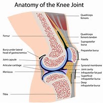
knee joint anatomy

You want to clamp on the synovial joint between the tibia and femur.    Michael has suggested that you should load lower on the epiphysis rather than above.  However, I think loading the synovial joint is important.  The bone there is weaker and it’s easier to cause deformation of the synovial joint then it is the bone itself.  There might be stimulus from the synovial joint that stimulates neo-growth plate formation.  The synovial joint is connected to the growth plate area after all.  You want to clamp more on the femur than the tibia as the clamp will eventually slip to be closer to the middle.  You always want to start clamping more on the bigger bone so it slips to the middle.  If it slips to be clamping on one bone then restart.  Essentially:  Make sure you are clamping the synovial joint.

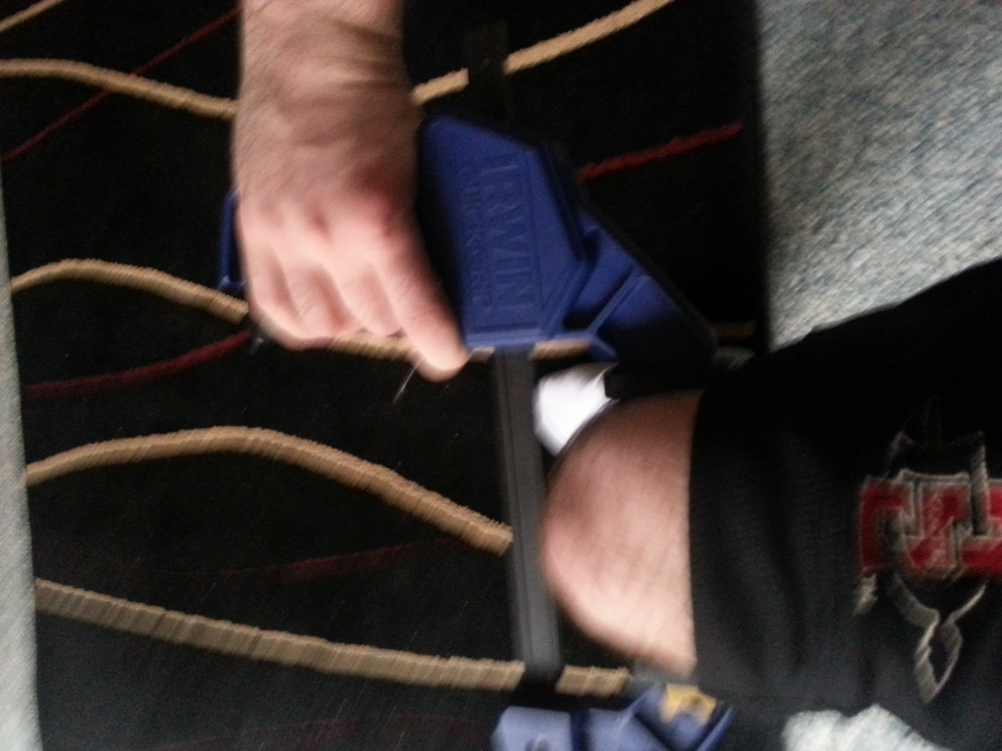
20140715_143030Now you want to use the Irwin Quick Grip(Irwin Industrial Tools 512QCN Next Generation 12-Inch Clamp and Spreader).  But any clamp that can generate enough pressure is sufficient as long as it avoids slipping.  Try to clamp as hard as possible.  Use both hands to get harder clamps.  Take breaks with the clamp still on and the pressure still applied and try to clamp harder.  This method is untested so I can not make any guarantees on the optimal pressure nor can I guarantee that you won’t suffer an injury.  Right now my goal is to generate enough pressure such that there’s a visual or feeling of increased blood flow.  The goal is to generate hydrostatic pressure to create a pro-chondrogenic microenvironment to encourage new growth plate formation.  With my fingers I could see visually increased blood flow and I could feel it.  The goal is to generate enough pressure in the synovial joint to encourage a pro-chondrogenic microenvironment.  Any clamping method that does that is sufficient.
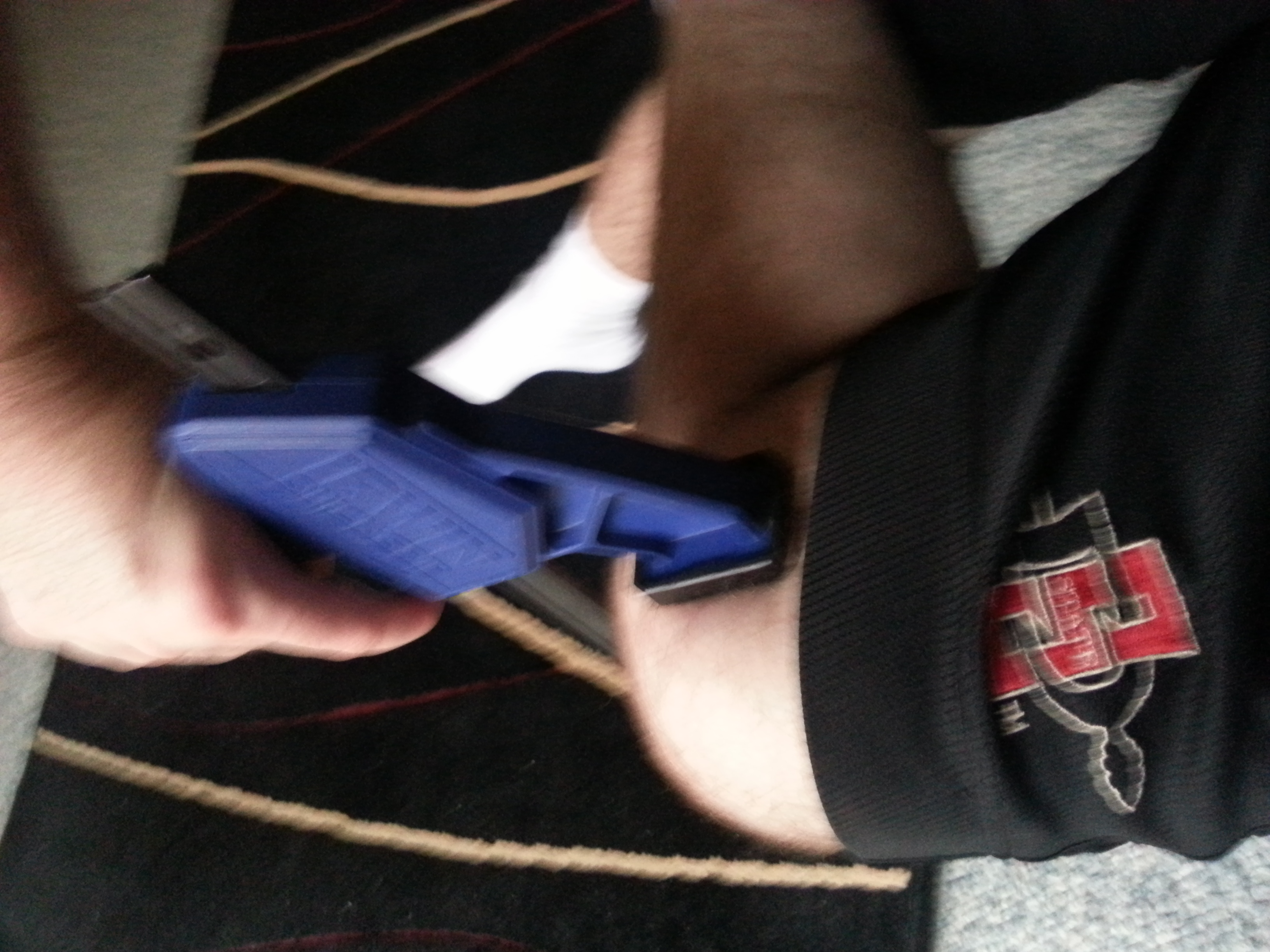
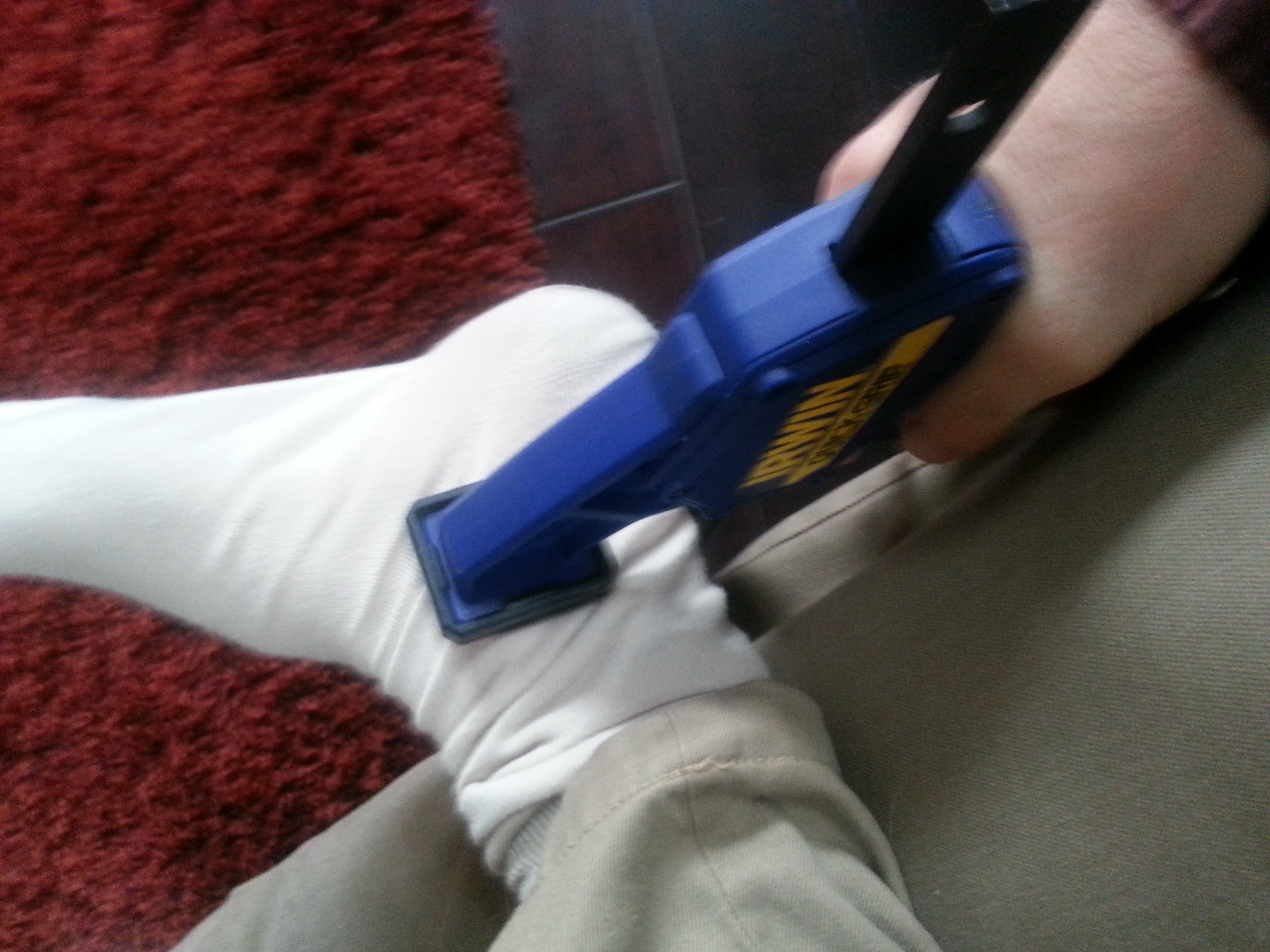
 

Here’s another angle:
20140715_143046

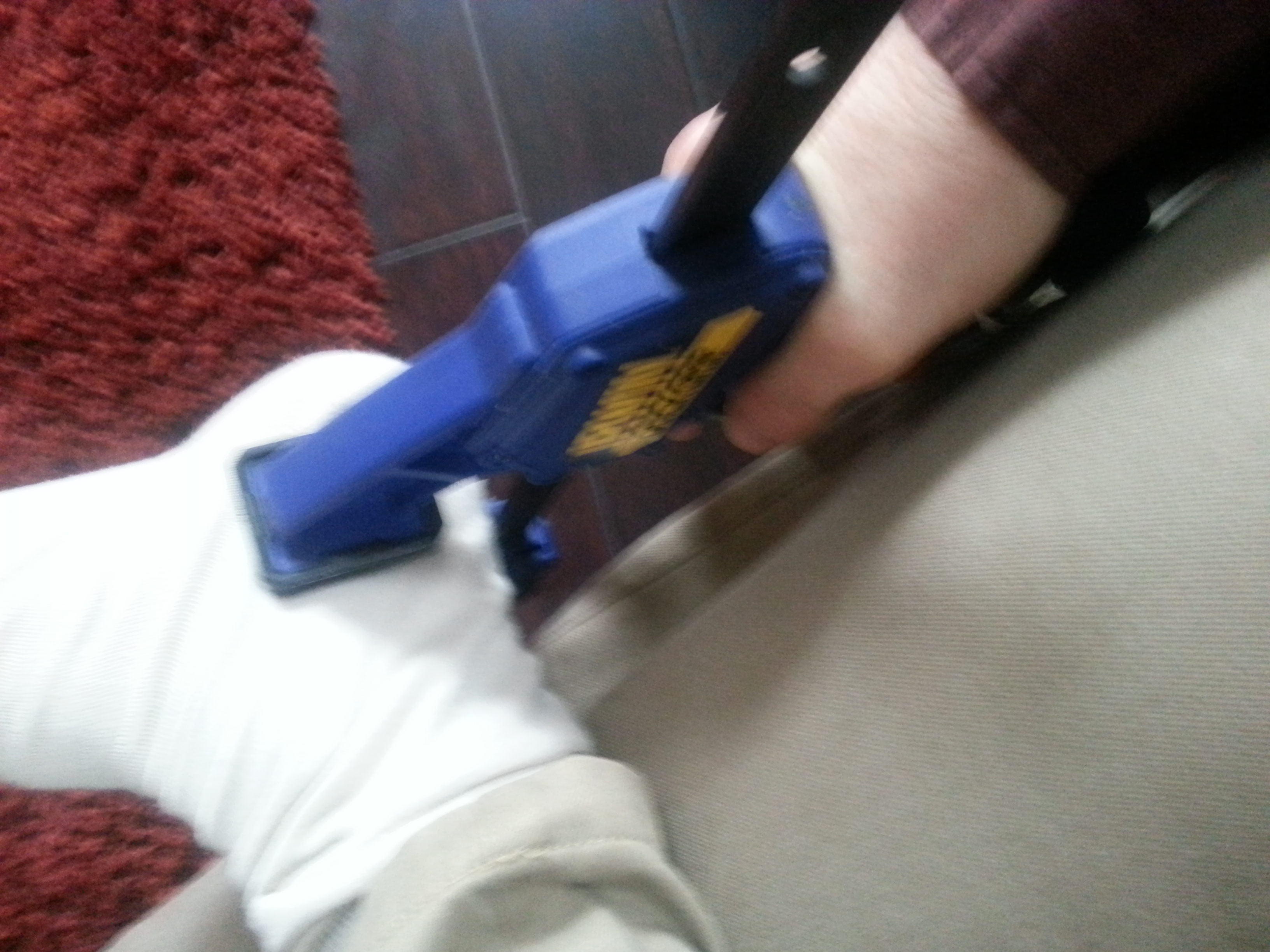
Me clamping between the tibia and talus:


20140715_100809

Me clamping between the talus and calcaneus:


20140715_100819

Please let me know if you have any questions.  I know people will want a video so I’ll work on that for Part III.

Michael: Here is what I would suggest. Get a 2nd clamp to clamp simultaneously for the knee area. We do have two hands, and the clamping area is on the legs. A easy position to get into. To get the frequency correct, squeeze both hands at the same time.

Fit the two clamps along side of each other, one on the angular part while the other is on the sides, which you suggest. Since load is just pressure (Force/area) to increase the load, we just get a 2nd clamp to double the amount. At this point, I would not suggest increasing the amount of force from one clamp, but put the clamps into series on the sides of the tibia.

LSJL knee method + Progress update Part I

Note:  This post will provide evidence that LSJL does not merely expand the synovial joints but directly lengthens the bone.

I provided what I consider to be fairly irrefutable proof that I’ve increased finger length.  The issue is that finger length doesn’t come into play in athletics that much.  Larger hands could help in places where increased surface area would come into play.  I also increased wingspan which is more helpful in athletics but I have no documentation and wingspan isn’t measured by doctors.

So I have to return to developing a better methodology for LSJL for the legs which would increase height which is measured by doctors and important in everything in contrast to more niche uses for finger length and wingspan.  Here’s some of the methods I’ve tried.   I got initial results but stopped over time.  One of the reasons could have been that the results were due to dumbell loading since I stopped doing dumbell loading over time but I doubt that the results were due to dumbell loading since 65lbs is so slow.  There might be some sort of adaptive mechanism that reduces effectiveness over time.

The question is:  Why am I gaining length in my finger and arms but not in my legs?

20140623_123745

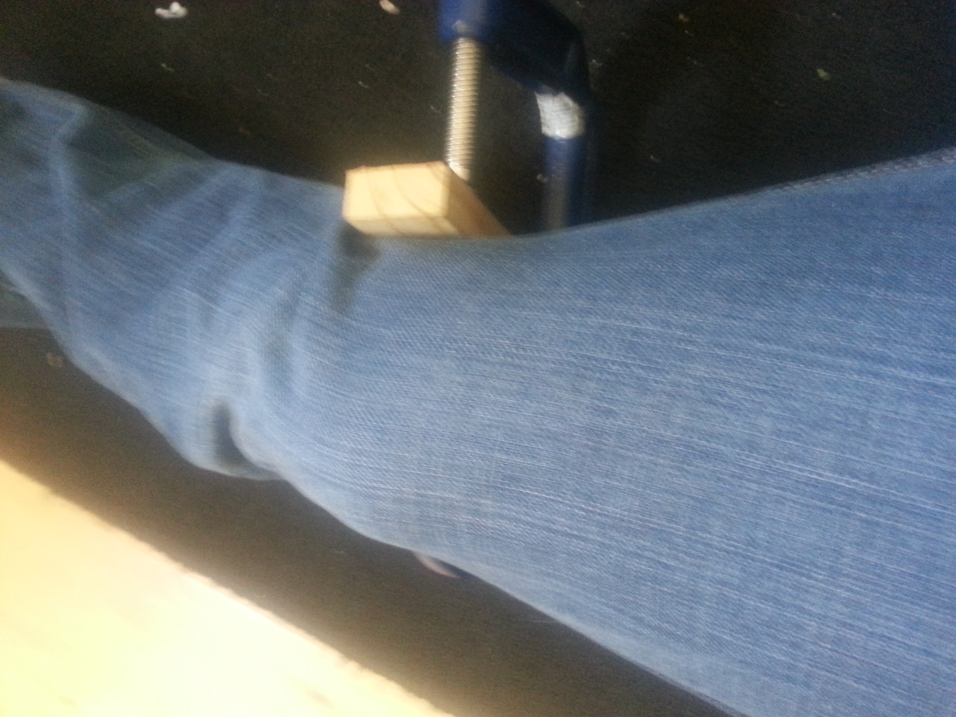
The answer may be related to angular loading.  When I clamp a bone it begins to bend at an angle. Before I was trying to keep it straight which resulted in the knee popping out in the clamp at the top.  By allowing the knee to tilt the knee becomes trapped within the clamp stopping it from popping out.  My finger and other arm joints always tilt when I’m clamping them and I gained length there so it makes that I can allow for the knee joint to tilt and still gain length in that region.

I’m using the block to increase surface area.  I have to constantly adjust the block and clamp though for keeping it from slipping.  Slipping is far more an issue for leg clamping still so there are further advancements to be made here.

After trying this method for a couple of weeks it was too inconsistent.  Sometimes I would get a really good clamp and generate a lot of pressure other times the clamp would slip off before decent pressure was generated.  So right now I’m using the Irwin Quick Grip and I’ll cover what I’m doing in part II.  But right now I’m mainly focusing on one intense clamp rather than a certain time duration as before but bearing in mind to avoid injury which is possible since I am using so much force when clamping.  Although the time duration method was working with fingers/wing span, I think maximizing intensity is working even better even for fingers/arms.

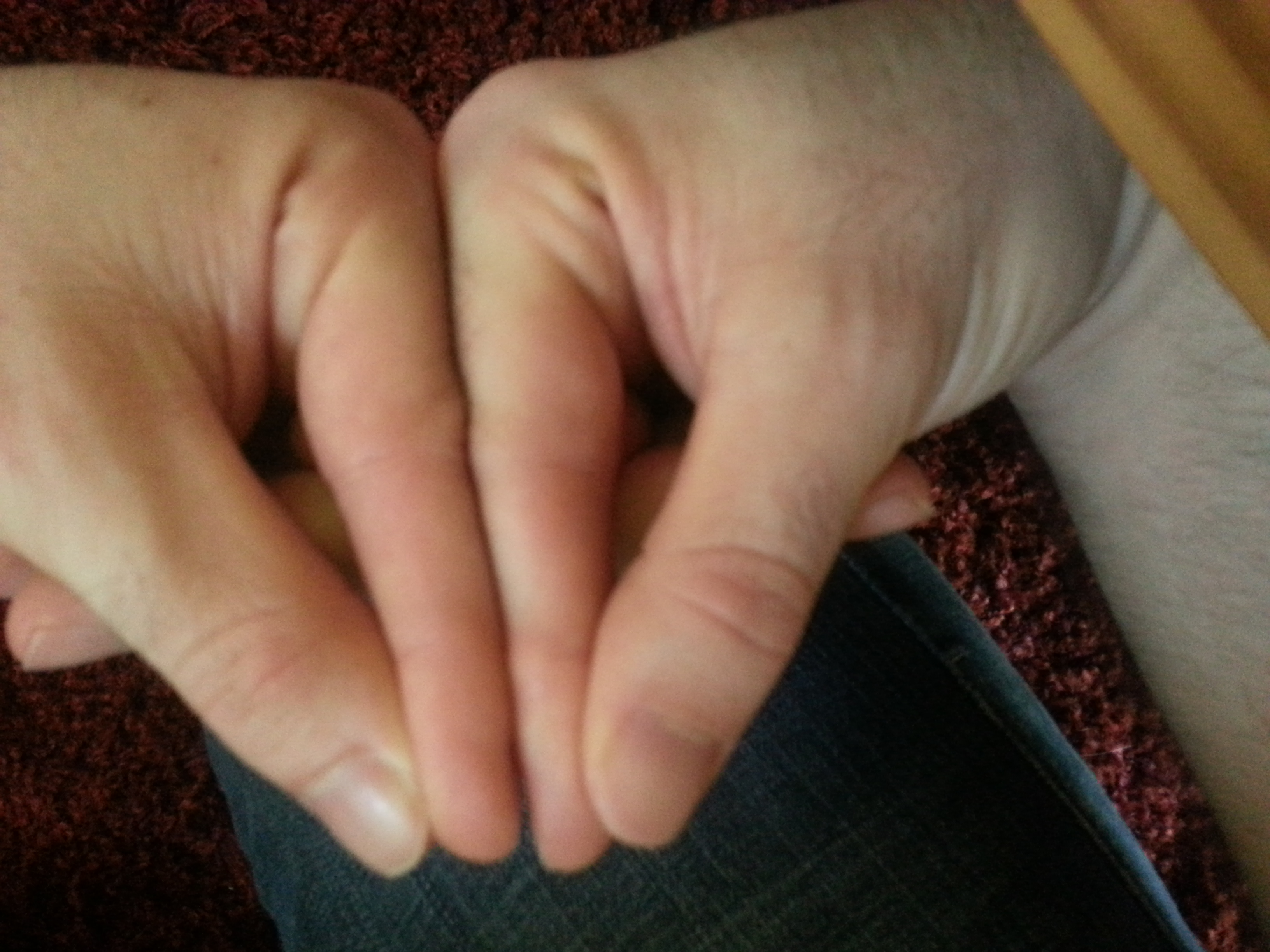
Finger ComparisonI got a better angle of the finger length comparison.  I am only clamping my right finger and not my left.  You can clearly see that the right finger is definitively longer than the left.  It’s longer than it appears in the picture because I wanted to make sure the right knuckle was higher than the left so people didn’t think I was just sliding the right finger down.

Here’s another angle:

20140709_123054In this one it’s harder to prove that there’s no manipulation to alter finger length but it’s still another perspective.  You can also see the osteophyte on my right finger.  I studied a little bit about osteoarthritis and although osteophytes are a symptom of osteoarthritis they can be caused by other forms of mechanical stimuli too.  So, just because I have osteophytes doesn’t mean I have osteoarthritis.

Now to prove that it’s not just enlarged joints and it’s actually the bone that’s longer.

20140709_123109Comparison of two bones in the finger only and clearly the right finger bone is longer.  So LSJL lengthens bones and does not just merely expand the synovial joint.

Now here’s a thumb comparison.  I’ve only been doing LSJL on my left thumb and not my right.  I figured it would be unlikely for someone to argue that my right hand has always had longer figures if my right index finger was longer then the left but my left thumb was longer than my right.

20140709_123240And it looks like the left thumb is longer but I can’t rule out measurement error since it’s hard to tell exactly when the thumb ends on the hand.

I have before pictures of each appendage but it’s much easier to compare side to side against the contralateral limb.

So here’s some more evidence of LSJL but hopefully also switching up the knee and ankle method will be able to prove LSJL there and that is the big ticket for proof.  So look for Part II soon that explains the current LSJL technique I’m using(I have pretty much finalized the technique but need to take the pictures) and hopefully more LSJL proof.

Michael: I answered your later posts before this one, so I did not realize you accounted for synovial joint expansion.  As for the one question you wanted answered, I will just go back to the fact that the fingers and the arms are not always being pushed upon. With the legs, since we are always walking, the effects of the clamping might be negated by the loading from just walking itself.

As for the osteophyte issue, it is a unique sign that something is not going correctly. Has osteophytes also developed in the finger bone segments which were not clamped?