Author Archives: Tyler

LSJL Progress Update 8-26-14

In my previous progress post I continued to establish that I had in fact grown due to finger clamping.  I have been using the new LSJL method which emphasizes clamping force rather than duration.  I’ve been clamping right index finger, left thumb, elbows, wrists, knee, ankle, calcaneous, and experimentally toes. Michael suggested using two clamps at the same time and I’ll have to try that.  I’ve been trying to find alternative ways to get X-rays but looks like Michael was right and the best bet is Urgent Care.  It’s going to be a pain in the butt but I don’t think I have a choice.

I’ve been gradually increasing the clamping force.  I think it’s key to be clamping on the synovial joint.  Since the bone is so strong, it’s likely that a lot of the LSJL affects are due to clamping the synovial joint.  If a clamp only the epiphysis of one bone than I can seemingly clamp forever but if I clamp more at the synovial joint I can tolerate much less.  It’s interesting to note that I did grow in the arms from a wingspan of 72.5″ to 74.4″ and the way I’m been clamping the elbow is more on the humerus side because the bone structure is so awkward.  So I can’t clamp exactly in the the middle of the humerus and ulna.  However, I have still gotten results there.   So I think it’s more important to clamp in an area where you get some sort of feedback from your body rather than to clamp in some specific spot.  If you can clamp an area forever, you’re doing it wrong.   Right now I’m clamping areas between 100 to 140 seconds and that’s after working up to it.

20140826_123611Here’s another finger angle with the tips aligned.  You can see that the knuckle is higher on the right finger.  But yeah, yeah, yeah,  you have to accept X-rays.  And I have to make sure I get both hands in the x-ray.  Although if there are neo-growth plates in the right finger that would be enough regardless of a comparison.  Although it’s possible that the growth was due to another mechanism say fibrocartilage, etc.

As for my legs and arms, there are signs that I am growing.  Last measured wingspan was 74.5″ but since arms can be stretched a little bit it’s not enough.

As for legs, they seem longer and my jeans and shorts seem to be a little higher but nothing definitive yet.  Now when I have a chance I’m clamping twice a day.  At least once a day seven days a week.

LSJL Update-8-18-2014

So I’m still working on getting a hand x-ray.  I’m contacting facilities to see if they take walk-ins.  No replies so far. I also read you may be able to get an x-ray at the airport(but that was in context of a full body scan)?  I’m looking for alternative medicine facilities that do x-rays but I’ve read they need a doctors script too.  I don’t really want to go do the Urgent Care but it looks like I may not have a choice.  If anyone has any experience let me know. Here’s the last update.

20140817_150801The right finger could be longer since last update but not sure.

Here’s a thumb pic:

20140817_150737I’ve been clamping my left thumb but not my right and from this picture you can see the left thumb is slightly but significantly longer.  The osteophytes are much less noticeable in my left thumb than my right index finger but they are there if I feel around the clamping region.

Interestingly, I clamp each of the three joints of the finger.

Here’s an image of the finger bones:

027 Bones of the hand (dorsal view)I clamp the two interphalangeal joints from side to side  but at the metacarpophalangeal joint I clamp from top to bottom.   The epiphysis near the MP on the index proximal phalanx of my right (clamped) finger is thicker than my left finger but not as enlarged as the epiphysis near the IPs of the distal and middle phalanx.  The IPs of the clamped joints are also rougher than the MP which is clamped overhead rather than side to side.  So some of the enhanced epiphyseal thickness may be due to LSJL growth stimulation and other effects may be due to irritation from the clamping surface on the joint.  If I can get an x-ray it will be interesting to compare the two IPs to the MPs.  For a period of time, I tried clamping the two IPs from top and bottom but didn’t get noticeable results from that.  Top to bottom clamping may be more effective in the MP than the IPs due to differences in joint structure.  You can feel much more pressure generated by top to bottom clamping of the MP than you can the IPs.

As for leg clamping, I am increasing the duration and intensity of the clamping.  I may be getting taller but I cannot yet rule out placebo(like I have with my thumb and index finger which are too noticeable and significant to be placebo) or measurement error.  I grew about 1 1/2 inches in wingspan for sure but I don’t have photographic evidence.  I’ll have to keep monitoring there.  In a week if I don’t have noticeable leg progress, I’ll bite the bullet and go to urgent care to get the x-rays.

Pressure plasma for height

Non-Thermal plasma is plasma that is not in thermodynamic equilibrium.  Atmospheric pressure plasma matches the pressure of the surrounding atmosphere.

Non-Thermal Atmospheric Pressure Plasma Enhances Mouse Limb Bud Survival, Growth and Elongation.

“appendage regeneration is dependent on reactive oxygen species (ROS) production and signaling. Mesenchymal cell stimulation by non-thermal (NT)-plasma, which produces and induces ROS, would (1) promote skeletal cell differentiation and (2) limb autopod development. Stimulation with a single treatment of NT-plasma enhanced survival, growth and elongation of mouse limb autopods in an in vitro organ culture system. Noticeable changes included enhanced development of digit length and definition of digit separation. These changes were coordinate with enhanced Wnt signaling in the distal apical epidermal ridge (AER) and presumptive joint regions. Autopod development continued to advance up to 144 hr in culture, seemingly overcoming the negative culture environment normally observed in this in vitro system. Real time qPCR analysis confirmed the up-regulation of chondrogenic transcripts. Mechanistically, NT-plasma increased the number of ROS positive cells in the dorsal epithelium, mesenchyme and the distal tip of each phalange behind the AER, determined using dihydrorhodamine. The importance of ROS production/signaling during development was further demonstrated by the stunting of digital outgrowth when anti-oxidants were applied. NT-plasma initiated and amplified ROS intracellular signaling to enhance development of the autopod. Parallels between development and regeneration, suggest the potential use of NT-plasma could extend to both tissue engineering and clinical applications to enhance fracture healing, trauma repair and bone fusion.”

“Vertebrate limbs grow through the coordinated activity of numerous growth factor signaling pathways, including the Wnt, fibroblast growth factor (FGF), bone morphogenetic protein (BMP), Notch, and transforming growth factor-β pathways. ROS play a primary role in mediating the activity of these factors and/or are integrally involved in regulating downstream signaling pathways. For example, a sustained increase in ROS levels is required for Wnt β-catenin signaling and the activation of one of its main downstream targets, fgf20. In addition, naturally occurring oxidative spikes are observed in most of the transition stages throughout developmental and regenerative processes. Furthermore, there is precedence for ROS production to both direct and enhance mesenchymal cell differentiation into either chondrocyte or osteoblast lineages”

“Since the NT-plasma discharge occurs in air, it is a highly complex mix of reactive chemical species (i.e., nitric oxide [NO], ROS, oxygen singles, and ozone), free ions, visible, ultraviolet, and near-infrared light, electric fields, and electromagnetic (EM) radiation.”

“one study showed that a 2 h treatment with EM field stimulated limb growth through increased proliferation and chondrocyte differentiation”

That is an absolutely huge elongation.

“Morphological formation of the joint region and alcian blue staining of proteoglycan appeared normal in sham and NT-plasma-treated digits. Chondrocytes in the proximal segment of the sham-treated digit were entering the prehypertrophic stage, based on the larger size, greater cellular spacing, and decrease in proteoglycan (white arrowhead). This typically precedes the formation of the primary center of ossification in the diaphysis. In contrast, chondrocytes from the NT-plasma-treated digit were immature and contained large amounts of proteoglycan. Even more striking was the much larger cartilage phalangeal segments in the NT-plasma-treated digit. A white line with double arrows marks the joint between the first and second phalanges in both sections. In the sham-treated digit, the adjacent joint spaces (white arrow) can be clearly observed; whereas in the NT-plasma-treated digits, there is no evidence of the next joint space, due to the increased segment length.”<-So maybe the plasma treatment increases stem cell differentiation to chondrocytes which is what we’d be looking for.

“Histology and quantitative real time-polymerase chain reaction were used to assess chondrogenesis and molecular events associated with the accelerated development. (A) A representative, hematoxylin and eosin (H&E) and alcian blue stained sections of middle phalanges from sham and NT-plasma-treated limb autopods. A white double arrow line marks the first joint space. The white arrows mark adjacent joint spaces, and the white arrowhead marks prehypertrophic chondrocytes in the sham control. (B) At 24 h post-NT-plasma treatment, alkaline phosphate (ALKP), catalase (CAT), endothelial nitric oxide synthase (eNOS), and bone morphogenetic protein 2 (BMP2) expression increased twofold above control. BMP4 and runt-related transcription factor 2 (Runx2) increased 12-fold and 8-fold, respectively, and fibroblast growth factor (FGF)-2 showed no increase. One hundred forty-four hours later, increases for ALKP (6-fold), CAT (14-fold), and BMP2 (3.5-fold). eNOS remained increased twofold above control at 144 h. BMP4 and Runx2 showed a relatively consistent elevated expression at 24 and 144 h. The results are expressed as the mean±standard deviation (n=3 [two limbs pooled per sample], *p<0.05). ”

“NT-plasma treatment promoted both growth and differentiation of cartilaginous elements within the embryonic mouse autopod. Based on the lack of limb growth after NT-plasma treatment in an antioxidant environment, and the increase in H2O2-positive cells in and under the epithelia throughout the 6 day culture period, we propose that NT-plasma induction of intracellular ROS also plays a role in the growth and differentiation observed in the autopod. In addition, H2O2-positive cells were observed in the distal digit tips behind the AER, concurrent with enhanced Wnt signaling. These findings are consistent with the ROS dependence of signaling factors (e.g., Wnt, BMP, and FGF) required for autopod development.”

“ROS production may not be the sole mechanism driving enhanced autopod growth. A second possible reason that NT-plasma enhanced autopod growth was its inhibition of epithelial overgrowth. ”

“In limb regeneration, a thickening of the epidermis has been shown to inhibit signaling to the blastema, halting the regeneration process.”

Hard to determine causality as there are so many potential causal factors as there are so many confounding variables in plasma.

Older mice still capable of undergoing endochondral ossification

Mice growth plates do not go through the fusion stage but they do cease eventually in longitudinal bone growth due to growth plate dysfunction.  If we can reverse growth plate dysfunction in mice than perhaps we can do so in humans and get more height from the growth plate.

This study however does not discuss reversing elderly growth plate dysfunction but rather states that older mice are still capable of undergoing endochondral ossification outside the growth plate.

Fractures in Geriatric Mice Show Decreased Callus Expansion and Bone Volume.

“Using a small animal model of long-bone fracture healing based on chronologic age, we asked how aging affected (1) the amount, density, and proportion of bone formed during healing; (2) the amount of cartilage produced and the progression to bone during healing; (3) the callus structure and timing of the fracture healing; and (4) the behavior of progenitor cells relative to the observed deficiencies of geriatric fracture healing.
Transverse, traumatic tibial diaphyseal fractures were created in 5-month-old (young adult) and 25-month-old (which we defined as geriatric, and are approximately equivalent to 70-85 year-old humans) C57BL/6 mice. Fracture calluses were harvested at seven times from 0 to 40 days postfracture for micro-CT analysis (total volume, bone volume, bone volume fraction, connectivity density, structure model index, trabecular number, trabecular thickness, trabecular spacing, total mineral content, bone mineral content, tissue mineral density, bone mineral density, degree of anisotropy, and polar moment of inertia), histomorphometry (total callus area, cartilage area, percent of cartilage, hypertrophic cartilage area, percent of hypertrophic cartilage area, bone and osteoid area, percent of bone and osteoid area), and gene expression quantification (fold change).
The geriatric mice produced a less robust healing response characterized by a pronounced decrease in callus amount (mean total volume at 20 days postfracture, 30.08 ± 11.53 mm3 versus 43.19 ± 18.39 mm3; p = 0.009), density (mean bone mineral density at 20 days postfracture, 171.14 ± 64.20 mg hydroxyapatite [HA]/cm3 versus 210.79 ± 37.60 mg HA/cm3; p = 0.016), and less total cartilage (mean cartilage area at 10 days postfracture, 101,279 ± 46,755 square pixels versus 302,167 ± 137,806 square pixels; p = 0.013) and bone content (mean bone volume at 20 days postfracture, 11.68 ± 3.18 mm3 versus 22.34 ± 10.59 mm3) compared with the young adult mice. However, the amount of cartilage and bone relative to the total callus size was similar between the adult and geriatric mice (mean bone volume fraction at 25 days postfracture, 0.48 ± 0.10 versus 0.50 ± 0.13), and the relative expression of chondrogenic (mean fold change in SOX9 at 10 days postfracture, 135 + 25 versus 90 ± 52) and osteogenic genes (mean fold change in osterix at 20 days postfracture, 22.2 ± 5.3 versus 18.7 ± 5.2; p = 0.324) was similar{so the deficiencies of older mice to undergo endochondral ossification in response to fracture may be related to things other than gene expression}. Analysis of mesenchymal cell proliferation in the geriatric mice relative to adult mice showed a decrease in proliferation (mean percent of undifferentiated mesenchymal cells staining proliferating cell nuclear antigen [PCNA] positive at 10 days postfracture, 25% ± 6.8% versus 42% ± 14.5%.
the molecular program of fracture healing is intact in geriatric mice, as it is in geriatric humans, but callus expansion is reduced in magnitude.
Our study showed altered healing capacity in a relevant animal model of geriatric fracture healing. The understanding that callus expansion and bone volume are decreased with aging can help guide the development of targeted therapeutics for these difficult to heal fractures.”

So older mice are still capable of chondrogenic fracture healing which is a lot like endochondral ossification.  Therefore, this provides evidence that older humans may be capable of inducing new endochondral ossification.

“At 10 days postfracture, corresponding to peak cartilage, young adult mice produced more total cartilage than geriatric mice (302,167 ± 137,806 versus 101,279 ± 46,755 square pixels”

“No difference was found in the percent cartilage content of the callus at 10 days postfracture (41.17% ± 10.13% versus 28.94% ± 8.74%) and in the percent of total cartilage that was hypertrophic at 15 days postfracture (88.76% ± 14.56% versus 70.21 ± 16.05%; p = 0.078). Osseous tissue formation did not reach high levels until 20 days postfracture with cartilage resorption nearly complete for both groups. Geriatric mice exhibit decreased total cartilage production and delayed resorption”

Cyp26b1

If you’re deficient in Cyp26b1 then having a Vitamin A deficient diet may be a way to grow taller.

Cyp26b1 Within the Growth Plate Regulates Bone Growth in Juvenile Mice.

“Retinoic acid (RA) is an active metabolite of vitamin A and plays important roles in embryonic development. CYP26 enzymes degrade RA and have specific expression patterns that produce a RA gradient, which regulates the patterning of various structures in the embryo.  Localized RA activities in the diaphyseal portion of the growth plate cartilage were associated with the specific expression of Cyp26b1 in the epiphyseal portion in juvenile mice. To disturb the distribution of RA, we generated mice lacking Cyp26b1 specifically in chondrocytes (Cyp26b1Δchon cKO). These mice showed reduced skeletal growth in the juvenile stage. Additionally, their growth plate cartilage showed decreased proliferation rates of proliferative chondrocytes, which was associated with a reduced height in the zone of proliferative chondrocytes, and closed focally by four weeks of age, while wild-type mouse growth plates never closed. Feeding the Cyp26b1 cKO mice a vitamin A-deficient diet partially reversed these abnormalities of the growth plate cartilage. Cyp26b1 in the growth plate regulates the proliferation rates of chondrocytes and is responsible for the normal function of the growth plate and growing bones in juvenile mice, probably by limiting the RA distribution in the growth plate proliferating zone.”

“Cyp26b1 is expressed in the distal region of developing limb buds, and mice that lack Cyp26b1 show severe limb malformation due to the spreading of the RA signal toward the distal end of the developing limb, causing abnormal patterning of limb skeletal elements”

“In juveniles, focal closures of the growth plate in the distal tibia, the proximal tibia, the distal tibia, elbow, proximal femur and distal femur are caused by the treatment of acne with retinoids or the treatment of hyperkeratinosis with cis-retinoic acid. In guinea pigs, the application of RA caused closure of the growth plates in the proximal tibia”

“Excess intake of vitamin A causes growth impairment and skeletal pain in juveniles”

Huge Breakthrough study-hypertophic chondrocytes transdifferentiate into osteoblasts

This is a study that could have massive ramifications on height increase but the the exact mechanism is unknown.  But that cells that have the genetic material of growth plate cells are retained in adult bone is huge news.

Hypertrophic chondrocytes can become osteoblasts and osteocytes in endochondral bone formation.

hypertrophic chondrocytes<-full study

“Chondrocytes and osteoblasts are considered independent lineages derived from a common osteochondroprogenitor. In endochondral bone formation, chondrocytes undergo a series of differentiation steps to form the growth plate, and it generally is accepted that death is the ultimate fate of terminally differentiated hypertrophic chondrocytes (HCs). Osteoblasts, accompanying vascular invasion, lay down endochondral bone to replace cartilage.  [Can] HC become an osteoblast and contribute to the full osteogenic lineage? Here we use a cell-specific tamoxifen-inducible genetic recombination approach to track the fate of murine HCs and show that they can survive the cartilage-to-bone transition and become osteogenic cells in fetal and postnatal endochondral bones and persist into adulthood. This discovery of a chondrocyte-to-osteoblast lineage continuum revises concepts of the ontogeny of osteoblasts, with implications for the control of bone homeostasis and the interpretation of the underlying pathological bases of bone disorders.”

This is huge because it means that the transdifferentiated osteoblasts retain some of the chondrocytic genetic material which means they could possibly dedifferentiate back into chondrocytes!

“The expression of preosteoblastic markers in LHs before the formation of the POC raises the possibility that these cells may transition to an osteoblastic fate”

“HC-Derived Cells Are Present in Fetal, Neonatal, and Adult Bone.”

“The HC-derived cells, morphologically resembling osteoblasts, were found close to the chondro-osseous junction, on the surface of trabeculae, and in the endosteum”<-that means they are in a good position to be involved in neo growth plate formation.

“HC[hypertrophic chondrocytes] Derivatives Transit to the Primary Spongiosa and Become Col1a1-Expressing Cells.”<-the majority of HC derivatives become osteoblasts.

“HC-to-bone transition occurs during postnatal bone growth and that HC-derived cells may be long-lived within the mature bone”

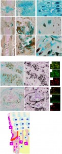
Here’s an image of the HC lineage:

hypertrophic chondrocyte lineage

“The reversion of HCs to a prehypertrophic- like state in response to endoplasmic reticulum (ER) stress suggests that hypertrophy is not an irreversible state in vivo”<-Could we revert HC-derived osteoblasts back to hypertrophic chondrocytes back to pre-hypertrophic cells to reform growth plates.

(Michael: That actually makes a lot of sense if one thought about it. It can’t be all dead inorganic bone ECM matter where the physeal cartilage turns into bones (aka Primary Spongiosa). How would bone cells even be able to travel to that middle-region later on if the chondrocytes completely died out and the lucanae was covered by calcium minerals. For the smooth gradual transition from the hypertrophic layer to the vascularization/calcification layer to the mineralization layers to work out, there would be some minority of chondrocytes which would change identities. If all the chondrocytes died out and the layer of the lucanae was completely mineralized, I would not be sure that osteoblasts would be able to reach the primary spongiosa layer.

Here is what I have found years ago. (Source: Chondrogenesis just ain’t what it used to be) Prehypertrophic chondrocytes secrete IHH while the chondrocytes from the perichondrium secrete PTHrP, which effect the PTH/PTHrP receptors which are on the prehypertrophic chondrocytes. In addition, we have to consider the effects of the VEGF. VEGF is secreted by hypertrophic chondrocytes, and that it acts as a major inducer of vascular invasion.

The big thing is the following…

“…demonstrated that Ihh is not only required for chondrocyte hypertrophy, but also for expression of Cbfa1, a transcription factor required for osteoblast differentiation”

If the molecular biologist researchers have been able to identify all of the  major players which causes the chondrocytes to go into apoptosis, and also transdifferentiation, then we at least know which transcription factors causes the changes. I suspect it is Cbfa1.

Refer also the seminal work “Indian hedgehog couples chondrogenesis to osteogenesis in endochondral bone development

That study proved conclusively that it is IHH that starts everything. IHH stimulates the Cbfa1, which causes the vascularization. I am going to make a guess that somewhere along the way, the two peptides also caused the transdifferentiation.

So how do we actually figure out what type of chemical would lead the osteoblast/osteocytes types to go in reverse and de-differentiate aka some type of reverse transdifferentiation?

We first have to assume that there is some type of external stimuli (mechanical, chemical, electrical) which would be able to even do the de-differentiation.

I propose this idea for Tyler…

Let’s assume that if we change the environment, the cell will start to change its identity. Remember the study which showed that the path which the MSCs will differentiate into can be determined by the shape of space that they are placed into? That suggest that if we change the environment that the osteoblasts/bone cells are in, maybe they will change as well.

Here is my first idea: I propose that we try to remove the calcium crystals/de-mineralize the area. To do that, remember that the PTH and the PTHrP balance from the parathyroid glands controls the level of calcium that is dissolved into the human blood (refer to “Chapter 5 – The parathyroid glands and vitamin D“)

In bone, within 1 or 2 hours, PTH stimulates a process, known as osteolysis, in which calcium in the minute fluid-filled channels (canaliculi/lacunae) is taken up by syncytial processes of osteocytes and transferred to the external surface of the bone and, thence, into the extracellular fluid. Some hours later, it also stimulates resorption of mineralized bone; a process that releases both Ca2+ and Pi into the extracellular fluid. The Pi is rapidly removed from the circulation because the most dramatic effect of PTH on the kidney is to inhibit reabsorption of Pi in the proximal tubule and markedly increase its excretion

Let’s increase the level of PTHrP in the local region, which is what I had proposed in a post I had written more than a year ago (The Connection Between Regenerating Deer Antlers and The PTHrP, PTH And IHH pathway for Cartilage Regulation, PTHrP Seems To Be The Answer (Big Breakthrough!)). Decrease the concentration of CA2+ from the ECM, and see what happens to the osteoblasts. Would they de-differentiate into chondrocytes if their environment changes?

It might be that the formation of osteoblasts and the increased levels of mineralization/vascularization/calcification is a positive feed back loop where each part feeds upon itself, which is initialized by VEGF. If we break the positive cycle at the process of mineralization, would the osteoblasts also go in reverse?
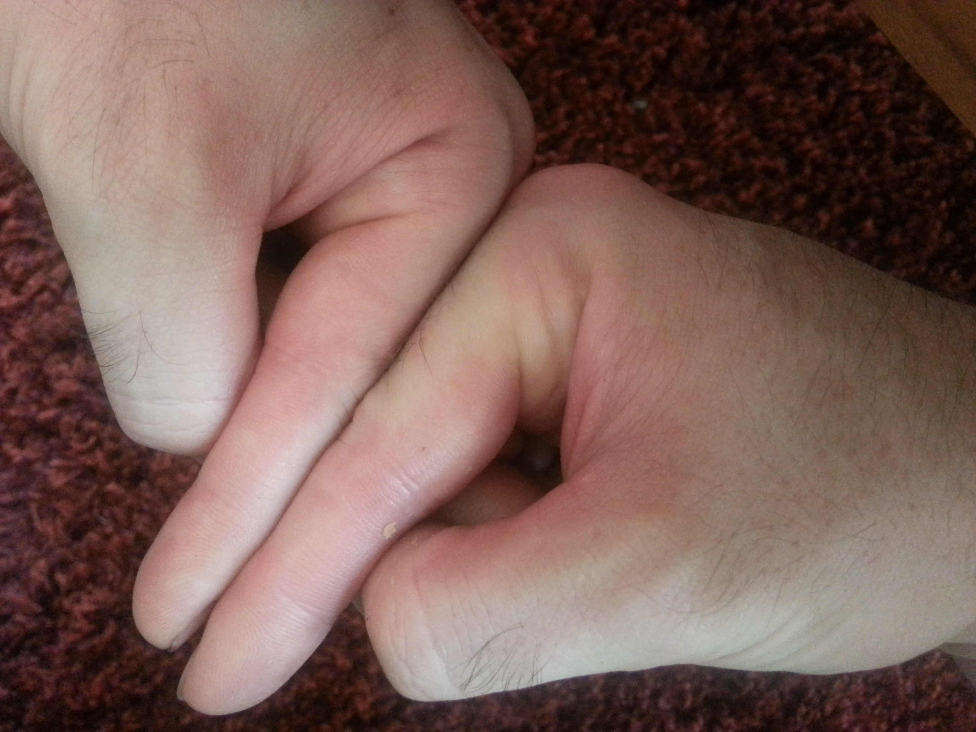
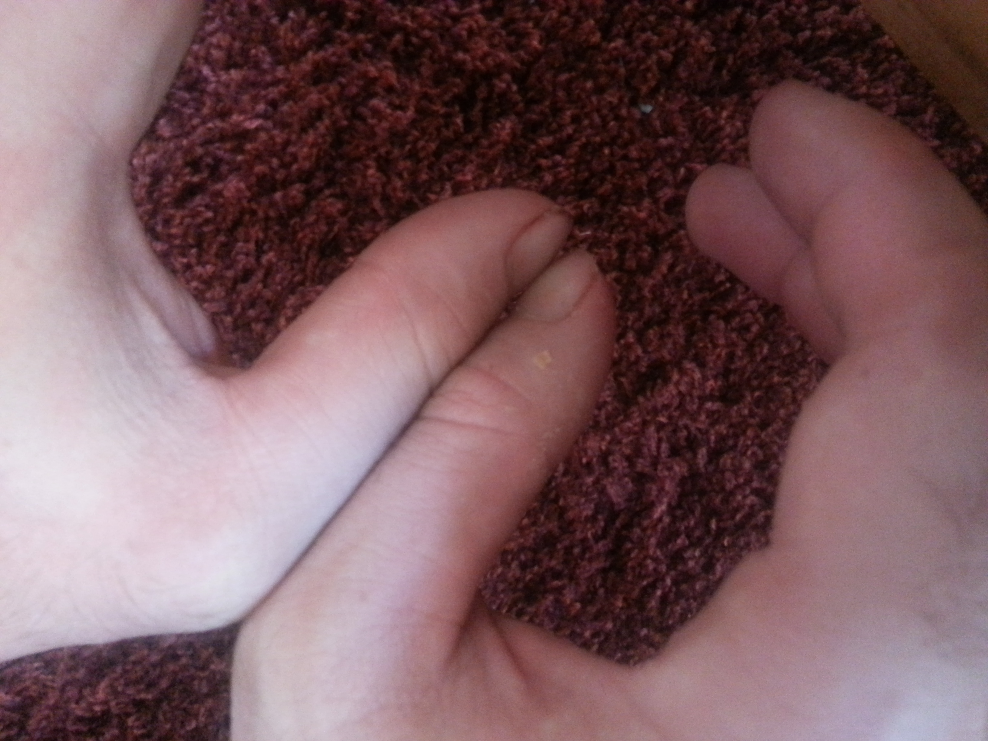
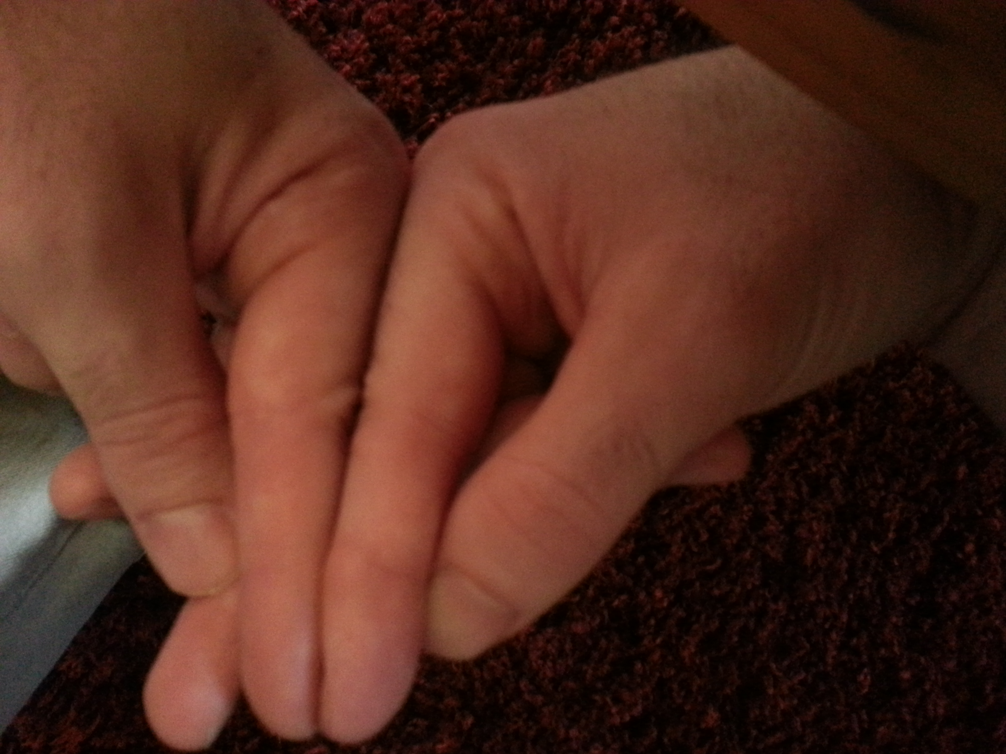