Author Archives: Tyler

Anti-epilepsy medication may increase height

P8 – 2228: The effects of anti-epileptic drugs on the growth plates of infant rats

“Epilepsy is among the most common neurologic disorders in childhood. Levetiracetam (LEV) and valproic acid (VPA) are widely used drugs in the treatment of children with epilepsy. The adverse effects of both drugs on bone metabolism and growth is defined in the literature. The present study aimed to investigate the effects of these two common anticonvulsant drugs on longitudinal bone growth in rats.

Twenty-four infant rats divided into three groups. The study groups received VPA (600 mg/kg/day) and LEV (150 mg/kg/day) for two weeks respectively. Normal saline was applied to the control group. The rats were sacrified after the appliance of the drugs and the rat femurs were removed. The thickness of the growth plates were measured by histopathologically.

All groups included four males and four females. The thickness of the growth plates were larger in VPA and LEV groups versus control group (p<0.05). In addition the measurement of the growth plate in VPA group was larger than LEV group (p<0.05).

We concluded that VPA and LEV both enlarge the growth plate on rats in this study. But this finding does not reflect all steps of endochondral bone growth. The hormonal factors and apoptotic processes also have role on longitudinal bone growth. Further investigations are needed to clarify the adverse effects of anticonvulsants on bone growth and influence on final height.”

Note though that enlargement of the growth plate does not always equal larger longitudinal bone growth.

Comparison of arthritis fingers to my finger results with LSJL

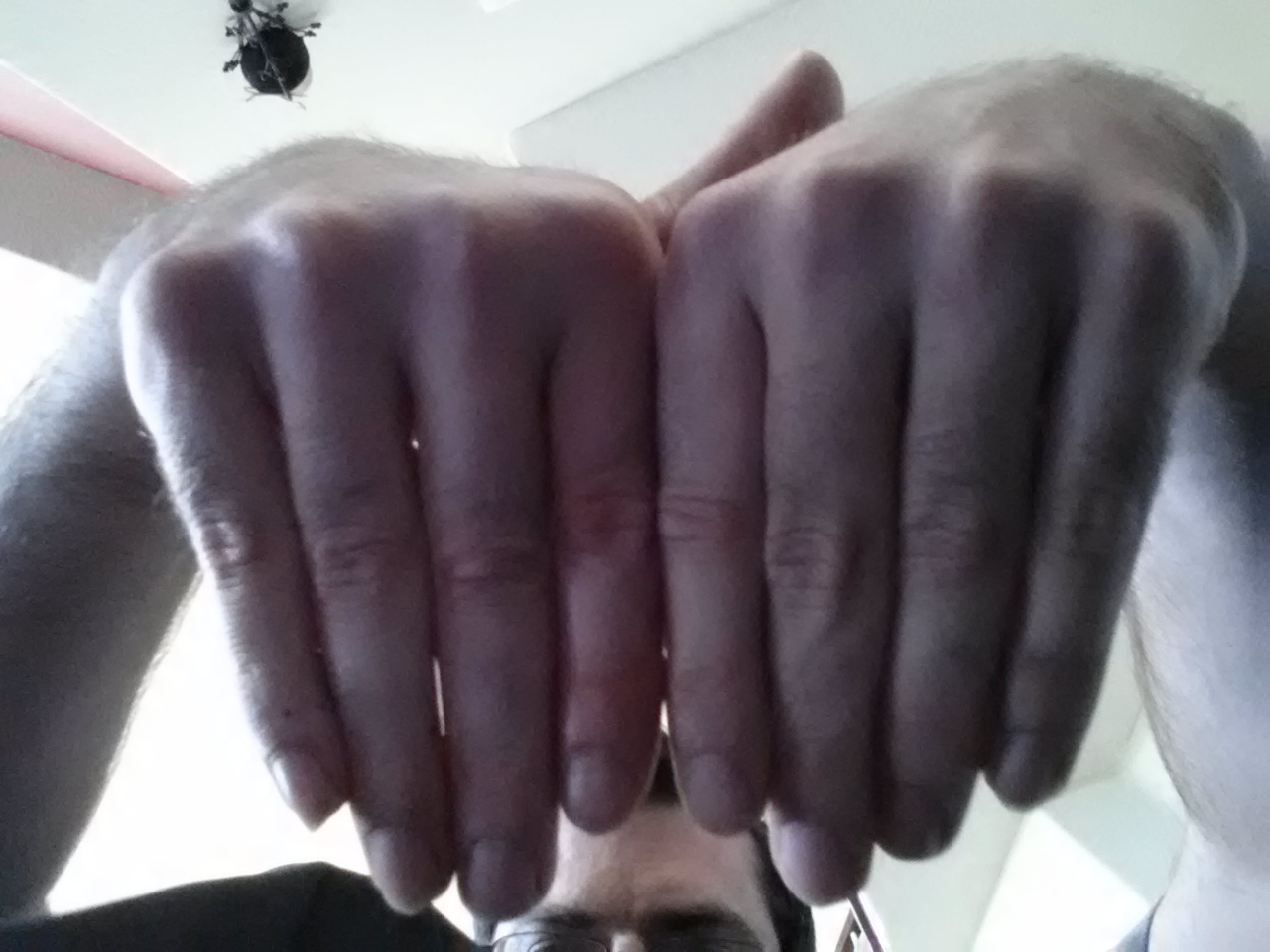
Here’s the pictures of my fingers(you can follow the link on the page to get more images).  I’ve been loading the right index finger with LSJL.

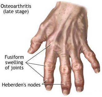
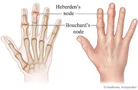
Here’s an image of osteoarthritis fingers:

osteoarthritis fingersIn contrast to heberden’s nodes my fingers are enlarged at the sides and there is no fusiform swelling of the joints.  Fusiform swelling is ” elongated and tapering at both ends; spindle-shaped  ”

“Heberden’s nodes are hard or bony swellings that can develop in the distal interphalangeal joints (DIP) (the joints closest to the end of the fingers and toes). They are a sign of osteoarthritis and are caused by formation of osteophytes (calcific spurs) of the articular (joint) cartilage in response to repeated trauma at the joint.

Heberden’s nodes typically develop in middle age, beginning either with a chronic swelling of the affected joints or the sudden painful onset of redness, numbness, and loss of manual dexterity. This initial inflammation and pain eventually subsides, and the patient is left with a permanent bony outgrowth that often skews the fingertip sideways. Bouchard’s nodes may also be present; these are similar bony growths in the proximal interphalangeal (PIP) joints (middle joints of the fingers), and are also associated with osteoarthritis.”

The question is, is my enlargement of the epiphysis of the fingers an osteophyte or is it a result of endochondral ossification.  Although there are lateral osteophytes.

Heberden’s and Bouchard’s nodes

Osteophyte nodes may be painless so lack of pain in my fingers is not an indication that is not an osteophyte.

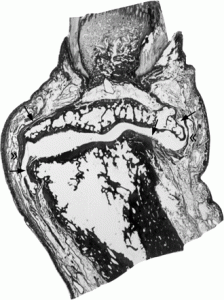
“The osteophytes beneath the lateral nodes arise lateral to the extensor tendon, and although the soft tissues have been distorted by the embalming process and have to be interpreted with caution, it appears that the adjacent capsule is displaced and attenuated.”

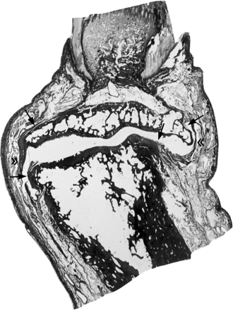
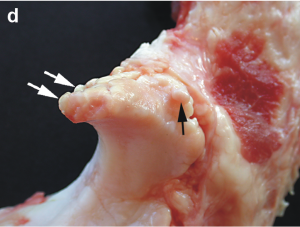
Here’s an image of a growing osteophyte:

growing osteophyteAnd admittedly this does appear to be like enlargement of the epiphysis.

“The true osteophyte, initially a chondrophyte, is intra-articular, developing from the synovium covered surface at the cartilage margin, either from metaplasia of existing synoviocytes or differentiation of precursor cells. The chondral hyperplasia that initiates it is not intrinsically vectored to grow in any particular direction, and in practice grows in the direction of least resistance, to an extent determined by that resistance. If the adjacent cartilage surface is unloaded as a consequence of subluxation or underuse, the osteophyte will grow centripetally over the cartilage surface. More usually it grows peripherally, forming a large pedunculated osteophyte if unopposed, as in the synovial recess of the shoulder joint. Where growth is constrained by strong capsules, as in the lateral ankle, by adjacent ligaments as at the tibial spine, or by tendon insertions as in the upper humerus, growth is inhibited and the osteophyte is small. In essence it will grow until the tension induced in the stretched soft tissues matches that induced by the growing osteophyte. It follows from these known growth characteristics of osteophytes that the regular location of osteophytic nodes at specific sites in these two joints must reflect a local area of low resistance to osteophyte growth unique to these joints. ”

Here’s another image that better shows lateral nodes:

osteoarthritis nodes

Here’s what an osteophyte looks like directly on the bone:

osteophyte on the bone

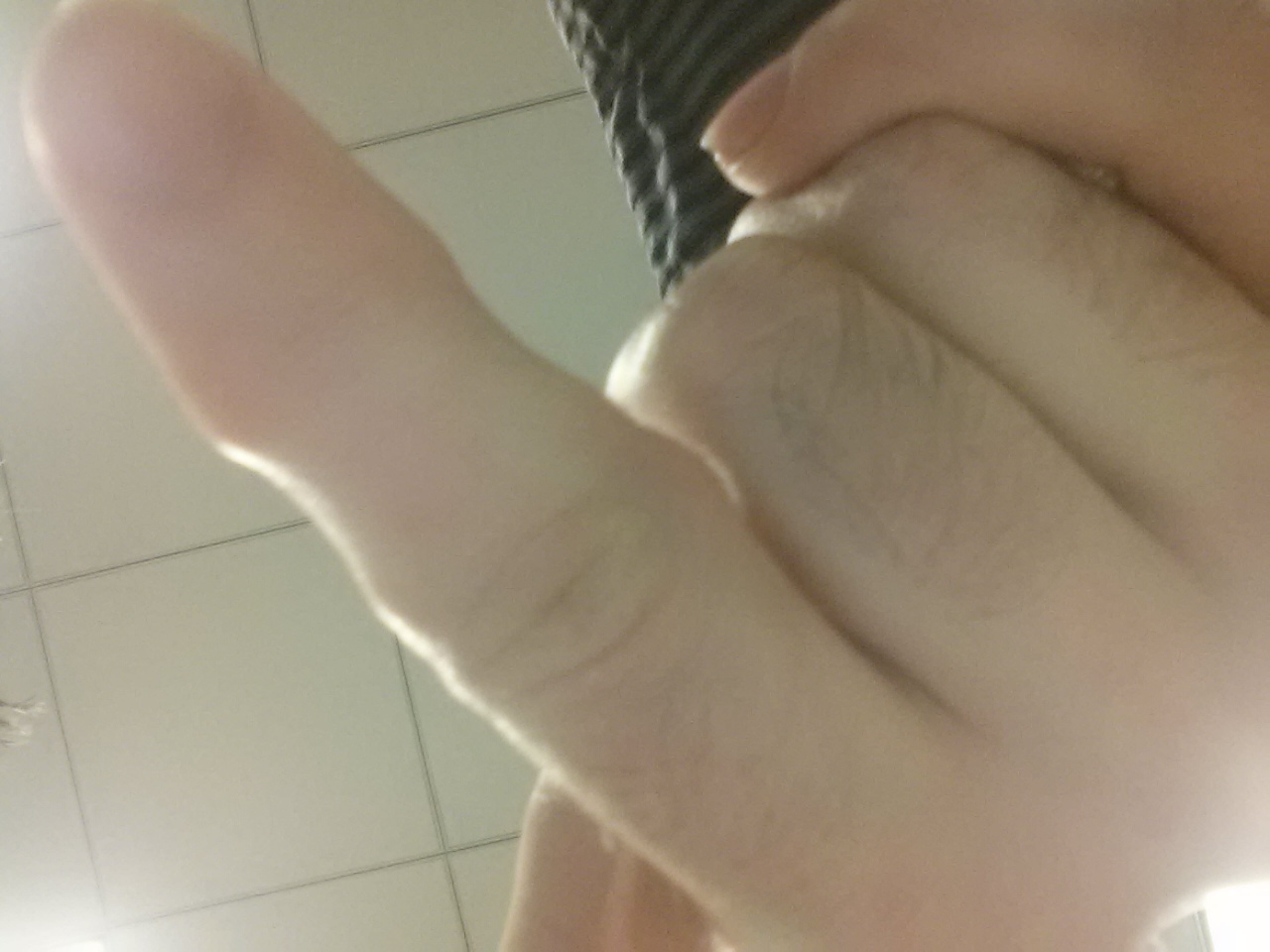
Now here’s another image of my loaded finger so you can see any potential osteophyte nodes:

20140529_132338Definitely node like.  I can’t see any evidence of swelling though.  I’ve seen young people report getting finger nudes at like age 14 so arthritis may cause nodes but arthritis may not be the only source of nodes.  And most evidence suggests that nodes aren’t painful and don’t limit mobility of the finger.

I couldn’t find any evidence upon searching that these nodes increase finger length although admittedly it is incredibly hard to use a search to find anything so that does not guarantee that there are not some anecdotal reports that people got longer fingers due to nodes.

So I conclude:  LSJL caused increase in bone length and osteophytes and these two effects may be independent.  I am using as much load as I can to get more finger results this may be overload and what is causing osteophytes.  However, less load may be possible to increase finger length without causing osteophytes.  I increased arm length with no evidence of osteophytes(unfortunately I did not document this).

LSJL update-Another finger angle and wingspan

Call to action: In the comments section, post anecdotal evidence of people increasing bone length due to tensile strain(stretching).  Example exercises would be inversion(If legs are stretched), hanging, or gripping weights.  Normal stretching would not be sufficient as that would put most of the load on the muscle and not the bone.

Also, please post suggestions on how to do a before and after finger(or toe) experiment that does not require expensive x-rays.  Preferably, a suggestion that only requires easy to take pictures.

Here’s a picture that proves that my right finger is now longer:

20140521_124303You can definately see that the right index finger is longer as it is aligned at the tips and yet the knuckle sticks out higher.  Also, the left thumb seems to be bigger too which I have also been loading with LSJL.

If you haven’t already click on the last post on the finger progress here.

Here’s another image very similar to one of the images on that page:

20140519_122107

This image may look similar to the image in the other but the other image was aligned at the knuckles with the right hand being slightly higher so that I can’t be accused of knuckle manipulation.  This picture is aligned by the tip of the finger tips.  You can see in this image that the right handed knuckle is above the left handled knuckle.  Meaning that the right finger(the one I’ve been performing LSJL on) is longer.

Also, I hadn’t measured my wingspan in a while and previous wingspan measurement was 72.5″(I like a lot of people have a longer wingspan than my height).  This was about 5 years ago.

Recently, I measured my wingspan again and it was 189cm or about 74.4″.  I haven’t really been measuring the wingspan as I didn’t have faith in the methodology of LSJL on the arms.  I use the irwin quick grip to clamp the elbows but the bone structure there is clunky.  I can get a good solid clamp on my wrists though.

This has given me faith that LSJL doesn’t need the extreme force that you can generate on your fingers.  However, unlike the legs it is much easier to generate tensile strain on the bones of the arms(although you can generate tensile strain on the legs via inversion).  Although, we’ve pretty much ruled out axial loading as a method to increase height we haven’t quite ruled out tensile loading.  So it’s possible that the increase is due to hanging or holding onto dumbells or barbells which would place a bit of a stretching force on the bone.

An increase in wingspan at adult age due to farmer’s walk or hanging is less likely to be reported than an increase in height and I don’t know for sure how many people did inversion for their legs and to what extremes.  So I can’t say for certain that the gain was due to LSJL and not tensile loading.

And I don’t have before pictures aside from just me standing but this does give me faith that LSJL can generate height at less extreme forces than one can generate on the finger.  Although 1 inch per arm over five years is pretty slow.

Breakthrough study about microgrowth plates

This study is important as although the scientists conclude that interstitial growth of bone is not possible(which would be a huge breakthrough for height growth) it provides a key term to describe the micro-growth plates that are the goal to be formed by LSJL(pseudoarthrosis).  Although this term refers to a fracture case by definition it does show that a microgrowth plate can make a bone longer.  The scientists suggest two possibilities as to how a bone can grow longer after spinal fusion:  either institial growth of the bone itself or psuedoarthrosis(microgrowth plates).  The scientists even state that microscopic areas of pseudoarthrosis may be be responsible for the lengthening in the conclusion.

Thus this study provides additional evidence that microgrowth plates can lengthen bone(as any amount of cartilage is more capable of interstitial growth(which increases bone length) unlike bone) and gives a new term to search for as a key to growing taller: pseudoarthrosis.  The key is to find instances of psuedoarthrosis in adults that are spontaneous and not a result of surgery.  If you want to help find a way to grow taller, helping find such studies would be a great boon.

Bone Growth after Spine Fusion A Clinical Survey

Full study is at the link below.  It should be noted that this study is quite old but it’s still important today because now the focus is more on the gene expression rather than the mechanics as in the old days.  And it’s easier for us to manipulate the mechanics rather than the gene expression.

bone growth after spinal fusion

“When a spine fusion is unquestionably solid and fairly massive, there is little increase in length of the fused area. The small increase that most of the cases we studied showed could be accounted for by magnification and other technical factors, but it is impossible to rule out a small amount of growth. The slight decrease in the kyphos in two of our cases suggests some bending of the fusion mass. No definite pseudarthroses could be seen on the roentgenograms but the presence of one or more pseudarthroses could not be ruled out. Microscopic and transient pseudarthroses are considered by us to be the most likely mechanism by which any real increase in length or any true change in angulation occurs. In our experimental studies, microscopic losses of bone continuity in transepiphyseal bone grafts in the distal femora of young rabbits were demonstrated. These defects were of such a nature that they could not be demonstrated by standard clinical or roentgenographic methods.

In our opinion, the end-result study of Hallock and his associates is valuable in that it indicates, from the practical, clinical point of view, what will happen to the average patient after spine fusion in early childhood. However, their data and the observations of the other authors previously mentioned convey an inaccurate and perhaps unintentional impression that considerable growth occurs in a solidly fused spine segment. It would be unfortunate, we believe, to allow this impression to persist since surgeons not familiar with all that is known about growth of the spine after spine fusion might be falsely encouraged on the basis of published data to perform longer and more massive spine fusions in young children. Our study suggests that a long, massive, and completely solid fusion in early childhood will impair spine growth to a significant degree.

We believe that growth of a fused segment of the spine can occur only at the ends of the segment or at the site of gross or microscopic defects in the fusion plate. Pseudarthroses in spine fusions in children are much more frequent, in our opinion, than is generally suspected because of the tension forces exerted by the growing epiphyseal cartilages, as well as the usual stresses caused by motion. These pseudarthroses or stress fractures may be microscopic or grossly visible; they may occur spontaneously at any time and heal spontaneously. The more massive the fusion plate, the less chance there will be that it will break down under the stress of growth and motion. Finally, we believe that the laws that govern bone growth in general apply to the bone of spine fusions. There is in our opinion no such thing as interstitial growth of bone.”

“all growth in [bone] length of the diaphyses of long boneses takes place at the epiphyseal cartilages, whereas growth of bones its other dimensions occurs through hyaline-cartilage proliferation as in the epiphysis or  through fibrous-tissue proliferation-as in the periosteum and flat bones.”

Scientists reported the apparent growth of fused spinal bones.  If this were to happen in the solidly fused spine two conclusions could be made: either the fusion plate broken down or there was interstitial growth of bone in the fusion plate.

Scientists also observed that  when interbody fusion was performed on the spine.  The bony bridge that was formed elongated in response to vertebral growth.  However, another study found that the fused area remained firm and did not increase in length in response to overall vertebral lengthening.

Another study with spinal fusion found that psuedoarthrosis occured at any interspace[Small hole surronded by bone].  An example of the interspace is perhaps the trabeculae.  Pseudoarthrosis occurs at fracture areas.  The scientists theorized that in their study the longitudinal bone growth was due to these areas of pseudoarhtrosis.

The study mentions Sincher’s law which states that: “increase of pressure or tension beyond the limits of tolerance leads to destruction of bone by resorption.”<-Perhaps LSJL needs to cause this destruction of bone in order to allow for microgrowth plates or psuedoarthrosis and that’s why so far LSJL on the finger has been more successful due to greater ability to increase pressure.

Next steps for proving LSJL

In my last LSJL progress update, the conclusion most people seemed to feel was that people tended to agree that the right index finger was indeed longer than the left index finger but that they weren’t positive that it was due to LSJL and that it wasn’t due to my right index finger being naturally longer than the other.

So here’s some things I’m going to try going further.  I’m going to continue to clamp my right index finger to try to increase the difference from the left index finger.  I’ve also been clamping my left thumb to see if I can get a difference there and there does seem to be a progression.

But this does not increase height which has a lot of social value.  For ankle clamping, I was clamping between the tibia and the talus.  Well, I realized that my talus grew bigger.  What if the calcaneus grew bigger than the whole body would be elevated higher increasing height in a manner akin to high heels or shoe lifts.


feet bones

The bone above the calcaneus is the talus(which has grown bigger and may in fact contribute to some of my height gains).  Getting enough force on the calcaneus to induce bone growth is easier as it doesn’t necessarily need to grow by the growth plate as there are no constraining factors on appositional bone growth like the joint does for the long bones.

I’ve done a few clamping sessions aiming for clamping the point between the talus and calcaneus and things seems to be going well.  It’s not as smooth as clamping the fingers as the bones are oddly shaped but I seem to be generating pressure and mechanical strain on the calcaneus.

I think showing calcaneus growth will be a way to inspire people to believe that growing taller is possible and it will also increase the height measurement too.  I’m still working on growing the legs evidence just points to that growth will be slow until a better clamp is developed that can generate more pressure.

Ischemia

Michael talked about Ischemia in the past here.  I found a study earlier that suggested that ischemia of cartilage canals may reduce height growth.  Since intense clamping transiently affects the blood vessels to the cartilage it is important to study this.

Chondro-osseous growth abnormalities after meningococcemia. A clinical and histopathological study.

“The cases of nine children who survived the acute stage of meningococcal septicemia and secondary disseminated intravascular coagulation were reviewed. All of the children had major orthopaedic problems as a result of the acute disease. Detailed histological studies were performed on specimens of bone and cartilage, obtained when these patients had either acute amputation for gangrene or subsequent revision for a chondro-osseous deformity. In the specimens that were obtained from the children who had acute gangrene, the histological changes included small-vessel thrombi[blood vessel clot], osteonecrosis, subperiosteal new-bone formation, cortical disruption, cellular disorganization in the physis, and medullary inflammation. These findings were compatible with a combination of inflammation (acute osteomyelitis) and ischemia. In the specimens that were obtained during revision of the amputation, three years or more after the initial infectious or ischemic process, the clinically relevant findings involved the epiphyses and physes. The growth plates showed variable permanent ischemic damage. Bone bridges connecting the epiphysis and metaphysis were observed in various stages of formation, including several early bridges with involvement of only the physis and metaphysis. Endosteal and cortical bone, in contrast, showed complete recovery with no evidence of permanent ischemic damage. We concluded that children who survive meningococcal septicemia are at high risk for complex orthopaedic problems, both acute and chronic. The disseminated intravascular coagulation and focal infections of the acute phase are primarily responsible for the vascular injuries to the growing chondro-osseous tissues. Ischemic changes also selectively involve the physeal circulation, but may take several years to adversely affect longitudinal and transverse growth of bone.”

Image of a growth plate clot:

clot in the growth plategrowth plate clot

Growth plate with abnormal “micro” growth plate like structure:

enchondroma