
Author Archives: Tyler

Osteoarthritis and Ligaments

Loading ligaments may be able to contribute to height.  Ligaments connect to bone via entheses.  Loading ligaments may be able to alter bone and in our case ideally causing formation of new growth plates.

How does bone and surrounding cartilage affect the surrounding ligaments?

High-resolution ultrasound evaluation of extrinsic wrist ligaments in patients affected by rheumatoid arthritis.

“Twenty-one consecutive patients affected by RA (12 men, 9 women; mean age 57 ± 14.6 years) were compared with 21 controls (12, 9; 54 ± 12.1, respectively). Wrists were evaluated using ultrasound on both palmar and dorsal sides along each ligament, using carpal bones as references. The following ligaments were studied: radioscaphocapitate, radiolunotriquetral, palmar ulnolunate, palmar ulnotriquetral, dorsal radiotriquetral, dorsal ulnotriquetral, and radial collateral ligament. Ligament number and thickness were noted. Echotexture was rated as fibrillar, fragmented, or heterogeneous; the surface was rated as smooth or blurred.

The number of palmar ulnolunate and palmar ulnotriquetral ligaments detected by ultrasound in patients was significantly lower than in controls. All ligaments had significantly more fragmented or heterogeneous echotexture and blurred surface and were significantly thinner in patients than in controls. No correlation was found between ligament thickness and RA duration or clinical parameters.

Extrinsic wrist ligaments were less detectable and thinner in patients affected by RA compared with healthy volunteers matched for age and sex. Ligament thinning did not directly correlate with RA duration and clinical parameters.”

“Damage in RA mainly originates from the synovium in the joints and tendon sheaths. During the course of the disease, inflamed synovial tissue may extend to bone, tendons, capsule, and ligaments”<-And possible the refuse may be true in which stimulus from the ligaments progresses into the bone.
What loads are generated on the bone during typical exercise?

The generation of loads in excess of the osteogenic threshold by physical movement.

“stresses in bones in excess of the osteogenic threshold will stimulate bone growth; however, protocols for the generation of these stresses had not been established. Two trial movements were examined in the study: the plié and a movement requiring the subject to move a leg sequentially to 45° displaced positions – the star excursion balance test. Using inverse dynamics and an optimisation approach, the loads in the muscles crossing the hip and knee joints and the corresponding joint contact forces were calculated. It was found that the osteogenic threshold was exceeded in both these trials identifying them as suitable exercises in the maintenance of bone health. In the order of increasing bone load at the hip, and hence increasing bone growth stimulation, are the following demi plié, star excursion balance test with maximum reach criterion, grande plié and star excursion balance test with maximum speed criterion. In the order of increasing bone load at the knee are demi plié, grande plié, star excursion balance test with maximum reach criterion and star excursion balance test with maximum speed criterion. However, due to the high loads encountered, these exercises are not recommended for subjects with advanced osteoporosis although the boundary between therapeutic bone loading leading to increase in bone mineral density and loads capable of causing fracture is unclear.”

“BMD fracture thresholds:110 mg/cm^3 in the distal femur and 70 mg/cm^3 in the proximal tibia”

“threshold of stress at which bone growth is stimulated corresponding to a stress of approximately 20MPa, which for a bone’s Young’s modulus of 20 GPa is 1000me.”

“As the lever arm at which the muscles act at the joint is typically relatively small compared to the segment length to which they are attached, the loads in the muscles and hence the compressive forces in the joint and bone can be very large”

“during a quasi-static rise from a deep squat, the tibio-femoral joint forces were 5.5–6.6 times the body weight.”

“a hip joint contact force of 2.38 times body weight occurred while walking at 4 km/h, 2.51 times body weight during stair ascent and 2.60 times body weight during stair descent.”

The star excursiion balance test generated a force around 6 times bodyweight in the knee.

What is the biomechanical method of bone formation?

If we understand how biomechanical bone formation is induced from mesenchymal stem cells we can induce new bone growth to reinvigorate longitudinal growth.

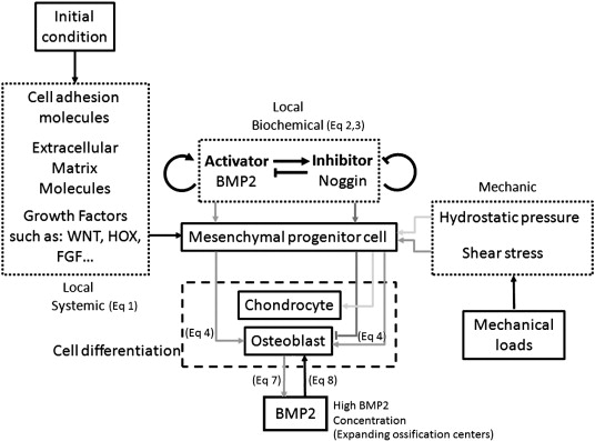
A computational model of clavicle bone formation: A mechano-biochemical hypothesis.

“Clavicle development arises from mesenchymal cells condensed as a cord extending from the acromion towards the sternal primordium. First two primary ossification centers form, extending to develop the body of the clavicle through intramembranous ossification. However, at its ends this same bone also displays endochondral ossification.  [These] embryological events [occur in] two serial phases: first formation of an ossified matrix by intramembranous ossification based on three factors: systemic, local biochemical, and mechanical factors. After this initial phase expansion of the ossified matrix follows with mesenchymal cell differentiation into chondrocytes for posterior endochondral ossification. Our model provides strong evidence for clavicle formation integrating molecules and mechanical stimuli through partial differentiation equations using finite element analysis.”

chondrocyte formation

“areas of low octahedral shear stress and high hydrostatic stress promote bone formation by endochondral ossification. On the contrary, areas of octahedral shear stress result directly in osteogenic induction.”

hydrostatic stress promotes mesenchymal cell into a cartilaginous differentiation pathway, whereas octahedral shear stress stimulates mesenchymal cells to differentiate into bone.”

Signaling pathways in normal chondrocytes versus LSJL gene expression

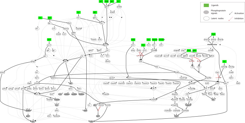
Modeling of signaling pathways in chondrocytes based on phosphoproteomic and cytokine release data.

“The signaling pathways downstream 78 receptors of interest are interrogated. On the phosphoproteomic level, 17 key phosphoproteins are measured upon stimulation with single treatments of 78 ligands. On the cytokine release level, 55 cytokines are measured in the supernatant upon stimulation with the same treatments. Using an Integer Linear Programming formulation, the proteomic data is combined with a priori knowledge of proteins’ connectivity to construct a mechanistic model, predictive of signal transduction in chondrocytes.
We were able to validate previous findings regarding major players of cartilage homeostasis and inflammation (E.g. IL1B, TNF, EGF, TGFA, INS, IGF1 and IL6). Moreover, we studied pro-inammatory mediators (IL1B and TNF) together with pro-growth signals for investigating their role in chondrocytes hypertrophy and highlighted the role of underreported players such as INHBA, DEFB1, CXCL1 and Flagellin, and uncovered the way they cross-react in the phosphoproteomic level.”

“up-regulation of the SOX9 transcription factor induced by TGFB or FGF stimulation leads to collagen synthesis. On the other hand, over-activation of NFKB induced by several pathways (E.g. Inflammation related pathways or bone development processes leads to the release of MMPs and collagen degradation.”

” a large number of stimuli raised a significant response in chondrocytes, activating at least one phosphoprotein signal. As positive control observations, well known players such as IL1B, TNF, EGF, TGFA, INS, IGF1 and IL6{up in LSJL} responded as expected from previous studies. The pro-inflammatory mediators IL1B and TNF activated IKB, HSP27{note this is reduced by Lithium}, MAPK14 (p38) and JUN{up in LSJL}, already known to promote inflammation in cartilage, together with pro-growth signals such as CREB, ERK, GSK3, IRS1 and MAP2K1 (MEK12), validating their role in chondrocyte hypertrophy”

” pro-growth stimuli such as EGF, TGFA and INS activated only anabolic pathways, leaving inflammation related signals unaffected.”

Stimuli also found to affect chondrocytes include “INHBA (Inhibin beta A), ADIPOQ (Adiponectin), DEFB1 (Defensin beta 1), BTC (Betacellulin){up in LSJL}, CXCL1{highly upregulated by LSJL}, HBEGF{up}, IL19, CXCL10, ODN2006 (TLR9 ligand), NOG (Noggin) and Flagellin.”

chfigure2_v2

“Major inflammatory mediators such as IL1B and TNF, signal through their receptors to IKB, MAPK14, HSP27 and JUN. IL1B also activates CREB and MAP2K1 (growth related signals) via TRAF6″

” Pro-growth stimuli such as TGFA, BTC, EGF, IGF1, INS and FGF2 signal through GRB2 to SOS, RAS and from there either to MAP2K1 (MEK12) via RAF1, or signal through PI3K to AKT and to CREB. IL6 activates mostly STAT3 via JAK1.”

“CXCL1, a small cytokine of the CXC family, binds to CXCR2 and activates RPS6KA1. HBEGF, a ligand of the EGFR, signals via the same pathways as EGF, BTC and TGFA. DEFB1, a TLR ligand, signals through TLR4 to RAC1 and from there to the MAPKs and finally activates HSP27 demonstrating pro-inflammatory action. Flagellin, also a TLR ligand, signals through TLR5 to MYD88 and then merges with the IL1 pathway activating major inflammatory signals, CREB and MAP2K1. INHBA, a ligand of the TGFBR, signals via the MAPKs to activate JNK and P53.”

Could what’s in the drinking water affect height?

Since drinking water contains Arsenic and Fluoride which can affect gene expression, it’s conceivable that drinking water could affect height growth.

Arsenic and fluoride co-exposure affects the expression of apoptotic and inflammatory genes and proteins in mononuclear cells from children.

“Humans may be exposed to arsenic (As) and fluoride (F) through water consumption.  Herein, the expression of cIAP-1, XIAP, TNF-α, ENA-78[also known as CXCL5 which is downregulated by LSJL], survivin, CD25, and CD40 was evaluated by RT-PCR. Additionally, the surface expression of CD25, CD40, and CD40L on peripheral blood mononuclear cells were analyzed by flow cytometry, and TNF-α was measured by western blotting. This study examined 72 children 6-12 years old who were chronically exposed to As (154.2μg/L) and F (5.3mg/L) in drinking water and in food cooked with the same water. The urine concentrations of As (6.9 to122.4μg/L) were positively correlated with the urine concentrations of F (1.0 to 8.8mg/L). The CD25 gene expression levels and urine concentrations of As and F were negatively correlated, though the CD40 expression levels were negatively correlated only with the As concentration. Age and height influenced the expression of cIAP-1, whereas XIAP expression was correlated only with age. Additionally, there was a lower percentage of CD25- and CD40-positive cells in the group of 6- to 8-year-old children exposed to the highest concentrations of both As and F when compared to the 9- to 12-year-old group (CD25: 0.7±0.8 vs. 1.1±0.9. CD40: 16.0±7.0 vs. 21.8±5.8). PHA-stimulated lymphocytes did not show any changes in the induction of CD25, CD69, or CD95. In summary, high concentrations of As and F alter the expression patterns of CD25 and CD40 at both the genetic and protein levels. These changes could decrease immune responses in children exposed to As and F.”

“Studies of As-exposed populations have demonstrated unusual gene expression profiles for cell cycle control-related factors, transcription factors, and inflammatory molecules”

“Using microarrays, our group determined that the IL-6, IL-1β, TNF-α, TGF-β, CD40, IL-2RA, CD40L, CD25, ENA78, SURVIVIN, XIAP, and IAP-1 genes are differentially expressed in adults chronically exposed to high concentrations of As (22.5–148.9 μg/L) and F (2.3–5.4 mg/L) compared to the control group (As: 0.3–1.4 μg/L; F: 0.1–0.7 mg/L)”<-Not strong height altering genes except possibly TGF-Beta.

“cIAP-1 expression was positively correlated with age and height”<-Can clAP-1(also known as clasp1) affect height?

XIAP and TNFalpha were also associated with height but not above statistical significance but closer than the rest of the genes.  p<0.15.  TNFalpha definitely affects height but there is it is likely that there’s an optimal quantity of TNFalpha for height growth rather than it just being good or bad.

This study could link Clasp1 to height growth:

Association of TALS developmental disorder with defect in minor splicing component U4atac snRNA.

“The spliceosome, a ribonucleoprotein complex that includes proteins and small nuclear RNAs (snRNAs), catalyzes RNA splicing through intron excision and exon ligation to produce mature messenger RNAs, which, in turn serve as templates for protein translation. We identified four point mutations in the U4atac snRNA component of the minor spliceosome in patients with brain and bone malformations and unexplained postnatal death [microcephalic osteodysplastic primordial dwarfism type 1 (MOPD 1) or Taybi-Linder syndrome (TALS); Mendelian Inheritance in Man ID no. 210710]. Expression of a subgroup of genes, possibly linked to the disease phenotype, and minor intron splicing were affected in cell lines derived from TALS patients. Our findings demonstrate a crucial role of the minor spliceosome component U4atac snRNA in early human development and postnatal survival.”

” The TALS phenotype includes marked intrauterine and postnatal growth retardation; short, bowed long bones with severe delay in epiphyseal maturation”

“The U4atac snRNA is located within intron 2 of the CLASP1 gene, –682 base pairs (bp) to –556 bp upstream of exon 3. Because of this, TALS mutations could, in theory, alter CLASP1 splicing and/or expression. In silico splice site predictions, real time–quantitative polymerase chain reaction (RT-qPCR) analysis of CLASP1 mRNA levels, and an RT-PCR study of CLASP1 exon 3 splicing in TALS patients revealed no effects of TALS mutations on CLASP1 expression or splicing”

However, the study seems to show a lack of connection of CLASP1 to dwarfism.

Idursulfase

Can Idursulfase affect normal individuals who want to grow taller?

The Effect of Recombinant Human Iduronate-2-Sulfatase (Idursulfase) on Growth in Young Patients with Mucopolysaccharidosis Type II.

“Mucopolysaccharidosis type II (MPS II; Hunter syndrome) is an X-linked, recessive, lysosomal storage disorder caused by deficiency of iduronate-2-sulfatase. Early bone involvement leads to decreased growth velocity and short stature in nearly all patients. [We] investigate the effects of enzyme replacement therapy (ERT) with idursulfase (Elaprase) on growth in young patients with mucopolysaccharidosis type II. Analysis of longitudinal anthropometric data of MPS II patients (group 1, n = 13) who started ERT before 6 years of age (range from 3 months to 6 years, mean 3.6 years, median 4 years) was performed and then compared with retrospective analysis of data for MPS II patients naïve to ERT (group 2, n = 50). Patients in group 1 received intravenous idursulfase at a standard dose of 0.58 mg/kg weekly for 52-288 weeks. The course of average growth curve for group 1 was very similar to growth pattern in group 2. The average value of body height in subsequent years in group 1 was a little greater than in group 2, however, the difference was not statistically significant. In studied patients with MPS II, idursulfase did not appear to alter the growth patterns. ”

So idursulfase therapy only seems to have an impact when used before 6 years of age.

“Mucopolysaccharidosis type II (MPS II, Hunter syndrome, OMIM# 309900) is caused by the deficiency of the enzyme iduronate-2-sulfatase (I2S; EC 3.1.6.13) that is responsible for breaking down heparan and dermatan sulfate (HS and DS) within the cells”

I don’t think idursulfase will have an impact on “normal” individuals.  And it’s only having above a certain threshold of the enzyme affects height.