
Author Archives: Tyler

How does hydrostatic pressure induce chondrogenesis?

A key theory behind LSJL is the use of hydrostatic pressure to induces mesenchymal stem cells into chondrocytes to form new growth plates.  Hydrostatic Pressure alters the expression of some of the genes altered by LSJL.  Although, there are many genes not shared which is not surprising considering that LSJL exerts other forces than hydrostatic pressure and the microenvironment is different between the rat bone and the cell lines used.  Hydrostatic pressure is one of the most consistent stimuli in inducing neo chondroinduction.

Hydrostatic pressure decreases membrane fluidity and lipid desaturase expression in chondrocyte progenitor cells.

“Membrane biomechanical properties are critical in modulating nutrient and metabolite exchange as well as signal transduction. Biological membranes are predominantly composed of lipids, cholesterol and proteins, and their fluidity is tightly regulated by cholesterol and lipid desaturases. To determine whether such membrane fluidity regulation occurred in mammalian cells under pressure, we investigated the effects of pressure on membrane lipid order of mouse chondrogenic ATDC5 cells and desaturase gene expression. Hydrostatic pressure linearly increased membrane lipid packing and simultaneously repressed lipid desaturase gene expression. We also showed that cholesterol mimicked and cholesterol depletion reversed those effects, suggesting that desaturase gene expression was controlled by the membrane physical state itself. This study demonstrates a new effect of hydrostatic pressure on mammalian cells and may help to identify the molecular mechanisms involved in hydrostatic pressure sensing in chondrocytes. ”

“Hydrostatic pressure (HP) is known to reduce lipid membrane fluidity. High HP triggers an adaptive mechanism, during which membrane fluidity is increased by raising the proportion of unsaturated fatty acids.”

“TDC5 cells responded to the change in their membrane fluidity under pressure by modulating the expression of Fads1, Fads2, Scd1 or Scd2. 10 or 20 MPa did indeed significantly inhibit the expression of all four genes after 24 h, while 5 MPa also significantly decreased Fads1 and Scd1 expression.”<-None of the genes were directly altered over significance by LSJL.  The genes were measured one hour after loading so it’s possible that the genes returned to baseline after one hour.
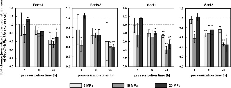
gene expression change to HPWith this  data it’s easy to see why there was no change in gene expression level by LSJL as most of the repression of these genes occurred after 6 or 24 hours under hydrostatic pressure.

“MβCD increased ATDC5 membrane fluidity (laurdan GP values respectively increased and decreased under cholesterol and MβCD treatment”

“Similar to HP, cholesterol increased laurdan GP and significantly inhibited Fads1 and Fads2 expression. By contrast, MβCD[methyl-β-cyclodextrin], which decreased laurdan GP, significantly increased the expression of all four desaturase genes. Together, this suggests that membrane fluidity itself may be the general modulator of desaturase gene expression.”<-Cholesterol has been implicated in controlling endochondral bone growth before.

“the beneficial effects of loading on cartilage are mediated by the transcription factor CITED2, which represses cartilage degradation by the matrix metalloproteinase MMP1.”

“In the human hip joint, loads typically reach 10 MPa during normal activity, with peaks of up to 18 MPa. Considering that interstitial fluid pressure supports about 70 to 90% of the applied load, HP within the cartilage can be expected to regularly exceed 7 MPa and peak at around 16 MPa. 20 MPa is therefore a relatively high pressure”

“How HP is actually sensed by the cell remains poorly understood, but the link between HP and cholesterol suggests at least two hypotheses: both HP and cholesterol may converge on the SREBP pathway, cholesterol by affecting SREBP maturation via SCAP, HP by affecting the membrane fluidity of the endoplasmic reticulum, where non-activated SREBPs reside and which is more sensitive to HP than the plasma membrane; the mechanosensing mechanism of HP could also reside in cholesterol-rich domains like lipid rafts or caveolae”

“membranes rich in unsaturated fatty acids may be compressible enough to deform significantly, and trigger signaling events, even under relatively small pressures. Finally, the plasma membrane is supported by the cytoskeleton, which is mostly incompressible; it is possible that pressurization of the compressible intracellular fluid leads to large deformations in membrane domains unsupported by the cytoskeleton, the change in bulk volume being focused onto a small membrane area.”
Cfm1 and Cfm2

LSJL upregulates Cftr.  Although other than the name I can’t find a connection between this gene and Cfm1 and Cfm2.

According to Hierarchical fine mapping of the cystic fibrosis modifier locus on 19q13 identifies an association with two elements near the genes CEACAM3 and CEACAM6., Cfm1 is located on the same gene as Tgfb1 a height increase gene.  “seven microsatellite markers on 19q13 spanning a 4.8-Mb genomic area encompassing both, TGFB1 and CFM1”

“Cystic fibrosis (CF) is an autosomal-recessively inherited disease transmitted by two defective copies of the cystic fibrosis transmembrane conductance regulator gene (CFTR). The disease manifests as a generalized exocrinopathy, affecting all tissues that express the chloride and bicarbonate channel CFTR ”

Cystic fibrosis can result in shorter height.

Filamin-interacting proteins, Cfm1 and Cfm2, are essential for the formation of cartilaginous skeletal elements.

“Mutations of Filamin genes, which encode actin-binding proteins, cause skeletal abnormalities. The molecular mechanisms underlying Filamin functions in skeletal system formation remain elusive. In our screen to identify skeletal development molecules, we found that Cfm (Fam101) genes, Cfm1 (Fam101b) and Cfm2 (Fam101a), are predominantly co-expressed in developing cartilage and intervertebral discs (IVDs). To investigate the functional role of Cfm genes in skeletal development, we generated single knockout mice for Cfm1 and Cfm2, as well as Cfm1/Cfm2 double-knockout (Cfm DKO) mice, by targeted gene disruption. Mice with loss of a single Cfm gene displayed no overt phenotype, whereas Cfm DKO mice showed skeletal malformations including spinal curvatures, vertebral fusions, and impairment of bone growth, showing that the phenotypes of Cfm DKO mice resemble those of Filamin B (Flnb)-deficient mice. The number of cartilaginous cells in IVDs is remarkably reduced, and chondrocytes are moderately reduced in Cfm DKO mice. We observed increased apoptosis and decreased proliferation in Cfm DKO cartilaginous cells. In addition to direct interaction between Cfm and Filamin proteins in developing chondrocytes, Cfm is required for the interaction between Flnb and Smad3, which [regulates] Runx2 expression. Cfm DKO primary chondrocytes showed decreased cellular size and fewer actin bundles compared to those of wild-type chondrocytes. Cfms are essential partner molecules of Flnb in regulating differentiation and proliferation of chondryocytes and actin dynamics.”

“Inhibitors of actin polymerization stimulate chondrocyte differentiation in cultured mesenchymal cells and murine embryonic stem cells”

“mouse Cfm2 transcripts markedly increased in ATDC5 cells upon differentiation.”

“The CR length and tibial length of Cfm DKO mice were significantly shorter than those of wild-type, Cfm+/−/Cfm2−/−, and Cfm1−/− / Cfm2+/− mice at 4 weeks”

“Cfm1 and Cfm2 bind to Filamins (Flna, Flnb and Flnc)”<-LSJL upregulates Filamin C.

“Flna and Flnb are expressed in chondrocytes”

“Cfm1 and Cfm2 regulate chondrocyte survival and proliferation, and interact with Filamins. Cfms interacted with Filamins to organize perinuclear actin networks and regulated nuclear shape”

“Cfm is required for the interaction between Flnb and Smad3 in chondrocytes, and the Flnb-Cfm-Smad3 complex may play an important role in chondrogenesis.”

“Flnb normally prevents excessive Smad3 phosphorylation.”

“TGF-β1 stimulation up-regulated Cfm1 protein expression, but Cfm2 protein and mRNA were below detection level in an epithelial cell line”

ADAMTS5

This study is by Hiroki Yokota of LSJL fame:

In Vitro and in Silico Analysis of ADAMTS5 Transcription in Human Chondrocytes

“Since Lrp5 is an important mechano-sensitive receptor in Wnt signaling, we examined its role in the mRNA expression of A Disintegrin and Metalloproteinase with Thrombospondin Motifs 5 (ADAMTS5), a major proteolytic aggrecanase that degrades extracellular matrix in articular cartilage. Using genome-wide expression data for C28/I2 chondrocytes with and without Lrp5-specific siRNA, we employed a systems biology approach and built a regulatory network model. Experimental data revealed that silencing Lrp5 significantly altered Wnt signaling gene expression and elevated the mRNA level of ADAMTS5 and several cytokines. A series of experiments using RNA interference showed that the expression of ADAMTS5 was at least in part stimulated by p38 MAPK and IL1β, while Lrp5 acted as a suppressor of their upregulation. Regulatory network analysis using an algorithm predicted the potential involvement of Wnt3a, Myc and CCAAT/Enhancer-Binding Protein β (CEBPB). A secretary factor such as Wnt3a might be involved in Lrp5-mediated homeostasis of ADAMTS5.”

“ow-density-Lipoprotein Receptor-Related Protein 5 (Lrp5) is a co-receptor in the canonical Wnt signaling pathway and global deletion of Lrp5 in mice as well as conditional deletion in osteocytes results in a deficiency in load-driven bone formation and presents a phenotype of low bone mass”

” Mice deficient in ADAMTS5 are protected from cartilage erosion in mouse models of osteoarthritis”<-however erosion of the cartilage is important for endochondral ossification.

“In response to treatment with Lrp5 siRNA, mRNA levels of to 6 interleukin genes (IL6, IL1β, IL12A, IL15, IL18, IL16) were elevated while IL8 mRNA was suppressed”

“Compared with Lrp5 siRNA, IL1β treatment caused a similar elevation to mRNA abundance of IL6, IL1β and ADAMTS5. However, administration of IL1β did not elevate the mRNA levels of IL12A, IL15, IL18, IL8 and IL16”

“both sequences of LRP5 siRNA elevated the phosphorylation of p38 MAPK. Applying p38 siRNA decreased total and phosphorylated p38 MAPK, while a double knockdown with LRP5 siRNA showed a slight recovery in the phosphorylated p38 MAPK level. ADAMTS5 mRNA levels decreased after p38 siRNA. Double knockdown of both p38 siRNA and LRP5 siRNA slightly decreases the elevation of ADAMTS5 mRNA caused by a single knockdown of just LRP5 siRNA.”

How growth plate structure affects longitudinal bone growth

Growth plate stress distribution implications during bone development: A simple framework computational approach.

“We found stress distribution during bone development established osteogenic index patterns that seem to influence locally epiphyseal structures growth and coincide with growth plate histological arrangement.”<-So how stress is applied to the growth plate affects longitudinal bone growth.

“Growth plate characteristics, such as location within the bone, morphology, and width, change according to bone type and age”

“the growth plate morphology of a proximal femoral epiphysis can resemble a concave meniscus at the age of four. With increasing age, at seven the growth plate becomes straight, as a ridged non-uniform line. Last, the growth plate assumes the form of an arch at the beginning of puberty”

“growth plate’s width changes through life, with a wider growth plate during early stages and diminishing progressively until its disappearance toward the end of adolescence”

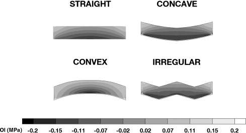
“Stress distribution pattern for all simulations demonstrated peak octahedral normal stress (P) beneath the loading area. Furthermore, highest values for octahedral shear stress (S) were observed in the central zone of the epiphysis.”<-Load was 1 MPa and applied on the top of the epiphysis.

“high OI values predict cartilaginous tissue ossification”

LSJL growth plates became more convex in response to load.

growth plate morphology

“prior to complete epiphyseal ossification OI patterns coincide with growth plate histological organization (reserve, proliferative, hypertrophic) which may be involved in stimulating longitudinal bone growth. Additionally, it was observed that complete epiphyseal ossification has an impact on growth plate stress distribution favoring its maintenance rather than ossification.”

” Since long bone growth results from ossification within the growth plate, we suggest OI values in this structure can be an indicator of expected growth rate. Within the growth plate OI values were affected by all variables studied: localization, morphology, and width”

“we obtained a decreasing trend in OI values from the epiphyseal to the diaphyseal end of the growth plate. Our results evidenced OI peak values in the reserve zone, with OI values comparable to those observed in the epiphysis. The reserve zone is in charge of providing cells to the proliferative zone assuring growth process continuity. Moreover, it has been proposed that resting chondrocytes may synthesize biochemical factors, responsible for growth orientation and inhibition of hypertrophy in the proliferative zone. These events favor growth plate histological organization in distinct zones”

“octahedral normal stress has been reported to linearly increase chondrocyte membrane lipid packing and lipid desaturase gene expression”

Thesis related to LSJL

APPLICATION OF QUANTITATIVE ANALYSIS IN TREATMENT OF OSTEOPOROSIS AND OSTEOARTHRITIS

“The math model of osteoporosis reveals a quick response to salubrinal and a delayed
but substantial response to knee loading”<-knee loading=LSJL

“the epiphyses of the proximal tibia and the distal femur are loaded, increasing intramedullary pressure in the adjacent bones. This pressure leads to interstitial fluid flow, which has been
shown to induce bone formation”<-interesting that the intramedullary pressure leads to the fluid flow and dismissing possible effects of the pressure itself.

Osteoperosis study:

C57BL/6 female mice (8 weeks old) were housed four to five mice per cage at the
Indiana University Animal Care Facility and fed with mouse chow and water ad
libitum.

“Loads were applied in the lateral-medial direction for 3 minutes/day at 15Hz, with peak-topeak
force of 0.5 N.”

“knee loading and salubrinal application elevated BMD via sclerostin and phosphorylated eIF2❛ (p-eIF2❛), respectively”

“Under 4-week salubrinal application, the BMD of ovariectomized mice decreased only 13%, with osteoblast activity increasing 136% while osteoclast activity decreased by 48%. After 4 weeks of knee loading, the BMD of ovariectomized mice decreased only 12%, with osteoblast
activity increasing 136% osteoclast decreasing 34%”

” loading’s suppression of p-eIF2❛ did not propagate to ATF4 and NFATc1.”

“ATF4 was modeled to increase the differentiation rate of osteoblast  precursors to active osteoblasts, while NFATc1 was modeled to increase the production of osteoclast precursors.”

” Loading was found to be more effective when applied earlier, as its effects are long term. Salubrinal seems to have a more immediate effect, thus being more advantageous when applied closer to the data collection time.”

PKDCC

Xenopus Pkdcc1 and Pkdcc2 Are Two New Tyrosine Kinases Involved in the Regulation of JNK Dependent Wnt/PCP Signaling Pathway

Protein Kinase Domain Containing, Cytoplasmic (PKDCC) is a protein kinase which has been implicated in longitudinal bone growth through regulation of chondrocytes formation. Nevertheless, the mechanism by which this occurs remains unknown. Here, we identified two new members of the PKDCC family, Pkdcc1 and Pkdcc2 from Xenopus laevis. Interestingly, our knockdown experiments revealed that these two proteins are both involved on blastopore and neural tube closure during gastrula and neurula stages, respectively. In vertebrates, tissue polarity and cell movement observed during gastrulation and neural tube closure are controlled by Wnt/Planar Cell Polarity (PCP) molecular pathway. Our results showed that Pkdcc1 and Pkdcc2 promote the recruitment of Dvl to the plasma membrane. But surprisingly, they revealed different roles in the induction of a luciferase reporter under the control of Atf2 promoter. While Pkdcc1 induces Atf2 expression, Pkdcc2 does not, and furthermore inhibits its normal induction by Wnt11 and Wnt5a. Altogether our data show, for the first time, that members of the PKDCC family are involved in the regulation of JNK dependent Wnt/PCP signaling pathway.”

” both Pkdcc and Gli3 [may] cooperate on the regulation of long bone formation by modulating the temporal kinetics of columnar and hypertrophic chondrocyte domains establishment”

“Pkdcc could also modulate Wnt signaling, since inactivation of Wnt5a also alters the transition between proliferating to hypertrophic chondrocytes. Since Pkdcc regulates protein export from Golgi, its inactivation may directly interfere with either the secretion of the relevant signals or cell-surface localization of receptors ”

“Cell movements are essential for the correct shape of body axis and organ formation during embryo development. These morphogenetic cell movements are not stochastic, they undergo extensive control by distinct signal transduction pathways. One of this pathways is Wnt/Planar Cell Polarity (PCP) signaling pathway that, for example, in polarised tissue, coordinate the morphogenetic processes of the cells in the epithelial sheets plane. A set of core proteins was identified to be involved in PCP pathway, in both vertebrates and invertebrates. In vertebrates this group include the transmembrane receptor Fizzled (Fz), the cytoplasmic molecules Dishevelled (Dvl), Diego (Dgo) and Prickle (Pk), the transmembranar protein VanGogh/Strabismus (Vang/Stbm) and the cadherin-like protein Flamingo/Celsr1 (Fmg/Clsr1). These core PCP components were identified as genes whose inactivation leads to cell polarity mis-alignment. The PCP is involved in the coordination of cells within a tissue sheet, either by direct cell-cell interaction or under the influence of a diffusible ligand-based signalling system [9]. This occurs because these proteins localize in different regions inside the cell: Fz, Dvl and Dgo are localized in the proximal region, Vangl2 and Pk in distal region and Clsr1 localize in both distal and proximal regions, which is essential for the proper establishment of polarization”

“Pkdcc1 and Pkdcc2 promote recruitment of Dishevelled to the plasma membrane through DEP domain”

” Pkdcc1 alone is able to induce the expression of Atf2-luc, and the activation of non-canonical Wnt signaling. Curiously and contrary, Pkdcc2 is not able to activate Atf2 expression, inhibiting the normal activation of JNK dependent non-canonical Wnt downstream of Wnt11 or Wnt5a”