Tag Archives: articular cartilage endochondral ossification

Study finds that vibration can induce articular cartilage endochondral ossification

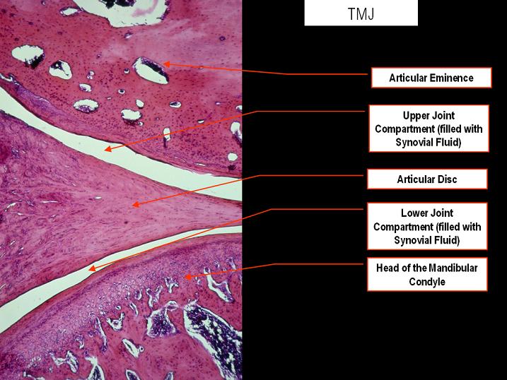
We’ve studied endochondral ossification of the articular cartilage in the condyle before. If vibration can induce endochondral ossification of the articular cartilage in the jaw it is theoretically possible that it could induce endochondral ossification in any joint unless the jaw cartilage is special. Which I don’t believe it is I believe that the jaw is more mobile than other joints and subjected to superior loading. If other joint regions were subjected to the same kind of loading as the jaw they could grow as well.

Effects of Mechanical Stimuli on Adaptive Remodeling of Condylar Cartilage

“Trabecular bone has been shown to be responsive to low-magnitude, high-frequency mechanical stimuli.
This study aimed to assess the effects of these stimuli on condylar cartilage and its endochondral
bone. Forty female 12-week-old C3H mice{this is pre skeletal maturity unfortunately, howver later in the study they say that the mice are “adult”} were divided into 3 groups: baseline control (killed at day 0), sham (killed at day 28 without exposure to mechanical stimuli), and experimental (killed following 28 days of exposure to mechanical stimuli).

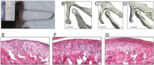
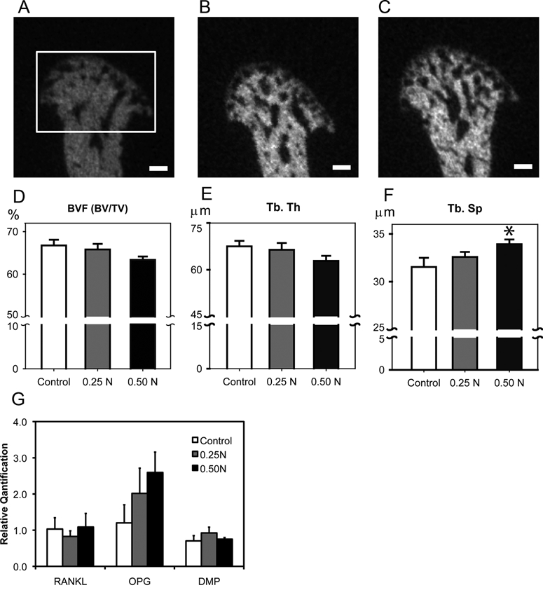
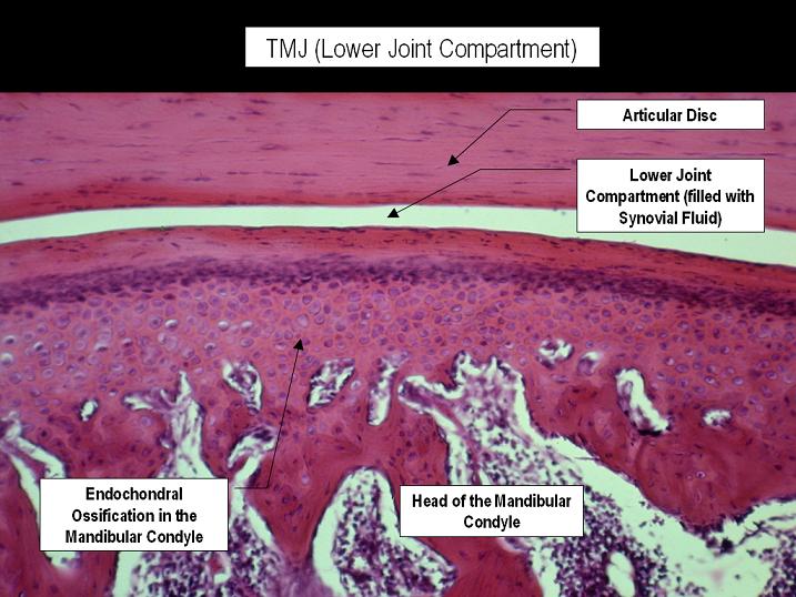
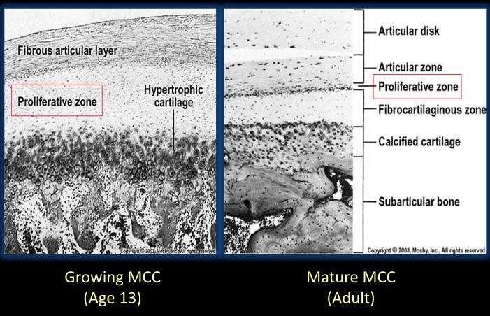
The experimental group was subjected to mechanical vibration of 30 Hz, for 20 minutes per day, 5 days per week, for 28 days. The specimens were analyzed by micro-computed tomography. The experimental group demonstrated a significant decrease in the volume of condylar cartilage and also a significant increase in bone histomorphometric parameters. The results suggest that the low-magnitude, high-frequency mechanical stimuli enhance adaptive remodeling of condylar cartilage, evidenced by the advent of endochondral bone replacing the hypertrophic cartilage{vibration can induce articular cartilage endochondral ossification, if this can occur in other joints with articular cartilage then you can get longer bones by that mechanism!}.”

“there is a linear association between bone-regenerative capacity and bone density”<-So could things that increase bone density increase bone regenerative capacity?

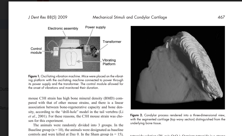
“We achieved mechanical stimulation to the animals by placing them, while still in their cages with bedding removed, directly on a device which generated vertical ground-based vibration. This machine generated 30-Hz pulses, creating peak-to-peak accelerations of 2.9 m/sec2, referred to as a fraction of earth’s gravitational field, 0.3 g (1g = 9.8 m/sec2). Based on Rubin’s previous research, it is believed
that this produces peak strains of approximately 5 μє. Animals in the Experimental group were subjected to 20 minutes’ vibration per day for 5 days per week for a total of 28 days,”<-so the mice were subjected to vibration not specific to the jaw and it still underwent articular cartilage endochondral ossification. It would be interesting if other bones changed as well.

“the LMHF stimuli induced osteogenesis, leading to adaptive growth of the condyle in adult mice.”<interesting that they call these adult mice! But we have to be sure they’re skeletally immature

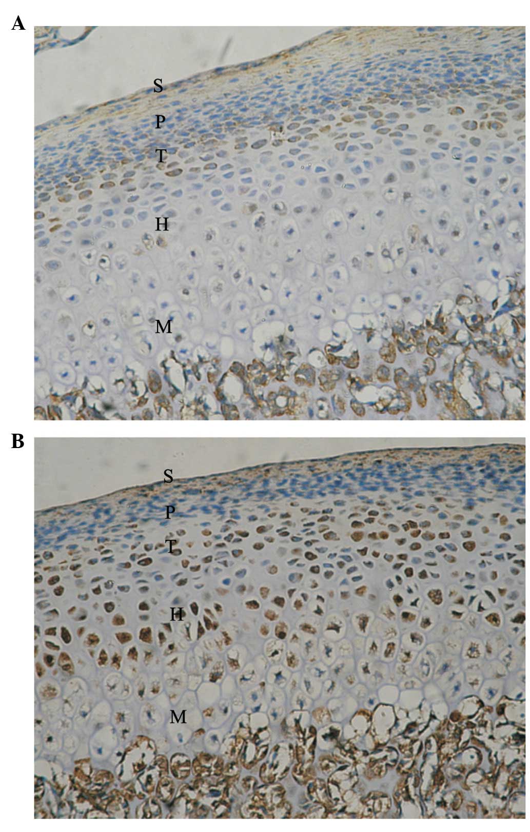
“the adaptive modeling of condylar cartilage is characterized by enhanced transition from chondrogenesis to osteogenesis. The vibrating mechanical stimulation in the present study was shown as a possible mechanism for the acceleration of the modeling.”

“A questionable aspect of this study design is the degree of transmission of the vertical ground-based oscillation, since it diminishes as the signal travels proximally through the skeleton. In a study measuring transmissibility in the hips and spines of humans standing on the oscillating plate, the authors showed
that approximately 80% of a 30-Hz ground-based signal reached the hip and spine”

This study here suggests that vibration can have benefit on growing bone:

Does Vibrational Loading Modulate the Effects of Radiotherapy on Growing Bone?

“[we evaluate] the possible beneficial effects of low-magnitude high-frequency mechanical vibration (LMHFV) stimuli on growing irradiated bone and the possibility for restoration of function of the epiphyseal plate”

ighteen 3-week old weanling male Sprague-Dawley rats were subjected to a standard radiation dose of 17.5 Gray applied to right hind limbs, with the contralateral leg serving as a non-irradiated control. Then, the animals were divided into three groups: A) rats subjected to (LMHFV) only at 45 Hz, 0.3 g for 20 minutes once per day, 7 days/week, for 3 weeks, B) rats subjected the same conditions of LMHFV plus an injection of spermine NONOate, a nitric oxide donor that that has shown weak positive results as post-irradiation recovery agent, and C) rats subjected to sham LMHFV. After euthanizing the animals, skeletal growth was measured by x-ray analysis, marrow mesenchymal stem cell osteoblastic potential was measured by CFU-F analysis, and bone morphology was measured by micro-CT analysis.

X-ray and CFU-F analyses show statistically significant differences between right and left limbs in all groups. No statistical significance was observed between vibration versus control groups, but trends suggest there could be some positive effect of vibration, although not statistically significant. Micro-CT results show a clear difference between right and left limbs in all groups. Regarding vibration versus control groups, micro-CT results are ambiguous, but do suggest that vibration may have altered local growth characteristics and stimulated local shape changes in the 20% region from the distal end of the femur, just above the growth plate.”

“Eighteen 3-week old weanling male Sprague-Dawley rats were obtained from Taconic Farms (Germantown, NY) and randomly divided into three groups: A) rats subjected to low-magnitude high-frequency vibration (LMHFV) only at 45 Hz, 0.3 g for 20 minutes once per day, 7 days/week, for 3 weeks, B) rats subjected to LMHFV with the same conditions as group A plus an injection of spermine
NONOate, a nitric oxide donor , and C) rats subjected to sham LMHFV, placed in cages
used for vibration but with no stimulus applied.”<-so we’re looking at group A versus group C as to whether vibration can increase bone length.

“there was possibly a difference, although not statistically significant, between right femora of different groups (A: 32.6±2.3 vs. B: 31.9±2.2 vs. C: 31.2±1.2, Figure 2), as well as total leg lengths of the femur
plus the tibia between different groups (A: 66.4±2.4 vs. B: 65.3±2.9 vs. C: 64.4±2.1, Figure 4).”<-So vibration group was longer than via group C.

“the ability of physical signals to influence bone morphology is strongly dependent on the signal’s magnitude, frequency and duration”

Pravilo Training Apparatus may help make you slightly taller

I’ve searched for something akin to bite jumping appliances that can induce articular cartilage endochondral ossification which has been shown to occur in the jaw to other joints of the body that can affect height..

There is a stretching apparatus promoted by Pravilo USA that may potentially work to induce articular cartilage endochondral ossification on height affecting joints.

I have stated in the past that the rack apparatus(and sleep stretching) is not likely to work to make you grow taller. First the load is not really on the bones or the articular cartilage. Second the load is not dynamic which has been to shown to be a key mechanism by which the bite jumping appliance in dentistry serves to induce articular cartilage endochondral ossification. Like dynamic movement is important to the bone(because of bones fluid contents and dynamic movement moves the fluid contents around) to stimulate growth, dynamic movement is important to the cartilage and the cells to stimulate the structure of the cells via fluid forces. Ultimately, a lot of the load is going to be on the tendons and ligaments but just be the constant moving around some of the load is surely going to applied to the articular cartilage.

I do not believe the load is going to be in such a way as to stimulate the bone directly(mostly via the enthesis and the articular cartilage). I do not think that applying load in a tensile fashion is necessary to stimulate the bone via torsional force. Torsion will stimulate the fluid contents of bone regardless of whether the torsion is applied in a compressive or tensile fashion.

I do however think this apparatus has possible potential in which to prove articular cartilage endochondral ossification. However, ACEO(articular cartilage endochondral ossification) is very slow so we’d need actual studies and actual going to the gym here and using the device is cumbersome. If we can prove that ACEO work then we can create easier to use devices.

Here’s an example of how Pravilo training apparatus can induce dynamic loading on the articular cartilage:

The twisting of the body is what makes it dynamic.

At about the one minute mark is where you can see the twisting applied:

In the store on the website there is parts to construct your own apparatus. I’m not sure if it’s worth it as ACEO is very slow. It’d be more for proof of concept. However, it could potentially work for your spine and there is no surgical mechanism that increases spinal height.

Ligaments constrain growth

The reason that the jaw can grow via stretching by forwarding positioning and bite-jumping appliances is that it is not as constrained by ligaments as other bones.  It’s possible that other movable joint regions like the wrists, fingers, and toes would also be stretchable.  The wrists would come under extreme stress due to the farmers walk so if this true there should be anecdotal evidence of longer arms due to the farmers walk.  It is possible that a new height increase method could be invented that causes enough tensile strain in the articular cartilage to activate endochondral ossification in that region.  The ligaments holding the bone together would constrain this.  This would also help explain why people with Marfan’s become taller as they have more flexible joints and therefore their growth is less constrained by ligaments.   Here’s another connective tissue disorder that may affect longitudinal bone growth.  It’s also possible that differences between condylar cartilage and articular cartilage allow this to happen in the mandible but not in other joints.

So if anyone could look for instances of longer arms due to heavy farmers walk.  This would be much easier to do than LSJL.  So any research on this would be a great boon to further the cause of height increase.

The adaptive remodeling of condylar cartilage—a transition from chondrogenesis to osteogenesis.

“Mandibular condylar cartilage is categorized as articular cartilage but markedly distinguishes itself in many biological aspects, such as its embryonic origin, ontogenetic development, post-natal growth mode, and histological structures. The most marked uniqueness of condylar cartilage lies in its capability of adaptive remodeling in response to external stimuli during or after natural growth. The adaptation of condylar cartilage to mandibular forward positioning{basically bringing your jaw forward} constitutes the fundamental rationale for orthodontic functional therapy, which partially contributes to the correction of jaw discrepancies by achieving mandibular growth modification. The adaptive remodeling of condylar cartilage proceeds with the biomolecular pathway initiating from chondrogenesis and finalizing with osteogenesis{so basically by stretching the articular cartilage you activate enchondral ossification enabling you to grow on the longitudinal ends of the bone}. During condylar adaptation, chondrogenesis is activated when the external stimuli, e.g., condylar repositioning, generate the differentiation of mesenchymal cells in the articular layer of cartilage into chondrocytes, which proliferate and then progressively mature into hypertrophic cells. The expression of regulatory growth factors, which govern and control phenotypic conversions of chondrocytes during chondrogenesis, increases during adaptive remodeling to enhance the transition from chondrogenesis into osteogenesis, a process in which hypertrophic chondrocytes and matrices degrade and are replaced by bone. The transition is also sustained by increased neovascularization, which brings in osteoblasts that finally result in new bone formation beneath the degraded cartilage.  The repositioning of the mandibular condyle in adult rats led to a reactivation of chondrogenesis in condylar cartilage which otherwise is at resting status, and finally results in increased bone formation{this is huge if as it means a bigger stronger jaw}

“chondrogenic activity of BMP-2 in vitro involves the action of the cell-cell adhesion protein, N-cadherin, which functionally complexes with beta-catenin the change of condyle position relative to the glenoid fossa constitutes an important trigger for [the endochondral ossification related adaptation of the mandible].”

The deviation of the condyle from the glenoid fossa by mandibular forward translation is the basis for orthodontic functional therapy, which aims to enhance condylar growth and therefore to eliminate the discrepancy between upper and lower jaws.

a decrease in compressive loading enhances condylar growth, whereas an increase in loading inhibits growth”

Note that they do say that condylar cartilage is distinct from articular cartilage.

Here’s another study:

Murine TMJ loading causes increased proliferation and chondrocyte maturation.

“The purpose of this study was to examine the effects of forced mouth opening on murine mandibular condylar head remodeling. We hypothesized that forced mouth opening would cause an anabolic response in the mandibular condylar cartilage. Six-week-old female C57BL/6 mice were divided into 3 groups: (1) control, (2) 0.25 N, and (3) 0.50 N of forced mouth opening. Gene expression, micro-CT, and proliferation were analyzed. 0.5 N of forced mouth opening caused a significant increase in mRNA expression of Pthrp, Sox9, and Collagen2a1, a significant increase in proliferation{These alone will not increase height except maybe the increase in proliferation}, and a significant increase in trabecular spacing in the subchondral bone, whereas 0.25 N of forced mouth opening did not cause any significant changes in any of the parameters examined. Forced mouth opening causes an increase in the expression of chondrocyte maturation markers and an increase in subchondral trabecular spacing.”

“The articulating surfaces of the temporomandibular joint (TMJ) are covered by fibrocartilage, which is composed of chondrocytes at various stages of maturation that are organized into four zones: (1) superior articular zone—cells in this zone express proteoglycan 4 (Prg4); (2) polymorphic zone—cells in this zone express SRY-box 9 (Sox9) and parathyroid-hormone-related protein (Pthrp); (3) flattened zone—the cartilage cells in this layer express collagen type II (Col 2a1); and (4) hypertrophic zone—the chondrocytes express collagen type X (Col 10a1)”

“Growth in the condylar cartilage proceeds from cells exiting the proliferative pool in the polymorphic zone and undergoing endochondral ossification in the inferior cartilage zones. The mandibular condylar cartilage is able to remodel its structure in response to mechanical strains

F and G are both forced mouth opening with F and G to a higher degree.  C and D have longer bones but there is less separation between the two bones with C and D.  The fear with articular cartilage endochondral ossification is that you’ll decrease the joint space.

“Functional appliance therapy is believed to place the mandible in a protrusive position, causing the condyle to be unseated caudally and ventrally from the articular fossa, while forced mouth opening is believed to cause a compressive force between the articular eminence and the mandibular condyle”

“A possible explanation for both functional appliance therapy and forced mouth opening causing similar effects in the mandibular condylar cartilage is that the mandibular condylar cartilage responds similarly to different types of loading compression for forced mouth opening and tension for functional appliance therapy{another possibility is that both activate the lateral pterygoid muscle and that’s what pulls on the cartilage}. Another possible explanation is that forced mouth opening and functional appliance therapy influence the masticatory muscles in such a way that the actual deformational strains within the mandibular condylar cartilage are similar for both.”

Forward mandibular positioning enhances the expression of Ang-1 and Ang-2 in rabbit condylar chondrocytes

“Functional appliances correct dental malocclusion, partly by exerting an indirect mechanical stimulus on the condylar cartilage, initiating novel bone formation in the condyle. Angiopoietin is involved in the angiogenesis associated with novel bone formation. This study aimed to determine the expression of angiopoietin (Ang)‑1 and ‑2 following forward mandibular positioning (FMP) in the condylar chondrocytes of rabbits. Sixty rabbits (age, 8 weeks) were randomly allocated to the experimental and control groups (n=30 per group). In the experimental group, FMP was induced by a functional appliance. Five rabbits from the experimental group and the control group were sacrificed following 3 days and 1, 2, 4, 8 and 12 weeks, respectively. The right temporomandibular joints (TMJs) were collected and the expression of Ang‑1 and -2 was evaluated by immunohistochemical staining. The expression of Ang-1 increased at day 3 and reached a peak at 2 weeks, whereas Ang‑2 reached maximal expression 4 weeks after FMP. Subsequently, the expression of Ang‑1 and ‑2 gradually decreased. Thus, FMP enhanced the expression of Ang‑1 and Ang‑2 in condylar cartilage, which is related to angiogenesis in the process of endochondral ossification.”

“Autocrine Ang-1/Tie-2 modulates blood vessel plasticity and contributes to vascular maintenance. In addition, Ang-1 enhances survival, migration and network formation of endothelial cells in vitro, and induces neovascularization in vivo. Ang-2 is a naturally occurring antagonist of Ang-1 that inhibits Ang-1-induced activation of Tie2. Ang-1 and -2 are located at sites of endochondral bone formation in the growing skeleton”

“The appliances were worn for 24 h to produce a continuous forward and downward positioning of the mandible.”

Look at all the cartilage growth.  There seems to be some endochondral ossification going on in the longitudinal ends of bone which is very good for height growth.

Here’s a normal jointNotice how they are connected strongly by a ligament(Li)

Here’s Mandibular Condylar Cartilage as a Juvenile and as an adult: