Monthly Archives: February 2014

Regenerate Articular Cartilage Using Silver Electrode and Direct Current Electrical Stimuli

Regenerate Articular Cartilage Using Silver Electrode and Direct Current Electrical Stimuli

This post will be a followup post to one of the most important posts I have ever written for this website “Electromagnetic Stimuli Will Increase Ossification and Make Bones Longer, Big Breakthrough!“. This is an idea which will definitely lead to at least 1-2 mm of extra increases in height, but it would probably be most applicable for people who are already suffering from some type of cartilage tissue degeneration.

As some of us might know, certain types of arthritis that older people suffer from is from the loss of thickness in the articular cartilage at the end of their long bones, at the joint area. The decrease in thickness means that bones will be rubbing against bone. There is no lubricant. That means that it is extremely painful to walk if your knees have all the cartilage gone.

Apparently if you take an electrode of at least a silver–platinum combination soldered with a resistor in the middle (to lower the current) and then apply a DC current, you can stimulate an area of articular defect to regenerate the hyaline cartilage back.

Regenerate Articular Cartilage

Dr. Becker with Bruce Baker showed in a standard experimental study using lab rabbits which they performed and then sacrificed, that you can regenerate hyaline cartilage back in using this method. Of course their approach was again similar to what they have been promoting for decades.

Here are the key points that they stated which we should remember

1. Use Silver

It turns out that silver is extremely effective in killing bacteria. The way that the silver ions diffuse, somehow they can disrupt the protein layers that form the outer membrane of bacterias (and even some viruses). Somehow Silver is also not poisonous to the human body and its cells.

2. Using a low level Current

You don’t need a very high current to stimulate the cells to do much. It is more likely that high currents would do more damage than good, since the current could cause the cells to go into rapid mitosis and turn malignant. Brighton and Friedenberg used high levels of current (10,000-20,000 nanoamps) but that might not be needed. Becker’s group found that you can use just around 100-200 nanoamps per centimeter of electrode and it would have some effect.

That is also the reason you see in the schematic above (taken from page 189 of his book) there is a resistor. The resistor is used to lower the current to a very, VERY low rate. The rating on the resister is at 10 MEG!!. We are talking about 10 MegaOhms. The voltage you need is less than 1 Volt. Using V=IR, and find for the current, we should understand why just a high level resistor is used. You want just a tiny bit of current, and that is enough apparently.

3. Stimulate close to either the periosteum and/or the marrow, but not the actual bone

Notice how there is a defect made on the joint area purposely on the lab rabbit. One of the electrode tips gets embedded into the bone layer underneath, which has the bone marrow in the epiphysis/ joint head.

Previously I had talked about the virtues of micro-fracture surgery “A Proposed Height Increase Method Using Microfracture Surgery Techniques With Fibrocartilage Formation“. However at the end of that post, I had lamented that it would be fibrocartilage we have to work with, instead of the hyaline cartilage. If you drill holes around the entire perimeter of the bone, you would theoretically for maybe a few days turn the defect into cartilage, which we can actually manipulate. If instead, we can use silver electrodes and small DC electrical stimuli to create our preferred hyaline cartilage, then we would be much, much closer in created a full plan of cartilage which we have work with.

We want the cells in the adult bone marrow (adipocyte derived stem cells) to turn into the type which we want to work with, which is either chondrocytes or cartilage which is stable.

The experiment that they did back in the 1970s-1980s showed that with such a small battery and simple device, you can regenerate hyaline articular cartilage almost completely back.

Something to remember: Bone tissue don’t actually grow.

It is the periosteum and the bone marrow which has the cells that can actually make the bone tissue expand in any way.

Low-Intensity Pulsed Ultrasound LIPUS Does Not Increase Longitudinal Growth Of Bone

Low-Intensity Pulsed Ultrasound LIPUS Does Not Increase Longitudinal Growth Of Bone

Update March 6th 2014: Tyler did leave a response to this post originally but there is now even more evidence found which tips the question of the effect of Low-Intensity Pulsed Ultrasound on bone longitudinal growth to the side of having no affect at all.

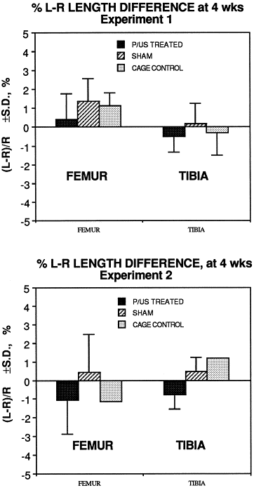
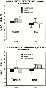
Study #1: Effects of therapeutic ultrasound on longitudinal growth of the femur and tibia in rats

The conclusion for this particular study was that using the three different intensities of LIPUS at 0.5, 1.0 W/cm2, and 1.5 W/cm^2 it was found that the emissions had no effect. There was no inhibitory effect and there was no stimulatory effect. If we were to be extremely accurate, there was a very slight stimulatory effect in the first two groups and an inhibitory effect in the third group, but the slight deviations were too small to be considered significant.

There have been some studies like the Chapter 5 “Low-intensity ultrasound stimulates endochondral ossification in vitro” which shows that using LIPUS is critical when going through the beginning stages of endochondral ossification though.

This recent news is slightly sad to hear but this is one study I did find by Joseph Spadaro, who collaborated with Dr. Robert Becker about 30 years ago, which shows that the LIPUS technology which we had previously believed to work in increasing bone growth (at least density wise) seems to do very little towards the longitudinal growth of bones “Application of low-intensity ultrasound to growing bone in rats.

Abstract Below

Low-intensity pulsed ultrasound recently has been shown to accelerate long bone fracture healing, but its effect on bone growth and development is unknown. The longitudinal growth and bone density of the femur and tibia in young rats was measured after application of an ultrasound transducer emitting 1.5-MHz pulsed ultrasound (30 mW/cm2, SATA) for 20 min/day. After 28 days, no length difference was detected (< or = 2%) compared to the sham-treated leg or to unexposed controls. Also, no significant difference in bone mineral density (BMD) of the femur or tibia was found (< or = 6%). In a repeated experiment in which a periosteal trauma stimulus was created in the femoral diaphysis, the ultrasound also had no effect on growth or BMD. This results suggests that physeal bone growth is far less sensitive to this level of ultrasound application than is fracture repair. This may be related to the cascade of cellular events and regulatory factors that are present after a fracture.

So what does this mean for us?

Recently I have been looking into the piezoelectric properties of the bone, as described by the book The Body Electric. Dr. Becker wrote that back in the 1975-1980 year range, he had found that from the arrangement of how the collagen fibers and apatite crystals are aligned next to each other, they created a PN Junction as well as being piezoelectric. The bones in our body are actually semi-conductors.

While the research group originally thought that the apatite, being a crystal, would be piezoelectric, it turned out that the large collagen fibers seemed to be the real compound.

The author of this paper was one of his colleagues, whom he collaborated extensively with. I’ve searched through the PubMed database to see what he has written in the decades that followed. This particular study done by the current professor emeritus of at SUNY suggest that even using young lab rats (which means that they have healthy growth plates available), the LIPUS technology of placing a transducer emitting Ultrasound at levels of 1.5-MHz pulsed ultrasound (30 mW/cm2, SATA) for 20 minutes every day.

If the transducer has no effect on young rats with clearly available cartilage to possibly stimulate, then it would most likely not have any effect on humans with no cartilage at all.

Note the following points made

  • There was no effect on longitudinal growth of the leg bones (femur and tibia) after 30 days
  • There was no effect on the bone mineral density of the same bones after 30 days.
  • When a load was tried on the diaphysis of the femur, there was no effect on the factors again

It seems that while LIPUS might work in somehow increasing the rate of bone non-union healing, it has almost no effect on bone physeal growth. It doesn’t even work on young lab rats.

At this point, I propose that we STOP going down this line of research, on the possibility of using the Low-Intensity Pulsed Ultrasound as a possible way to increase height, especially for adults with no cartilage to work with.

Tyler-I looked over this full study

“Four-week-old Sprague–Dawley male rats (approximately 90 g) were treated for 20 min/day with ultrasound applied to the left leg during four weeks, while the right leg served as internal control”

leg differenceExperiment 2 is with periosteal abrasion.

Exposure time (days) Treatment group Experiment 1 (ultrasound only)


Experiment 2 (ultrasound + periosteal trauma)


n Left femur (treated) Right femur (control) Left tibia (treated) Right tibia (control) n Left femur (treated) Right femur (control) Left tibia (treated) Right tibia (control)
0 Active 6 22.00 ± 0.50 21.82 ± 0.39 25.80 ± 0.87 25.90 ± 0.79 6 19.24 ± 0.80 19.11 ± 0.81 26.75 ± 0.70 26.89 ± 0.79
Sham 6 21.47 ± 0.45 21.93 ± 0.60 25.65 ± 0.47 26.13 ± 0.90 6 19.64 ± 0.23 19.89 ± 0.75 27.25 ± 0.79 27.20 ± 0.93
Normal 4 22.32 ± 0.25 21.55 ± 0.89 24.29 ± 0.78 26.07 ± 0.92
14 Active 6 28.39 ± 0.71 28.55 ± 0.65 32.17 ± 0.47 32.35 ± 0.87 6 25.89 ± 0.70 26.36 ± 0.62 33.32 ± 0.62 33.49 ± 0.71
Sham 6 27.91 ± 0.97 28.00 ± 0.60 31.64 ± 0.45 31.53 ± 0.62 6 26.77 ± 0.80 26.93 ± 1.09 34.14 ± 0.80 34.20 ± 0.17
Normal 4 28.69 ± 0.47 28.52 ± 0.77 33.42 ± 1.53 32.84 ± 0.28
28 Active 6 31.25 ± 0.64 31.07 ± 0.64 35.36 ± 0.41 35.34 ± 0.69 6 27.58 ± 1.43 28.72 ± 1.33 36.82 ± 0.59 36.83 ± 0.58
Sham 6 31.02 ± 0.67 31.18 ± 0.97 34.49 ± 0.72 34.45 ± 0.81 6 28.13 ± 1.68 27.87 ± 1.35 37.20 ± 1.11 37.17 ± 1.01
Normal 4 32.16 ± 0.73 32.03 ± 0.41 36.03 ± 0.62 35.89 ± 0.40

In one study I mentioned on this post on ultrasound, Pulsed Wave Ultrasound on the metatarsal was able to increase longiudinal bone growth rate.

Looking at this study:

Effects of growth hormone and ultrasound on mandibular growth in rats: MicroCT and toxicity analyses.

“Mandibular growth can be enhanced by the systemic administration of recombinant growth hormone (rGH) and/or local application of therapeutic low intensity pulsed ultrasound (LIPUS). The purpose of this study was to determine if local injection of rGH and application of LIPUS to the temporomandibular joint (TMJ) would synergistically enhance mandibular growth. In an animal study, the effect of rGH, LIPUS, and combination of rGH and LIPUS on male Sprague-Dawley rats was observed. Mandibular growth was evaluated by measuring total hemimandibular and condylar bone volume and bone surface area as well as condylar bone mineral density (BMD) after 21 days on dissected rats’ mandibles using micro-computed tomography (MicroCT). The expression of c-jun mRNA extracted from the liver of each of these rats was also quantified by real-time polymerase chain reaction to evaluate possible systemic effect of local rGH administration. Significant growth stimulation was observed in the mandibular and condylar bone of the animals treated with rGH, LIPUS, and rGH/LIPUS combined when compared with the control group. Bone volume, surface area, condylar bone mineral density, and c-jun expression were also compared between the treatment groups and the control in the liver. The results suggest that mandibular growth may be enhanced by injection of rGH or LIPUS application. The current study although showed synergetic effect of rGH and LIPUS application in increasing mandibular condylar head length, there was no significant changes in mandibular bone volume using both treatments together when compared to the two individual treatments. Moreover, combined rGH and LIPUS decreased condylar bone mineral density than each treatment separately. Future research could be directed to investigate the effects of different rGH doses and/or different LIPUS exposures parameters on lower jaw growth. ”

Both LIPUS and HGH increased c-Jun expression but HGH did it to a far greater degree.

” 8-week-old male Sprague–Dawley rats weighing 200 g”

Mandibular Condyle seems to respond to different things than other bone types(like femur and tibia) respond to stimuli though.  It’s been shown that LIPUS can affect stimuli involved in height growth like the growth plate directly and mesenchymal stem cells.  Maybe just specific application of LIPUS is needed.  Different frequencies for example could be required to induce longitudinal bone growth.

Update #11 – Decalcification of Bone Layer – February 1, 2014

Update #11 – Decalcification of Bone Layer – February 1, 2014

Decalcification of Bone LayerIn a seminal post I wrote about half a year ago (Why Growing Taller With Closed Growth Plates Is So Difficult To Figure Out And Impossible To Almost All People), I was talking about why it is close to impossible to stretch out bones to make them longer. Bones are not designed to be easily stretched out. From billions and billions of years of natural evolution, the way that the individual units inside the bones are aligned next to each other means that it would be rather difficult to mold the bones into a certain size or shape. (and yes, we are perfectly aware of Wolff’s Law). The problem is not just one problem, but multiple problems. For a long time, I was not sure how to tackle this problem, since it was a nested layer of problems we have to resolve. The way that the bones are actually structured, with the osteons, lamellar layers, and lacunae of osteoblasts, suggest that it would be very hard with today’s technology to say lengthen the bones or regrow growth plates in any way.

In recent months, we have been inundated with quite a few people who commented and said that we need to search for real solutions to the problem. Move away from talking about the problem and actually search for a real solution. Well, I can only say that we are trying our best with the resources that we have at our disposal at this time.

If you add in the fact that we all have our own lives to live, with other responsibilities, it means that the research is going at a snail’s pace. However, that doesn’t mean that we can’t get and find breakthroughs. So far, I would say that we have found at least half a dozen discoveries which can open more doors and paths for us to look into.

How to reopen growth platesA lot of people have been finding this website through searching for terms like “How to reopen growth plates” on Google. Well, we are not sure that you can using some type of normal pill or chemical reopen those growth plates. A common argument we hear is that there is still a thin line called the epiphyseal growth plate line which stays around and never goes away. Maybe we can do something to turn that line back. Well, from the research Ive done, I suspect that even that epiphyseal line would eventually go away after maybe 10 years after full one maturity. After I had to get an X-Ray on my knee about 4 years ago to check for bone spurs from knee pain, I looked very closed at the X-Ray of my tibia/fibula bone couplet and did not see any trace of even the epiphyseal line.

What I would be willing to propose at this point is a two pronged approach, if not a 3 pronged approach.

1. We first have to use something to change the adult adipocyte stem cells in the yellow bone marrow to start to differentiate into the chondrogenic lineage.

Two PubMed studies I haven’t read over yet suggest that it might be possible, if we keep up with the research for another 3-5 years and then create our own lab to test on ourselves.

2. We than have to figure out how to remove the crystals in the cortical layer of the bone which is the main reason we can’t stretch out the bone. Yes, we realize perfectly that the hydroxyapatite is the main component that gives the long bone the ultra-high compressive strength and the collagen is the main component that gives the long bone the nearly as high tensile strength. I however believe that if we can remove enough calcium crystals, but still keep the collagen, the range of a externally loaded Pressure (force/area) at which the bones can stretch, without causing a major fracture would be lowered. Not only that, the range at which you can use the pressure  (as drawn on a stress-strain curve) would be widened. You would be less likely to get a major fracture.

3. The third option I propose is to use a type of high intensity vibration amplifier to get the resonance frequency of the bone, place a sharp edge to the bone, and make micro-fracture cracks on it. The marrow seeps through the cortical bone layer, and turns into cartilage tissue. Of course we have not considered the trabecular bone area at all though, and that could be a big issue.

It might be worth looking over the University of California, Berkeley’s paper The effect of matrix stiffness on the differentiation of mesenchymal stem cells in response to TGF-Beta

The Main Issue

Thee main problem (in my opinion) have always been the fact that the bones are just too strong in material strength. They are as strong as stainless steel. I personally think that while the first strategy can be figured out in say 3-5 years, using the right type of electrical stimuli or chemical compound, the 2nd strategy is something that will perplex even the highly trained orthopedic surgeon.

There is the idea of using a series of needles with some type of weak acid (like Apple Cider Vinegar) to slowly diffuse through the skin and react with the calcium crystals of the bone. In theory the calcium crystals are bases and the acid mixed with the bases, mixed with whatever enzyme and catalyst is in the human body, might be able to dissolve the crystals into the blood stream, making a band on the lone bone just weak enough to allow for pulling of the bone.

The last month has been focused on me trying to look up on Google Scholar and Google Patents ideas that people have proposed on how to decalcify or reverse the calcification of bone. I’ve only found mostly quack ideas by people on how to decalcify the pineal gland related to ancient indian medicine, so the search has been elusive.

What I would love to figure out now is what compound is safe enough for us to diffuse into the bone to remove the crystals (aka decalcification of bone layer) and make the bones weaker. Of course, based on how high the material strength is, this idea would not be practical. If a small band on your long bones are weak enough to be stretched out, your body weight pushing down on it due to gravity would shorten it as well. That would be the other problem.

As for My Own Height Increase Progression

I recently spent the half of January in Osaka, Japan for a business trip so there was no way for me to check my height and see whether it has increased. I have been taking the glucosamine sulfate 1000 mg, Vitamin K3, and started to do about 10 minutes of daily stretching so that is fine for now.

I realize now that instead of keeping people on track on the progress of my height increase and whether anything really works, these monthly posts are a good way to tell people thoughts, ideas, and research that I have been getting into. This is good enough for me.