Author Archives: Tyler

Why a CNP supplement would help you grow taller.

We know that CNP is important for height growth but we don’t know why it affects height so strongly.  This study provides us with the information that elevated systemic levels of CNP increase longitudinal bone growth meaning that a CNP supplement to increase height in the growing has great promise.  If only cartilage specific CNP increases height, than a CNP supplement would not help.

The Local CNP/GC-B system in growth plate is responsible for physiological endochondral bone growth.

“C-type natriuretic peptide (CNP) and its receptor, guanylyl cyclase-B (GC-B) are potent stimulators of endochondral bone growth. As they exist ubiquitously in body, we investigated the physiological role of the local CNP/GC-B in the growth plate on bone growth using cartilage-specific knockout mice. Bones were severely shorter in cartilage-specific CNP or GC-B knockout mice and the extent was almost the same as that in respective systemic knockout mice. Cartilage-specific GC-B knockout mice were shorter than cartilage-specific CNP knockout mice. Hypertrophic chondrocyte layer of the growth plate was drastically reduced and proliferative chondrocyte layer, along with the proliferation of chondrocytes there, was moderately reduced in either cartilage-specific knockout mice. The survival rate of cartilage-specific CNP knockout mice was comparable to that of systemic CNP knockout mice. The local CNP/GC-B system in growth plate is responsible for physiological endochondral bone growth and might further affect mortality via unknown mechanisms.”

“we developed transgenic mice with an elevated plasma concentration of CNP under the control of human serum amyloid P component promoter and exhibited that these mice showed prominent skeletal overgrowth phenotype, indicating that CNP can humorally[relating from a hormone] affect endochondral bone growth”

“CNP and GC-B exist in nonhypertrophic and prehypertrophic chondrocyte layers of the growth plate, respectively. Together with the fact that mice with systemic depletion of CNP or GC-B exhibit severely impaired growth of bones formed through endochondral ossification, we could suppose that the local CNP/GC-B system in the growth plate is a physiological stimulator of endochondral bone growth. Nevertheless, it remains possible that the CNP/GC-B physiologically regulates endochondral bone growth via mechanisms other than the local effect on the growth plate; CNP secreted from a tissue other than growth plate cartilage might influence or stimulate endochondral bone growth. In fact, CNP is capable of humorally stimulating endochondral bone growth, as demonstrated by the observation that transgenic mice with elevated plasma concentrations of CNP exhibit skeletal overgrowth phenotype

“the extent of impairment of endochondral bone growth observed in cartilage-specific CNP or GC-B knockout mice is almost the same as in systemic CNP or GC-B knockout mice, respectively. Thus, the autocrine/paracrine effect of the CNP/GC-B system in the growth plate is the primary physiological stimulator of endochondral bone growth in body.”<-It doesn’t matter if you stimulate CNP or GC-B directly in the growth plate.  As long as you do it systemically you will grow taller while you’re actively growing.

A paper mentions restoring bone growth after cessation

I haven’t gotten all the required information on this post but it’ll be in this post(I’ll bump it when I get more).

A Review of the Epiphyseal Plate in Long Bones

“The human skeleton is a complex organ involved in movement, support, and protection. The epiphyseal plate is the mediator of longitudinal bone growth in long bones. The various layers of the plate function to facilitate growth, and a disturbance to any layer has the potential to cause growth cessation. The Salter-Harris classification system is used frequently to diagnose epiphyseal fractures according to the location of various separations. Youth sports, especially American football, are the leading cause of epiphyseal injuries. Future research is focused on developing a gene therapy to restore bone growth after cessation.”<-Interesting that they feel a gene therapy is sufficient to restore bone growth after cessation without any implants or fracture to make room for the growth plate.  Even if we don’t have access to gene therapy methods, finding a way to stimulate longitudinal bone growth without mechanical intervention would be a boon to supplement only methods of increasing height.

It was written by Cameron Shegos at Kalamazoo College.  So if you have any info please share.

He has a more detailed version

Here

“The human skeleton is a complex organ involved in movement, support, and protection. During a child’s adolescent years, this skeleton is rapidly growing. The epiphyseal plate, better known as the growth plate, is the mediator of longitudinal bone growth in long bones. The various layers of the epiphyseal plate function uniformly to facilitate growth, and a disturbance to any layer has the potential to cause growth cessation. The Salter-Harris classification system is used frequently to diagnose epiphyseal fractures according to the location of various separations. Modern technology has further improved this system by developing a comprehensive classification of pediatric long bong fractures as well as additional classifications to the Salter-Harris scheme. Youth sports, especially American football, are the leading cause of epiphyseal injuries, along with many other sports and recreational actives such as skate boarding and bicycling. Reasons for concern and possible countermeasures to prevent an epiphyseal injury are provided and suggested. The idea of developing a gene therapy to restore bone growth after cessation as well as surrounding damaged tissues due to injuries is the future of medical research with regards to the epiphyseal plate.”

Enthesis LSJL May 21st, 2015 Update #1

Here’s the things that I’ve been keeping in mind while performing LSJL loading.  First, is that bone adapation itself can not make you taller.  Bone adapts to the stimulus provided to it but not always in the way you want.  If you stretch the bone it won’t get longer but rather stronger and more resistant to stretching(unless you stretch it with enough force as to get into the plastic deformation range).

Since the loads to cause plastic deformation would be extreme and difficult to apply properly the goal is to affect other types of stimulus within the bone to encourage longitudinal bone growth like the stem cells, attached ligament enthesis’, the periosteum, and other surrounding soft tissue.

The previous method of LSJL involved clamping the synovial joint region whereas enthesis LSJL involves clamping two bones against each other.  The enthesis attaches into the bone so it would be a potential region where a new growth plate could form.  The enthesis structurally resembles the zone of ranvier where stem cells reside to provide for the growth plate.

This was the regime I was using.

I noticed that I was getting better results with my arms than my legs as my wingspan had increased from 72.5″ to 74.5″.  The arm method I use with the enthesis method is the same as the standard LSJL method but the leg method is different.  The structure of all the joints is different so the loads are applied differently.  In the LSJL studies, the scientists found that the amount of bone deformation induced by LSJL was actually very small and they theorized that the results(including partially longitudinal bone growth) due to lateral knee loading could be due to the creation of a pressure gradient.  As fluid flows more easily from a lateral direction than an axial one.  However, it’s also possible that the LSJL results were due to the loading of bone against each other(which are attached by ligaments).  The screw that was used to load the rats knee and ankle was huge relative to the size of the knee and ankle so the screw would’ve easily been loading the bones against each other.  This is in contrast to our method where the clamps are small relative to the knee and ankle.

With this new enthesis LSJL method I have not been getting definitive results on my arms and legs.  However, it was a significant amount of time before I noticed that my arms grew longer by about 1 inch each.  With the old method I loaded my arms by about a count of 500(with instances of stopping) with a clamp every other day.  Now I do it for about 30-40 counts several times a day.

It’s possible that the sustained session was more effective than the intermittent sessions.  I don’t think mechanosensitivity is a big issue.  Mechanosensitivity is the decreased response of a tissue to load. We know that increased longitudinal bone growth is not going to come from bone directly.  All cells have mechanosensitivity but not to the degree that osteocytes seem to have in bone.  Osteocytes are more mature cells than stem cells and chondrocytic like cells so they would likely be less prone to adaptation.

So, I don’t think that cycling off of loading would be an issue for enthesis loading.  And you probably need a minimum amount of time for it to be effective.

So I’m going to try to sustain the load on the sites for longer.  I know for sure that I gained about an inch of arm length it’s just a matter of reproducing it.

Found a good paper about mechanical factors on longitudinal bone growth

I recommend reading this because it is more informal than typical scientific papers and has some good insights.

The mechanical factors which influence bone growth

“the uptake of dye ‘madder’ by bone [is] only deposited where osteoblast activity was present”

“the shaft of the bone actually expanded, so that the bone cells grew apart as interstitial bone formed. ”

” in a deformed bone the internal structure was radically altered as a response to the static forces working on it. A normal bone will alter to meet a change in its function{But that doesn’t mean that the change will be what you want.  The body will adapt to you playing basketball but not the desired effect of growing taller}.  If such change in mechanical environment is rectified, the bone will resume its former shape and structure.”

” the assumption of the right posture will be associated with a change of cranial length as the centre of gravity of the cranium is required to rest over the feet.”<-So the predominant poster that a child uses will affect his cranium length.

“The hydroxy-apatite crystals are the pressure absorbing component of bone. The collagen fibres give the bone its tensile strength and elasticity. Compression and elongation give rise to electrical charges at the boundaries of the crystals and seem to produce appositional growth and reabsorption by triggering off the chemical processes leading to the formation of the metabolites which control these processes. Bone thus responds in its structure to the different forces such as compression, tension and torsion. The torsional strength of bone is about a third of its compressive strength, so it is not surprising to find that the tibia and humerus have a spiral arrangement of collagen fibres to protect them from the torsional forces to which they are particularly subjected. The bones of infants have a much lower modulus of elasticity than those of children, as though walking only becomes possible once the bone has stiffened.”<-So if one method to grow taller involves decreasing bone age then it might suggest that the bone age has decreased via weaker bone.

” the individual apatite crystals rather than the long glass rods enable the cracks, which always develop before a material fails under stress, to remain isolated rather than spreading rapidly as they do in fibreglass. Thus any lack or excess of muscle pull or body weight will have a significant effect on bone growth.”

“Sustained pressure on the growth plates of the distal femur and proximal tibia may compress the anterior portion and distract the posterior portion of the upper tibial growth plate”

” a simple piezo-electric effect on the apatite bone crystals [responds] to unusual pressure just as a crystal gramophone needle does. The effect is to stimulate growth on the side compressed by body weight until spontaneous correction occurs.”

“The medial femoral condyle tends to force the upper tibial epiphysis laterally so that the cells which bud off from the epiphysis, on which all longitudinal growth depends, will not only be excessively compressed but also subject to lateral sheer. The result may be a cessation of growth in the area where the abnormal stress is greatest. As the lateral half of the growth plate continues to grow, the result is a steadily increasing deformity and all the changes characteristic of tibia vara develop. The medial portion of the growth plate will become grossly abnormal and the histology shows not the normal beautifully ordered vertical columns of cells but total disorganization. I personally believe that this has a purpose, because the tongue of disordered cartilage and bone which is readily palpable, acts as a claw and, rather like a climber’s hands, holds the epiphysis in place and prevents any further lateral shift.”<-So would it be possible to avoid this and get a longer femur?

Here’s another good more traditional technical paper that’s related but still possible for a layman to understand:

The Developing Bone: Slave or Master of Its Cells and Molecules?

“A large number of molecular, cellular, and epidemiologic factors have been implicated in the regulation of bone development. A major unsolved problem is how to integrate these disparate findings into a concept that explains the development of bone as an organ. Often events on the organ level are simply presented as the cumulative effect of all factors that individually are known to influence bone development. In such a cumulative model it must be assumed that each bone cell carries the construction plan of the entire skeletal anatomy in its genes. This scenario is implausible, because it would require an astronomical amount of positional information. We therefore propose a functional model of bone development, which is based on Frost’s mechanostat theory. In this model the genome only provides positional information for the basic outline of the skeleton as a cartilaginous template. Thereafter, bone cell action is coordinated by the mechanical requirements of the bone. When mechanical challenges exceed an acceptable level (the mechanostat set point), bone tissue is added at the location where it is mechanically necessary. The main mechanical challenges during growth result from increases in bone length and in muscle force. Hormones, nutrition, and environmental factors exert an effect on bone either directly by modifying the mechanostat system or indirectly by influencing longitudinal bone growth or muscle force. Predictions based on this model are in accordance with observations on prenatal, early postnatal, and pubertal bone development. We propose that future studies on bone development should address topics that can be derived from the mechanostat model.”

“the desired effect of bone homeostasis is to keep the mechanically induced deformation of bone (in biomechanical terminology called “strain”) close to a preset level, or set point. The deformation of a bone is a surrogate measure of its strength, because a strong bone will deform less than a weak bone when a mechanical challenge is applied. Bone deformation generates canalicular fluid flow”<-A bone’s goal is to aid strength and not length.  So we have to increase bone length in other ways at the stem cell level.

“Longitudinal growth increases lever arms and bending moments and therefore leads to greater bone deformation. Greater muscle force will also increase bone deformation during muscle contraction. Body weight alone puts relatively small loads on bones, but the effect of weight is amplified by muscle action”

More info about the enthesis

Since the new proposed LSJL modality, involves loading sites where bones attach to each other specifically at the enthesis, due to the similarity of the enthesis of the zone of ranvier which is where the pool of stem cells resides to help formulate the growth plate.

Here’s some images and quotations from the orthopaedics blog:

Note although this article mainly refers to tendon enthesis, a majority of the info should apply to ligament enthesis(which tend to be attached closer to the regions near where the growth plate used to be).

tendon enthesis

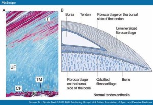
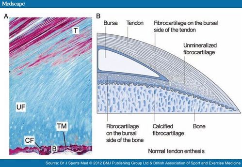
This next image shows in much more detail into how the enthesis really integrates into the bone and how enthesis stem cells could be stimulated to form a mini growth plate.  Bone erosion could occur allowing this growth plate to extend across the bone.  In fact, in arthritis which is associated with thickening of the enthesis’ bone erosion does occur.:

detailed view tendo enthesis
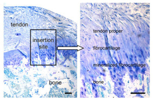
Note how the mineralized fibrocartilage integrates into the bone.  The thickening of the enthesis and the erosion of bone in some forms of arthritis, I could only find citations of it occuring in tendons and have not found instances of it resulting in increased length.  But that is likely a result of tendons being more subject to mechanical loading and this new method of LSJL being a novel way to mechanical load the ligaments.  Also ligaments in contrast to tendons are in a better position to contribute to longitudinal bone growth.  In fact, most of the studies I could find on mechanical loading on ligaments referred to loading on the ligament cells themselves rather than loading of the ligaments in the body.

Ligament injuries tend to occur due to heavy impact or overstretching of the ligament.  Pressing of the bones against each other is not a common way of ligament injury indicating that this method of LSJL loading is in fact a novel way of loading the ligaments.

“There is physiological thickening of the fibrocartilage with stress.”<-This tends to happen more with tendons as they are attached to muscle but with LSJL we can encourage it to happen at the ligament fibrocartilage.

” there are other components adjacent to the enthesis proper which also share the stress forces and are termed the “Enthesis organ”. These include the Periosteal fibrocartilage, the Sesamoid fibrocartilage, the Fat pad and Bursa. The Synovial entheseal complex is a concept that the adjacent bursa or joint lining share stress forces, especially compressive forces and are an integral part of the enthesis organ”<-These will be different for the ligament.

Here’s some info from another site that could be pertinent:

Bone Erosion at Normal Insertions

“Bone erosion is a process whereby the surface of a bone (the bone cortex) is degraded or eroded and is most typically seen in the setting of inflammation. However, the normal skeleton appears to be riddled with microscopic erosions. The enthesis is a highly mechanically stressed site which leads to microtrauma to the immediately adjacent bone. This is the basis for small erosions in the normal non-diseased skeleton which likely repair spontaneously.”<-Can we cause sufficient bone erosion as to allow for a new growth plate?

“The early phases of erosion may start to damage or loss of the shock absorbing fibrocartilage that covers the bone. “<-We don’t want this however as this would be the foundation of the neo growth plate.

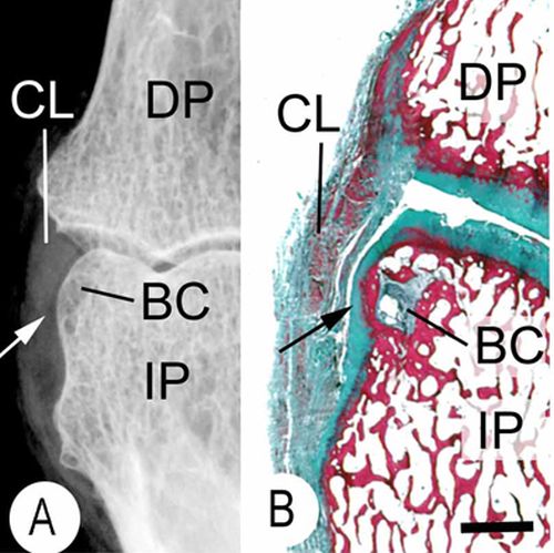
“Normal small joints tend to develop microscopic erosions at sites where the ligament immediately adjacent to the enthesis compresses the bone. This is because the shape of the bone leads to the forces being spread over a wide area that contributes to damage. This occurs at a structure termed a synovio-entheseal complex.”

synovio epiphysis complex

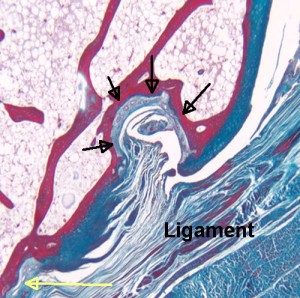
“The black arrows show a microscopic erosion over a knuckle joint. The overlying ligament is shown. The yellow arrow shows the point of ligament attachment closest to the joint cavity. Small blood vessels in the base of the erosion are likely linked to attempted repair. ”

“Sometimes the bone compression by the enthesis organ transmits stresses to the underlying bone and this initially manifests as a small cyst. Later on the roof of this may cave in leading to erosion. ”

bone cyst enthesis

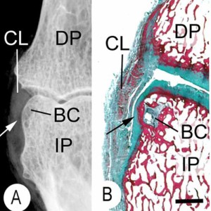
“This is an X-ray (A) and a corresponding tissue section (B). It shows a small bone cyst (BC). This is underneath the cartilage lining the side of the bone. (black arrow). Damage occurs here because the ligament (CL) presses against the bone as it runs between the joints. “<-We’d need more information about the bone cyst to see how promising it is in terms of neo growth plate formation.

Here’s another paper about the enthesis:

Bone Surface Micro-Topography at Craniofacial Entheses: Insights on Osteogenic Adaptation at Muscle Insertions

“Macroscopic details of the bone–muscle interface are represented by a mosaic of calcified features inclusive of fossae, tuberosities, crests, and ridges. These features are in part of an adaptive osteogenic response to dissipate forces of localized mechanical loading{how much can they be influenced by mechanical loading?}. In an osteoarchaeological or paleontological context, these features are interpreted as “musculoskeletal stress markers” to infer habitual behaviors. Microscopic surveys of bone surface topography of the enthesis can reveal localized osteogenic topologies. These features illustrate the developmental mechanisms that produce these bony forms and contribute to an evidential basis to read these structures. Microscopic osteogenic topographies at sites of gnathic muscle attachments located in the craniofacial skeleton were explored in reference to extrapolated loading vectors in an ontogenetic series of craniofacial skeletons of the primate (Procolobus verus). Epoxy resin replicas of bone surfaces were made, and micro-topographical detail viewed with Scanning Electron Microscope. Osteoclastic bone remodeling was found at entheses associated with presumptive net tensile loading. Mineralized fibrocartilage was present at entheses, associated with presumptive net compressive loading{which would be more beneficial for height growth mineralized fibrocartilage or osteoclastic bone remodeling?  I’d say osteoclastic bone remodeling because bone is not capable of interstitial growth and by allowing for osteoclastic bone remodeling we allow for tissues that it is}. Collectively, these outcomes suggest that entheses develop through adaptive osteogenic activity in response to differential vectors of local mechanical loading. However, the presence of mineralized fibrocartilage also suggests that proliferative cartilage has a role in the development of bone eminences providing functional processes. This study concludes that the vector of muscle loading at entheses as well as proliferative fibrocartilage is influencing the form of bony eminences in the primate craniofacial skeleton defining functional and species defining morphologies.”
“The majority are considered to be of the fibrocartilaginous type where zones of uncalcified and calcified fibrocartilage are interposed between the dense fibrous tissue of the tendon and a bone matrix”<-since they are intermixed stimuli that affects bone should benefit the enthesis like interstitial fluid flow.
“The bone-penetrating extrinsic fibers known as Sharpey’s fibers are considered as the defining feature when these fibrous tendons are directly anchored to the bone”
“There are emerging reports that contradict the above assertion, for example, zones of mineralized and unmineralized fibrocartilage have been identified at the deltoid insertion into the diaphysis of the humerus. This fibrocartilaginous zone can act as a mini growth plate to produce mass for the deltoid tuberosity. Furthermore, these cells are likely to be derived from a different source to those that form long bone analgen as they express the transcription factor Scleraxis (Scx) consistent with a syndetome origin, the precursors for tendon cells”
” muscle action of this enthesis aligned with extrapolated vectors of net tension at this locality result in osteoclastic activity”
Above is the osteoclastic activity mentioned.
These cellular based osteological functions can produce gross shape change of bone processes through micro anatomical bone surface modeling and remodeling modulated by the loading vector“<-so the enthesis can alter the bone this is huge!
“Loads extrapolated to be perpendicular to bony substrate exhibited osteogenic responses that adaptively opposed the loading vector. When in net tension, osteoclastic remodeling topographies were observed contributing to the formation of fovea and depressions on the bone surface. These forms redistribute the loading vectors to the periphery and increase the surface area of attachment. This differs from entheses that are subject to net compression where topographies notably consistent with mineralized fibrocartilage. In these regions, gross bony eminences and processes form that provide mechanical leverage to muscle contraction.”<-by manipulating compression and tension how can we manipulate the bone?
Above image is the direct impact of the enthesis on mandible shape.
“Schematic of bone form changes in the mandibular ramus associated with masticatory muscle enthesis. The inner tracing depicting infant mandibular outline followed by adolescent to adult (outside tracing) depicting hypothesized growth trajectories developed from surface micro topographies indicative of differential osteogenic activity. The large arrows depicting the growth vector at the angle, coronoid, and condyle of the mandible. Bone mass facilitated by mineralized fibrocartilage deposits indicated by (0) at sites of net compression. Subtle shape changes to the condylar neck associated with remodeling indicated by minus sign (−) facilitated migratory pathway consistent with the appositional growth vector of the condyle. Where the masseter muscle belly lies on the ascending ramus a remodeling field can be observed indicated by minus (−). The ramus of the mandible in particular is modulated by the muscle anatomical units where intrinsic and extrinsic modulated proliferative fibrocartilage associated with entheses establishes species and functional defining forms.”
” locations of these chondron dense fields are found perpendicular to the growth and coincident with load vectors. This provides an avenue for ossification and progressive bony growth at these muscle attachments where the entombed fiber become destined to become fields of the bone penetrating Sharpey fiber”
“Entheses transmit force from tendons and ligaments to the skeleton. Regional organization of enthesis extracellular matrix (ECM) generates differences in stiffness required for force transmission. Two key transcription factors co-expressed in entheseal tenocytes, scleraxis (Scx) and Sox9, directly control production of enthesis ECM components.{perhaps gene therapy of Scx and Sox9 could be used to stimulate the enthesis and enhance limb lengthening recovery} Formation of embryonic craniofacial entheses in zebrafish coincides with onset of jaw movements, possibly in response to the force of muscle contraction. We show dynamic changes in scxa and sox9a mRNA levels in subsets of entheseal tenocytes that correlate with their roles in force transmission. We also show that transcription of a direct target of Scxa, Col1a, in enthesis ECM is regulated by the ratio of scxa to sox9a expression. Eliminating muscle contraction by paralyzing embryos during early stages of musculoskeletal differentiation alters relative levels of scxa and sox9a in entheses, primarily owing to increased sox9a expression. Force-dependent TGF-β (TGFβ) signaling is required to maintain this balance of scxa and sox9a expression. Thus, force from muscle contraction helps establish a balance of transcription factor expression that controls specialized ECM organization at the tendon enthesis and its ability to transmit force.”
” Specialized areas at the tendon-bone interface called entheses accommodate these physical constraints using a unique fibrocartilaginous extracellular matrix (ECM) that varies in composition along the length of the enthesis “
Scx and Sox9 are expressed in opposing gradients along the length of more mature entheses in mice, with much higher levels of Sox9 closer to the bony insertion”

Akt1 related overgrowth

Akt1 overgrowth tends to create proportional overgrowth whereas CNP overgrowth tends to create lankier overgrowth

Mosaic overgrowth with fibroadipose hyperplasia is caused by somatic activating mutations in PIK3CA.

“The phosphatidylinositol 3-kinase (PI3K)-AKT signaling pathway is critical for cellular growth and metabolism. Correspondingly, loss of function of PTEN, a negative regulator of PI3K, or activating mutations in AKT1, AKT2 or AKT3 have been found in distinct disorders featuring overgrowth or hypoglycemia. We performed exome sequencing of DNA from unaffected and affected cells from an individual with an unclassified syndrome of congenital progressive segmental overgrowth of fibrous and adipose tissue and bone and identified the cancer-associated mutation encoding p.His1047Leu in PIK3CA, the gene that encodes the p110α catalytic subunit of PI3K, only in affected cells. Sequencing of PIK3CA in ten additional individuals with overlapping syndromes identified either the p.His1047Leu alteration or a second cancer-associated alteration, p.His1047Arg, in nine cases. Affected dermal fibroblasts showed enhanced basal and epidermal growth factor (EGF)-stimulated phosphatidylinositol 3,4,5-trisphosphate (PIP(3)) generation and concomitant activation of downstream signaling relative to their unaffected counterparts. Our findings characterize a distinct overgrowth syndrome, biochemically demonstrate activation of PI3K signaling and thereby identify a rational therapeutic target.”

Proteus syndrome, a progressively deforming regional overgrowth syndrome that affects bones, adipose, and other mesenchymal tissues, is caused by a somatic p.Glu17Lys AKT1 mutation which constitutively activates PI3K/AKT signaling”

The Overgrowth can be local:

akt1-overgrowth

Interesting that in one case ” Muscle is replaced by fibrous and adipose tissue with occasional residual muscle fibers (arrow)”.  So muscular tissue basically dedifferentiated.  Maybe the same thing could happen with bone and allow neo growth plate formation.

“somatic occurrence of both AKT2 and AKT3 p.Glu17Lys mutants, paralogous to the Proteus-associated AKT1 mutation, have been described.”

Muscular loading may influence Akt1 signaling in bone:

The masticatory contractile load induced expression and activation of Akt1/PKBalpha in muscle fibers at the myotendinous junction within muscle-tendon-bone unit.

“The cell specific detection of enzyme activation in response to the physiological contractile load within muscle-tendon-bone unit is essential for understanding of the mechanical forces transmission from muscle cells via tendon to the bone{Are these mechanical forces transmitted via fluid flow or by some other means?}. The hypothesis that the physiological mechanical loading regulates activation of Akt1/PKBalpha at Thr308 and at Ser473 in muscle fibers within muscle-tendon-bone unit was tested using quantitative immunohistochemistry, confocal double fluorescence analysis, and immunoblot analysis. In comparison to the staining intensities in peripheral regions of the muscle fibers, Akt1/PKBalpha was detected with a higher staining intensity in muscle fibers at the myotendinous junction (MTJ) areas. In muscle fibers at the MTJ areas, Akt1/PKBalpha is dually phosphorylated at Thr308 and Ser473. The immunohistochemical results were confirmed by immunoblot analysis. We conclude that contractile load generated by masticatory muscles induces local domain-dependent expression of Akt1/PKBalpha as well as activation by dually phosphorylation at Thr308 and Ser473 in muscle fibers at the MTJ areas within muscle-tendon-bone unit.”

“The muscle-tendon-bone unit contains myocytes, fibroblasts, nerve fibers, blood vessels, osteoblasts, osteoclasts, osteocytes, and extracellular matrix.”

“Tendons transmit forces generated from muscle cells at the muscle-tendon-junction (MTJ) to bone cells.”

“Full activation of Akt1/PKBα requires phosphorylation of the enzyme at Thr308 and at Ser473”

“Thr308 is phosphorylated by 3-phosphoinositide-dependent kinase-1 (PDK1). The phosphorylation of Akt1/PKBα at Ser473 is mediated by both mammalian target of rapamycin-rictor complex (mTORC2) and DNA-dependent protein kinase (DNA-PK) depending on type of stimulus.”

“muscle fibers apply forces to the bone cells via tendon cells, which arise from a specialized region called the MTJ. In the MTJ, myofibrils and collagen fibers overlap, forming longitudinal infoldings”

According to The role of Akt1 in terminal stages of endochondral bone formation: angiogenesis and ossification., Akt1 may be involved in regulating MMP14 levels and Akt1 deficient mice had a delay in the formation of secondary ossificiation centers.

According to Adiponectin inhibits osteoclastogenesis and bone resorption via APPL1-mediated suppression of Akt1., Akt may reulate osteoclastgenesis.  So Akt1 may play a role in growth by increasing bone turnover.

akt1-pathway

So Akt1 increases Osteoclastogenesis and decreases Osteoclast Apoptosis.

Homocysteine may be an Adiponectin inhibitor according to Inhibition of adiponectin production by homocysteine: a potential mechanism for alcoholic liver disease.