Category Archives: Uncategorized

How much growth hormone is needed to cause gigantism?

If gigantism was only caused by excess HGH levels than why is gigantism so rare.  It is likely that extremely high levels of HGH or other factors are needed to cause gigantism.  This study will help us determine how much HGH is above the norm in cases of gigantism.

Gigantism caused by growth hormone secreting pituitary adenoma.

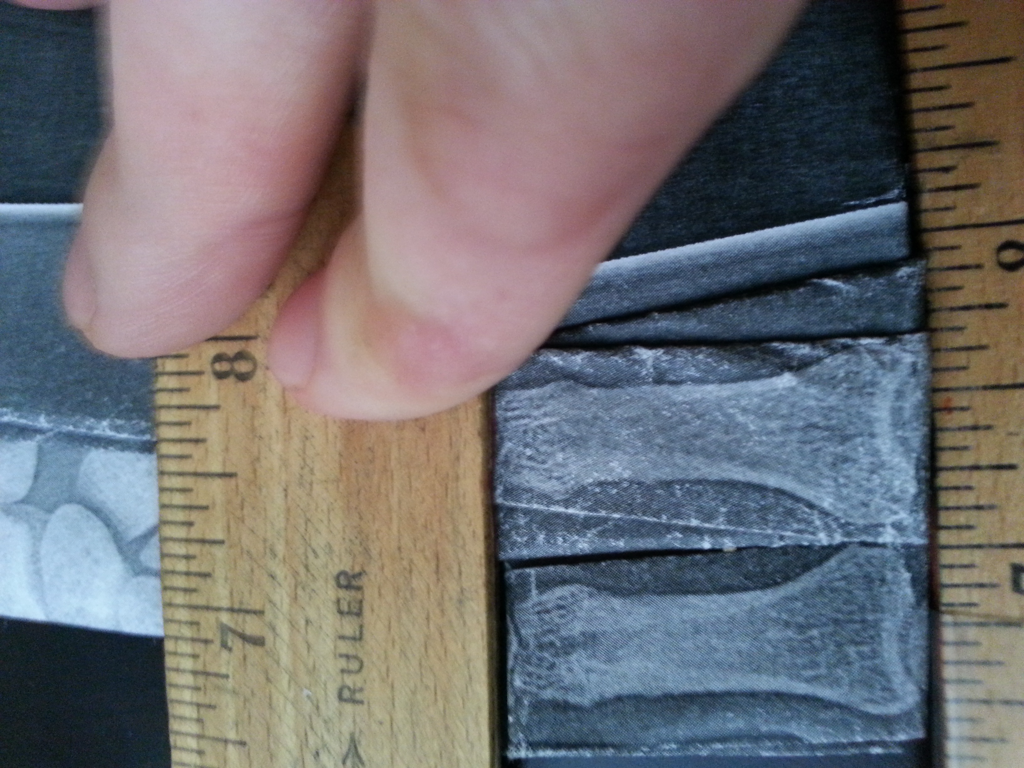
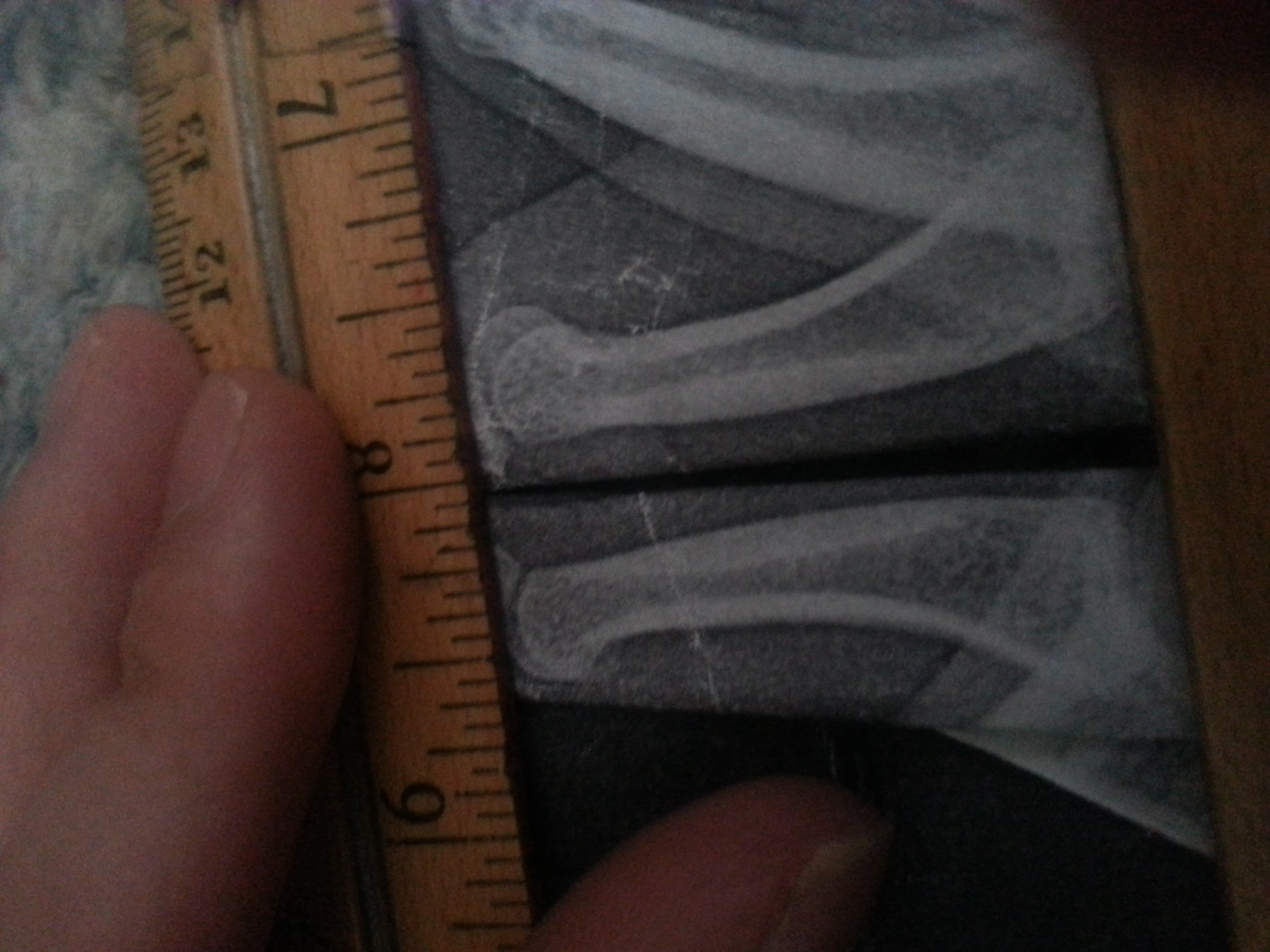
“Gigantism indicates excessive secretion of growth hormones (GH) during childhood when open epiphyseal growth plates allow for excessive linear growth. Case one involved a 14.7-year-old boy presented with extreme tall stature. His random serum GH level was 38.4 ng/mL, and failure of GH suppression{Resistance to GH surpression may be another factor needed for gigantism to be caused} was noted during an oral glucose tolerance test (OGTT; nadir serum GH, 22.7 ng/mL). Magnetic resonance imaging (MRI) of the brain revealed a 12-mm-sized pituitary adenoma. Transsphenoidal surgery was performed and a pituitary adenoma displaying positive immunohistochemical staining for GH was reported. Pituitary MRI scan was performed 4 months after surgery and showed recurrence/residual tumor. Medical treatment with a long-acting somatostatin analogue for six months was unsuccessful. As a result, secondary surgery was performed. Three months after reoperation, the GH level was 0.2 ng/mL{So the GH level was higher by a factor of 192 which is exceptionally high} and insulin-like growth factor 1 was 205 ng/mL. Case two involved a 14.9-year-old boy, who was referred to our department for his tall stature. His basal GH level was 9.3 ng/mL, and failure of GH suppression was reported during OGTT (nadir GH, 9.0 ng/mL). Pituitary MRI showed a 6-mm-sized pituitary adenoma. Surgery was done and histopathological examination demonstrated a pituitary adenoma with positive staining for GH. Three months after surgery, the GH level was 0.2 ng/mL{His GH was elevated by a factor of 46.5} and nadir GH during OGTT was less than 0.1 ng/mL. Pituitary MRI scans showed no residual tumor. ”

So key factors involved in Gigantism involve HGH much higher than normal and a resistance to HGH surpression.  According to this steriods forum, some experienced elevated serum HGH levels as high as 18ng/mL which is within the range of the serum levels of the two cases of gigantism.  Which means in terms of serum GH level, HGH injections may be sufficient to cause gigantism.  However, resistance of the HGH to surpression is one thing that is not conferred by the injections as the HGH injections have to be cycled.

“True gigantism is extremely rare”<-Which is why again it is not likely to be caused by elevated HGH alone.

“normal range, 0-5 ng/mL [For GH]”

In the 38.4ng/ml case for Gigantism, his IGFBP-3 was additional elevated.  IGF-1 levels were slightly above normal range.  Prolaction was within normal range.

The other boy with 9.3ng/ml case for Gigantism, had prolaction, IGFBP-3 and IGF-1 all in normal range.  But this boy was actually taller than the other case beside having about a 1/4 less HGH.

“Approximately 100 cases of children with pituitary gigantism have been reported”<-But yet gigantism was caused with levels of only about twice the maximum the normal range.

“Hyperprolactinemia is a common finding in GH excess presenting in childhood, undoubtedly related to the fact that mammosomatotrophs (GH and prolactin-secreting cells) are by far the most common type of GH secreting cells involved in childhood gigantism. However, gigantism caused by a pituitary tumor comprised of somatotropes (GH-secreting cells) show a normal prolactin level”

Why Stretching Does Work To Gain Permanent Height Increase

Usually the email to the website gets just the exact same type of messages from the same type of people. Maybe 75% of the emails we get are people asking me to upload and give them for free the E-Book PDF for the GT4I, but I have felt after dealing with too many potential legal problems that uploading that book will do more harm than good in the long term.

Yishan WongSometimes something useful actually comes along, and that happened today. An email showed me this post written on Quora by a Yishan Wong reveals that a correction of posture through stretching and maybe also yoga has let the poster increase his height by half an inch, going from 5’8″ to 5’8.5″. The person who emailed me mentioned that this Yishan Wong guy is supposed to be the CEO of reddit, but I would not know about that too much. A Quick check does reveal that this Wong guy does have a Quora account and has a similar phrase used in his profile. A Google search on this guys’s height comes up with 5′ 7.5″, which is close enough to his claims on the post about growing taller.

So in light of the new developments, it seem that even a multi-millionaire, super rich tech entrepreneur like Yishan Wong has himself noticed changes in his own height after puberty, through just the normal application of the principles of stretching and yoga.

Now, I personally don’t think there is any need for a person as rich as the CEO of Reddit to ever need to lie to others about his changes in stature. So I am going to just trust in this guy’s claims and say that somehow he increase his height by 1/2 an inch. If we go on the older claim that this guy was 5′ 7.5″ before, then his year of posture correction led to a full inch in height increase for him.

This type of change is NOT that uncommon. I remember years ago watching this televised golf tournament where the announcer noted that one of the professional golfers recently told in an television interview that he increased his height from 6′ 3″ to 6′ 4″ after spending 3 months doing stretching at a very intensive level. Why would anyone need to do that much stretching is beyond my comprehension but that professional golfer got results. Who was this golfer, or what is his name? I don’t remember, but I do remember that segment extremely well in my mind.

The point is that there are too many documented cases of people who took action to change their posture (for cosmetic or health reasons) and started doing stretching as adults and noticed height changes which never went away.

The most obvious and simple way to increase height, and not just temporary height, is to stretch, and stretch a lot. Just this last month I was at a Goodwill and found this old guide to Yoga book and it was showing Indian Hindu yogis who were like 80 years old and had amazing flexibility doing postures that teenage ballerinas can’t even do. In my opinion, stretching should be the first place we should start in our endeavors. and Join a yoga class.

Melatonin may play a role in scoliosis pathology

Scoliosis causes height reduction by causing a curvature of the spine thus it is relevant to height increase.

Abnormal Response of the Proliferation and Differentiation of Growth Plate Chondrocytes to Melatonin in Adolescent Idiopathic Scoliosis.

“Abnormalities in the melatonin signaling pathway and the involvement of melatonin receptor MT2 have been reported in patients with adolescent idiopathic scoliosis (AIS).  In this cross-sectional case-control study, growth plate chondrocytes (GPCs) were cultured from twenty AIS and ten normal control subjects. Although the MT2 receptor was identified in GPCs from both AIS and controls, its mRNA expression was significantly lower in AIS patients than the controls{Will increasing MT2 receptor levels make you taller(or shorter)?}. GPCs were cultured in the presence of either the vehicle or various concentrations of melatonin, with or without the selective MT2 melatonin receptor antagonist 4-P-PDOT (10 µM). Then the cell viability and the mRNA expression of collagen type X (COLX) and alkaline phosphatase (ALP) were assessed by MTT and qPCR, respectively. In the control GPCs, melatonin at the concentrations of 1, 100 nM and 10 µM significantly reduced the population of viable cells, and the mRNA level of COLX and ALP compared to the vehicle{Whether this would increase or decrease height is unclear}. Similar changes were not observed in the presence of 4-P-PDOT. Further, neither proliferation nor differentiation of GPCs from AIS patients was affected by the melatonin treatment.

“[The] abnormality [that] is manifested during the peripubertal period in patients with AIS in that they tend to be taller, leaner and have a longer arm span than their healthy peers”<-So perhaps MT2 receptor makes you shorter?  So an MT2 receptor inhibitor would make you taller or something that inhibits melatonin?

“Melatonin failed to inhibit the increase of 3′,5′-cyclic adenosine monophosphate (cAMP) induced by forskolin in osteoblasts from AIS patients when compared with cells from normal control subjects”

“melatonin inhibited both proliferation and differentiation of rat vertebral body growth plate (VBGP) chondrocytes with the involvement of MT1 and MT2 receptors”

“After incubation for 24 h in medium containing melatonin, the cell proliferation, gene expression of collagen type II and aggrecan, as well as protein expression of proliferating cell nuclear antigen (PCNA), Sox9 and Smad4 were significantly reduced. Moreover, it was found that the effects of melatonin could be reversed by the melatonin receptor antagonist luzindole, indicating the involvement of membrane melatonin receptors in these functions”

“AIS patients with a low level of expression of MT2 receptor in osteoblasts showed a longer arm span than those with a normal expression level of MT2 receptor”

Thus Luzindole may be a way to inhibit melatonin and grow taller at the expense of possibly inducing scoliosis.

{w/ Tyler’s Rebuttal}My Second Analysis On The Index Finger and Thumb X-Rays

{Tyler’s Notes-Since these measurements are critical to the validity of LSJL I wanted to make a rebuttal right away.  Here’s some images that provide evidence that there is in fact a difference between the right and left fingers:
20140929_110258

 The right finger is at the top.  This is the index finger proximal bone.  You can very clearly see that the right bone is longer than the left.  You can identify that these are the right and left bones on the x-ray page.

Here’s the middle bone:

20140929_110817

I apologize for the paper folds confounding where the bone ends and begins but if you look closely you can identify it again.  The middle proximal bone is clearly longer.  Again the right bone is on the top and the left at the bottom.  Michael discusses the protrusion on the bone and suggests it might indicate some type of arthritis and that it might cause bone.  What I could do is post a video of myself playing the piano which would provide evidence that my fingers have healthy mobility.

Here’s the proximal part:

20140929_110942Again right bone is on the top but a length difference isn’t really clear.  I’d say this length difference isn’t very significant.

I also did some lateral analysis:

20140930_105705


I aligned the bones at the top(proximal) end of the bone.  This is the lateral view of the proximal bone of the index finger.  Right bone is at the top and left bone is at the bottom.  The right proximal index finger bone is longer mainly via the protrusion at the bottom(distal) end.  I couldn’t find a significant difference in the middle or top part of the index finger via the lateral view.  I also tried comparing the thumb bones and couldn’t find anything noticeable which is odd because my left thumb is now noticeable taller than my right.  I’ll have to try to get more accurate measurements there.

How to tell who is right myself or Michael?  Print out the x-rays yourself.  Cut out each individual bone and compare them against each other.

The main difference and main and Michael’s results is that the left index finger is slanted at about 85 degrees whereas the right index is tilted 75 degrees.  Basically, the right index finger is slanting making it shorter.  Michael’s theory is that LSJL may have made my finger slanted.  However in the X-RAY office the x-ray tech asked me to spread my fingers apart and doing so adjusted the finger tilt.

I know that this proof isn’t optimal(once again) but I wanted to put in some reasonable doubt into Michael’s assertion that my fingers have not grown with LSJL.

If Michael had posted pictures and measurements that shown that I had not grown I would ACCEPT THAT LSJL HAD NOT INCREASED length.  Without pictures I can’t analyze his measurement technique.  Michael’s conclusion that I have not grown suffers from confirmation bias where he accepts the conclusion that I had not grown from LSJL because that is closest to his beliefs.  When I first saw the pictures I too measured that my left index finger was longer until I noticed that the right finger was slanted and had to find a way to adjust this in the measurements.

But I accepted this conclusion because I suffer from confirmation bias that I grew from LSJL.  And there’s a definitive and noticeable increase in finger length which is consistent with finger growth(but does not rule out alternative sources).  Michael states that he doesn’t have a camera and can’t take pictures of direct measurements.  So I’m going to have to do it even if I’m not as skilled(my hands were shaking a little bit when I was trying to take a picture with my camera which made things very difficult).

So hopefull these pictures prove that I noticeably and definitavely now have a right index finger longer than my left and soon I’ll have to step up and take pictures of the measurements.

Read Michael’s analysis below, it is essentially correct you don’t account for that the right finger is more slanted due to the x-ray tech asking me to spread my fingers.}

So in the last week Tyler has sent me a flurry of emails asking that I recheck the blown up X-ray pictures again and measure the entire thing over. Obviously I understand that we need to be extremely accurate on this particular set of X-rays since they would either validate or invalidate his theory LSJL. Here is the set of emails he has sent…

Finger X-Ray

“Did you also do the lateral view for comparison?  And did you factor in that the fingers are tilted at different angles?  How did you determine the end point of the bone?  I would’ve drawn a line at the highest point of each individual bone and then measured between then….Also remember that the right finger is slanted by 10 degree over the left finger.”

“Also you stated you measured the center of the finger which doesn’t take into account the height of the epiphysis of the proximal bone.  You can draw a line from the end of that bone and then measure to the end of the finger….And it’s much harder to adjust to the differences in angle for the whole finger if you measure each bone separately it’s easier….To give you an example, I measure the proximal bone of both fingers highest point of epiphysis to lowest point.  For left I get 401.0 pixels, in a straight 90 degree line.  For right I get 407.0 pixels at a 90 degree angle and the right finger is more slanted….Measure this way and see if you get similar results and we can see if we’re on the same page there at least.”

“Actually what you can do is cut out the bones individually and then it would be easier to align.”

“You stated that you placed the finger bones right next to each other.  How did you correct for the 10 degree difference in angle?  When you folded how did you align the two papers?  I found it was impossible to correct for the angle this way. That’s why I suggested cutting the bones out…Sorry for so many emails but this is obviously very important.”

“Flipping the image may help.  I flipped the image with GIMP.  Enlarge that image then you can lay them on top of each other and get a better comparison”

“I just did some more measuring.  I folded up the papers until only the proximal bones of the fingers were visible.  I compared the two bones side to side and used a ruler to align the bones at the bottom.  The right proximal bone was noticeable taller than the left.  The difference in angle between the two fingers must be partly responsible for the different results.  And it seems that each of the three finger bones are aligned a different way so you can’t really compare them accurately side by side because they all slant differently.”

—————————————————-

Here is what I can say after looking over the X-rays this morning. The differences in the lengths, whether I am looking at the most proximal phalanges, or the most distal phalanges all have extremely close/similar lengths, at least for the index finger.

There is a total of 5 pictures, two showing the hands, two lateral pictures of both of the index fingers, and one picture that has the hands side by side.

I have not been able to figure out some type of standard or exact reference point to measure all of these bones by. Bones are irregular shaped and there are no sharp or smooth surfaces to use.

When I tried to measure the epiphysis ends from tip to tip, the measurements came out to be either the same or too close to tell.

Tyler keeps on talking about the fact that the right index finger is at an angle, and I know that. Human bones don’t grow completely straight up.. There will be bound to be a little bit of bending or slanted angle. The question that is probably more important to ask is whether the clamping actually caused the slanting of the right index finger.

This is what I did.

I decided to take the slanted factor out of my analysis. I asked myself just what is going on.

Here is what the X-rays did reveal. It turns out that if you really tried to measure the bones, the most distal phalanges of the left index finger is actually longer than the right one! That would be the opposite of what we are hoping for.

However, the most interesting thing is that the right index finger’s middle phalanges has a completely different epiphysis shape than the left. The distal epiphysis protrudes out twice. That are two pronounced humps on the epiphysis. On the left one, there is just one.

It seems that by clamping that joint for many years, the bone shape of the epiphysis/head has completely changed. It is not smooth with a nice layer of articular cartilage. It is rugged.

That is where the extra length in the finger is from! When I measured the right finger’s middle phalanges, from the most protruded head on the distal epiphysis to the bone’s most proximal edge, it comes out to be substantially longer than if I tried to do it on the left hand. In addition, I did not notice the articular cartilage layer on the epiphysis head in that region. It seems that the clamping might have damaged the articular cartilage layer. In its place is the bulging bone area.

As for the lateral viewpoint of the index fingers, those pictures were the most pronounced. The right index finger had most noticeable thicker bones in the index fingers for the right one. So the bones are indeed thicker. That is where I noticed that the epiphysis head was irregular shaped.

As for the thumbs, I did measure them and could not see any differences that were substantial.

There is a 2nd issue – When I looked at the articular cartilage layer of the most proximal phalanges, it looked like that the right index finger’s one was much thicker than the one on the left. So if you are trying to make the bones thicker, it has noticeable effects.

Conclusion

There are no lateral X-rays pictures of the thumbs to view. The pictures I do have are for the index finger. When measuring the thumbs, I could not find the difference in the lengths of the thumbs.

I conclude now that the reason why the right index finger is longer than the left one is because the clamping has changed the shape of the epiphysis aka the middle phalanges head. The epiphysis is supposed to be thin and mushroom-like. The bone for the right hand has started to bulge out creating two bulges, unlike the one which you see in the X-ray for the left hand. The bulging of the epiphysis on the side is what is elevating the most distal phalanges.

Medical Note: There are 2 things which I should warn people who are potentially going to try the clamping.

Note #1: I don’t see a nice layer of articular cartilage on the distal epiphysis on the middle phalanges of the right index finger. It might indicate that the clamping destroyed the cartilage layer.

Note #2: The articular cartilage for the distal end on the most proximal phalanges of the right index finger looks to be somewhat thicker than the left one. That could mean that the cartilage layer has become inflamed, or maybe it is not.

To conclude the post, I would like to ask Tyler whether he can easily & comfortably bend the tip of his right index finger without pain, since joints without cartilage, aka bone on bone contact, is usually quite uncomfortable, since that is just osteoarthritis. I should know since my grandmother had to have total knee replacement surgery when her knees were just bones rubbing against bones.

For me, even though I have never done the clamping on the index fingers, I can’t bend the most tip part of my right finger very well. It would seem that maybe Tyler hasn’t noticed that it has become maybe progressively more uncomfortable or painful to bend that right index finger tip.

LSJL seems to have proven that it can make bones thicker, and can even change the head of long bones aka epiphysis into different shaped. Technically, LSJL does increase the length of the bones, when measured from the end points tip to tip, but it does the lengthening by altering the epiphysis to a shape which is not really normal. The surface of the top of the epiphysis is rugged/no longer smooth. The articular cartilage seems to become thicker, and become ossified. Whatever is below that joint obviously gets pushed in the distal direction. If we are talking about clamping the knees, the tibia/fibula/calcaneus/other feet bones will all be pushed downward. Technically the net result is an increase in height.

Many people who have been critics of the LSJL theory (like those on the Sceptic Forums, available here) claim that the clamping will cause the cartilage to swell up and lead to osteoarthritis later on. At this point, the X-Rays seem to validate their concerns.

Korean Red Ginseng Inhibits Articular Cartilage Degeneration

Recently, I read on a forum about these young Korean kids who are given some type of small green type pill by their mothers to help them grow taller. It is interesting that there is such stuff being sold for at least the last 30 years which supposedly would help young Asian kids grow taller.

Maybe the competitive nature of these East Asian cultures makes mothers try all sorts of rather bizarre things to help give their children any type of edge in life.

In the last two years of research, I personally have found at least 2 other different compounds which have been a part of Traditional Korean Medicine who has been believed to work in helping kids grow taller. Not only that, a group of Korean scientists filed a patent on a type of Ultrasonic-LED Combo type device which was supposed to stimulate the epiphyseal plates. It seems that South Korea, which has taken much of the older medical knowledge from Traditional Chinese Medicine, might have figured out a few natural ways to help its people end up slightly taller.

All this talk about Eastern Mysticism and the Esoteric made me wonder whether the Oriental Superpower Plant known as the Ginseng might have any type of chondrogenic or cartilage enhancing properties. After taking some time to look over PubMed, there were at least a couple of studies which validates this idea.

The active ingredient in all of the varieties of Ginseng, is this compound known as ginsenosides, which is in a category of compounds known as the saponin ,or more specifically the triterpenoid saponins. (For more information on Ginsenosides, refer here.) By last count, there is over 150 different kinds of Ginsenosides. From thousands of years of trial and error, humans found that they can orally consume the Ginseng and get the active ingredient inside the Ginseng to work. The Ginsenosides work in the stomach through acid hydrolysis and in the gastrointestinal tract by the reaction bacterial hydrolysis.

It seems that at least three of the over 150 different kinds of ginsenosides have some type of cartilage enhancing ability. I am quite sure that in the last century, multiple Asian researchers have validated the idea that whatever is in ginseng have anti-inflammatory effects. Inflammation is really the start of almost all types of cartilage degradation. If Inflammation is prevented, then the chances of the onset of osteoarthritis is going to be severely reduced.

In the first study, 11 different types of ginseng saponins were used. There were 2 which had some really powerful effects, ginsenosides F4 and Rg3. It is F4 which is really interesting. With increased dosage/concentration of the F4, the inhibition of the MMP-13, which is a collegenase, was increased, almost linearly in fashion. at 50 microMolars, the inhibition of not just the MMP-13, but also the p38 MAPK Pathway was also inhibited.

In the third study, it was found that the saponin was able to cause the number of chondrocytes in deficient mediums to proliferate and have the precursor stem cells to differentiate into chondrocytes{I believe that it was stating that it encourages differentiation of chondrocytes as in chondrocytes into hypertrophic chondrocytes not differentiation of stems into chondrocytes-Tyler}.

This is really interesting because for the longest time, many “grow taller pills” that were sold in places in South Korea (and maybe also China, Taiwan, Singapore, etc.) did have extracts of at least a little bit of ginseng in them. Is it really that far fetched for people to believe that the mystical ginseng plant would have some “special” ingredient that would also make them taller?

In the article “South Korea Stretches Standards for Success” published back in Dec. of 2009 in the New York Times, there was a really popular clinic called Hamsoa, which supposedly had already 50 clinics around South Korea, and they gave kids a special type of tonic which was supposed to be taken twice a day. Inside that tonic, was deer antler, ginseng, and other chemical compounds at a much lower concentration. Notice that deer antler and ginseng were mentioned.

It might be possible to use the ginseng to stimulate the activity in the growth plates of young kids, as well as even use the deer antler, but I am not sure how pronounced the stimulation would be. What can be well substantiated is that ginseng seems to help prevent cartilage degeneration and regular chondrocyte apoptosis.

Is it possible to increase disc height by stretching?

If it is possible to gain disc height by stretching. that could potentially explain the height gain of programs like agrobics.  Unfortunately, this batch of research I found, does not produce a strong link between stretching and height gain.  Mainly due to the nucleus pulposus being mechanically fragile.

The structural basis of interlamellar cohesion in the intervertebral disc wall.

“The purpose of this study was to investigate the structural mechanisms that create cohesion between the concentric lamellae comprising the disc annulus.”

” Additional bulk samples of annulus were fixed while held in a constant, radially stretched state in order to investigate the potential for interlamellar separation to occur in a state more representative of the intact disc wall. ”

“[IVD] tissues generally exhibit highly non-linear stress–strain responses, with the low-stress phase being a direct consequence of large-scale reversible alterations occurring in their fibrous architecture.”

hydrated lamellar section

“Fully relaxed, hydrated interlamellar section showing adjacent lamellae as both in-plane (IP) and cross-sectioned (CS) arrays. Note the compartmental division between the cross-sectioned bundles at Z.”

disc stretchng effectsHere you can show the possibility that stretch can potentially lengthen the IVDS.
intramellar section subjected to radial stretching

“(A) Interlamellar section subjected to radial stretching and revealing various modes of interconnection; (B) detail of radial bridging element passing between the cross-sectioned bundles; (C) detail of more uniformly distributed linking elements between adjacent lamellae.”<-This is radial stretching which should increase disc width rather than length.

“Interlamellar section radially stretching [causes] progressive fragmentation of cross-sectioned bundles ”
interlamellar tangential stretching“Interlamellar section subjected to tangential stretching. Selective fibre bundle pullout at grip ends has induced a substantial degree of shear between the in-plane arrays, thus revealing further the extent to which bridging elements (BE) pass between the cross-sectioned bundles and connect the neighbouring in-plane arrays (IP).”<-So fibre bundles pull out and bridge elements form resulting in possible overall lengthening in response to stretching.

“With increased stretching the forces transmitted by these same interconnections resulted in a progressive fragmentation of the cross-sectioned bundles involved.  [Fragments  separate] from [their] parent cross-sectioned bundle (CS).”

“the overall morphology of the permanently stretched samples reveals a radial elongation of the cross-sectioned bundles”<-but this is disc width and not height.

This study illustrates a possible mechanism of increasing interverterbral disc height although it’s possible that disc height could be still limited by mechanisms not investigated in this study.

Here’s a study that investigates the effects of twisting directly on the entirety of the spine:

Low back pain development response to sustained trunk axial twisting.

“The trunk axial twisting was created by a torsion moment of 50 Nm for 10-min duration.”

“The results showed that there was a significant  twist creep with rotational angle 10.5° as well as VAS increase with a mean value 45 mm{how would this effect spine height?}. The erector spinae was active in a larger angle during flexion as well as extension after trunk axial twisting.”<-creep implies a change in shape but whether that change involved a longitudinal increase is unclear.

“the elastic forces generated by the passive component of muscles are the main sources of passive resistance at the initial twisting motion, and then toward the end of ROM lumbar posterior ligaments and IVD will start to generate elastic forces and become the main contributor. This finding suggests that prolonged trunk axial twisting could also generate passive tissue creep and cause an alternation in the synergy between lumbar active and passive tissues.”

” The shear forces and moment created by spinal twisting within discs might elicit a shrinkage on spine by making the nucleus pulposus loose some fluid just like twisting a cloth full of water.”<-however this could also make the spine adapt by developing methods to absorb and retain more water.

Here’s a look of individual IVD cells response to loading regimes:

Region specific response of intervertebral disc cells to complex dynamic loading: an organ culture study using a dynamic torsion-compression bioreactor.

“We applied four different loading modalities [1. control: no loading (NL), 2. cyclic compression (CC), 3. cyclic torsion (CT), and 4. combined cyclic compression and torsion (CCT)] on bovine caudal disc explants”<-combined torsion and compression would be most akin to stretching as when you stretch one way you’re compressing another.

“In the CCT group, less than 10% nucleus pulposus (NP) cells survived the 14 days of loading, while cell viabilities were maintained above 70% in the NP of all the other three groups and in the annulus fibrosus (AF) of all the groups.”

“Gene expression analysis revealed a strong up-regulation in matrix genes and matrix remodeling genes in the AF of the CCT group”<-maybe developed of the extracellular matrix could increase height?

“Daily cyclic loading is important for disc health, as it assists in the transport of large soluble factors across the disc and from its surrounding vascular supply and applies a direct and indirect stimulus to disc cells.”<-This would increase height but does stretching apply a stimuli further than that?

” Characteristics of DD include increased cell death, a decrease in disc height due to a loss of essential matrix components which can also be reflected by an increased matrix catabolic gene expression (MMP-3, MMP-13, ADAMTS-4) but decreased anabolic gene expression (collagens and proteoglycans), increased inflammatory response (TNF-a, IL-1b, IL-6) and changes of mechanical properties of the disc (increased stiffness)”<-Although some of these things could also be involved in a anabolic protocol but the main thing we should watch is loss of essential matrix components in terms of reducing height.

“During the day, the disc experiences a pressure range from 0.1–1.1 MPa . However, studies have shown that dynamic compressive loading of >0.8 MPa could induce early DD; dynamic loading of physiological magnitude (1 MPa) at a frequency of 0.2 Hz was suggested to be the best in preserving disc metabolism while a frequency of 0.01 or 1 Hz could stimulate catabolic gene expressions ; signs of mild disc degeneration were seen when loading was applied in a longer term of 8 weeks (8 h/day) even at a physiological magnitude (1 MPa). The complex loading of side bending (in the form of asymmetric compression) and cyclic compression induced a greater structural disruption to the disc than simple cyclic compression”<-This would suggest that the best way of being as tall as possible would be to avoid excessive stimulus.

” torsional[twisting] injury is one of the initiators of disc degeneration, as evidenced by a decrease in disc height and a drop in disc proteoglycan content”<-so getting injured while stretching could possible reduce height.

“cyclic torsion could cause injury to the disc, provoking increased inflammatory (TNF-α and IL-1β ) and altered elastin gene expressions. An increase in elastin content in the AF is one of the observations in degenerated human discs and an alteration in the elastin fiber network might render the AF more susceptible to micro failure under torsion and bending”

“asymmetric dynamic compression (bending with compression) caused annulus fibrosus (AF) delamination and cell apoptosis”

“Discs used in this study had a mean dimension of 16.63±1.55 mm diameter and 9.58±1.22 mm height at day 0. By the end of the experiment, disc volume was increased by 10±5.76% for NL[no load], but increase in disc volume were less than 2% in all the other groups with loading”

“There was a slight increase in mean disc height of around 3% in the NL and CT[cyclic torsion/twisting] groups, while disc height was decreased by about 2% in the groups with cyclic compression (CC and CCT).”

“In the NP, collagen 1 expression was significantly up-regulated in CT. ADAMTS-4 was increased over 1000 fold in both CT and CCT, where its inhibitor TIMP-3 was also increased more than 10-fold”

” In the transition zone between the cartilaginous endplates (EP) and the nucleus pulposus (NP) , cells stayed as chondrocyte-like cells (indicated by black arrows) in the CC and CT groups with a round cell nucleus surrounded by lacunae. However, in CCT, very few cells stayed as chondrocyte-like cells in the cartilaginous endplate and cells right across the endplate region changed to spindle-shaped  and the cell lacunae and the cell boundary were lost.”<-This could be a key to height growth.  Maybe one way to restore growth plate is to remove either compressive or torsion forces in the bone.

CCT cartilage lossYou can definitely see the loss of cartilage but is it an irreversible loss?

“torsion-compression loading has caused micro-damage to the collagen, therefore disc cells have been activated to compensate for the destruction. As shown in the gene expression result, groups with torsion (CT and CCT) showed a larger increase in both anabolic and catabolic gene expression by AF cells as compared to no loading or pure compression, indicating that AF cells were more sensitive to torsional loading stimulation. Therefore they responded by increasing some matrix production and matrix destruction enzymes to remodel the matrix environment.”

“One possible reason for the difference in response between the NP and AF to the same loading is due to the fundamental difference in the matrix component and structure between NP and AF. AF collagen fibers are aligned in an angle that can withstand shear force but the disorganized gel-like matrix of the NP cannot withstand a high shear force under combined compression and torsion. The NP, which is mainly composed of water, proteoglycans and collagen 2, is more resistant to compressive force than direct shear force as in compression and torsional load. A uniform torque applied to the disc will result in a hoop strain within the tissue, which increases with the distance from the center of rotation. It might be that the reaction of the annulus cells to the applied torsion stress is also different between the outer annulus and the inner annulus fibrosus as the inter-lamellar angle decreases radially from the periphery to the center from 60° towards 40°. Moreover, the elastic fiber arrangements in intra-lamellar and interlamellar zones were shown to be architecturally distinct, suggesting that they perform multiple functional roles within the AF matrix structural hierarchy”