How does hydrostatic pressure induce chondrogenesis?

A key theory behind LSJL is the use of hydrostatic pressure to induces mesenchymal stem cells into chondrocytes to form new growth plates.  Hydrostatic Pressure alters the expression of some of the genes altered by LSJL.  Although, there are many genes not shared which is not surprising considering that LSJL exerts other forces than hydrostatic pressure and the microenvironment is different between the rat bone and the cell lines used.  Hydrostatic pressure is one of the most consistent stimuli in inducing neo chondroinduction.

Hydrostatic pressure decreases membrane fluidity and lipid desaturase expression in chondrocyte progenitor cells.

“Membrane biomechanical properties are critical in modulating nutrient and metabolite exchange as well as signal transduction. Biological membranes are predominantly composed of lipids, cholesterol and proteins, and their fluidity is tightly regulated by cholesterol and lipid desaturases. To determine whether such membrane fluidity regulation occurred in mammalian cells under pressure, we investigated the effects of pressure on membrane lipid order of mouse chondrogenic ATDC5 cells and desaturase gene expression. Hydrostatic pressure linearly increased membrane lipid packing and simultaneously repressed lipid desaturase gene expression. We also showed that cholesterol mimicked and cholesterol depletion reversed those effects, suggesting that desaturase gene expression was controlled by the membrane physical state itself. This study demonstrates a new effect of hydrostatic pressure on mammalian cells and may help to identify the molecular mechanisms involved in hydrostatic pressure sensing in chondrocytes. ”

“Hydrostatic pressure (HP) is known to reduce lipid membrane fluidity. High HP triggers an adaptive mechanism, during which membrane fluidity is increased by raising the proportion of unsaturated fatty acids.”

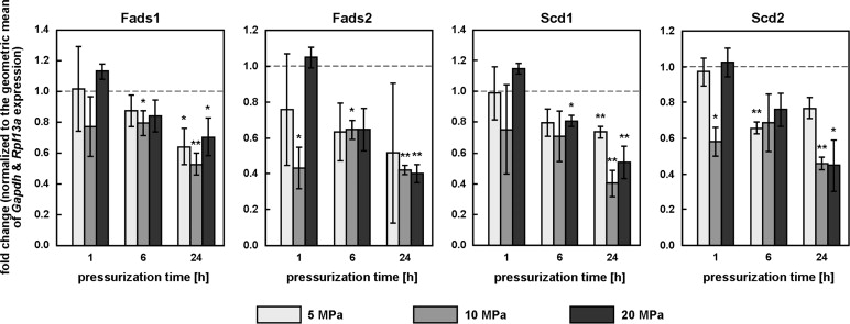
“TDC5 cells responded to the change in their membrane fluidity under pressure by modulating the expression of Fads1, Fads2, Scd1 or Scd2. 10 or 20 MPa did indeed significantly inhibit the expression of all four genes after 24 h, while 5 MPa also significantly decreased Fads1 and Scd1 expression.”<-None of the genes were directly altered over significance by LSJL.  The genes were measured one hour after loading so it’s possible that the genes returned to baseline after one hour.

gene expression change to HPWith this  data it’s easy to see why there was no change in gene expression level by LSJL as most of the repression of these genes occurred after 6 or 24 hours under hydrostatic pressure.

“MβCD increased ATDC5 membrane fluidity (laurdan GP values respectively increased and decreased under cholesterol and MβCD treatment”

“Similar to HP, cholesterol increased laurdan GP and significantly inhibited Fads1 and Fads2 expression. By contrast, MβCD[methyl-β-cyclodextrin], which decreased laurdan GP, significantly increased the expression of all four desaturase genes. Together, this suggests that membrane fluidity itself may be the general modulator of desaturase gene expression.”<-Cholesterol has been implicated in controlling endochondral bone growth before.

“the beneficial effects of loading on cartilage are mediated by the transcription factor CITED2, which represses cartilage degradation by the matrix metalloproteinase MMP1.”

“In the human hip joint, loads typically reach 10 MPa during normal activity, with peaks of up to 18 MPa. Considering that interstitial fluid pressure supports about 70 to 90% of the applied load, HP within the cartilage can be expected to regularly exceed 7 MPa and peak at around 16 MPa. 20 MPa is therefore a relatively high pressure”

“How HP is actually sensed by the cell remains poorly understood, but the link between HP and cholesterol suggests at least two hypotheses: both HP and cholesterol may converge on the SREBP pathway, cholesterol by affecting SREBP maturation via SCAP, HP by affecting the membrane fluidity of the endoplasmic reticulum, where non-activated SREBPs reside and which is more sensitive to HP than the plasma membrane; the mechanosensing mechanism of HP could also reside in cholesterol-rich domains like lipid rafts or caveolae”

“membranes rich in unsaturated fatty acids may be compressible enough to deform significantly, and trigger signaling events, even under relatively small pressures. Finally, the plasma membrane is supported by the cytoskeleton, which is mostly incompressible; it is possible that pressurization of the compressible intracellular fluid leads to large deformations in membrane domains unsupported by the cytoskeleton, the change in bulk volume being focused onto a small membrane area.”

Carrying Weapons And Looking Formidably And Dangerous Causes Height Increase Perception

In another thread from the Make Me Taller forums a poster named upinthesky would post a link to a story (HERE) which made me wonder whether it might be just easier to sometimes look taller and be perceived to be taller.

The article he cites is an editorial from The Economist on a study that came out from UCLA by a group of researchers and Daniel Fessler.

The study seems to have had over 600 subjects have pictures shown to them with people holding difference objects and having them judge how formidable they seemed to be. Apparently the pictures where a person was shown holding a .45 handgun were had been judged that the person in the picture was taller than a person holding something else.

The implications of this Economist article is to show that sometimes it is not how tall we might be objectively that is important, where the measurement of height is with a measuring tape and stadiometer but how tall we seem to be from the perception of other people. There have been many cases where a person’s height is perceived very differently when they are placed in different situations and settings. I remember once talking to a person who told me about the study of a height of a person being judged with the variable of authority or prestige being looked at. It would turn out that when a group fo students were asked to judge the height of a single person while their professional title was changed, from professor to graduate student to undergraduate student, the perception by the height guessers changed. The professor was judged on average to be around 2 inches taller than if the height guessers were told that the person was “just” a college student.

So….perception matters. When a person commands any type of authority which leads to either respect, admiration, or fear, their height is increased. For this case, when the person is holding a gun, their height increased by 2 inches. This shows that we can alter other people’s perception of our height through either acting in a formidable or threatening manner. (Not that I advocate acting violently just to make oneself appear bigger.)

The article is below…


The evolution of risk assessment

Big men with guns

Apr 11th 2012, 22:01 by J.P.

GUN-TOTING individuals intimidate unarmed folk because they tote guns. If that were not scary enough, the weapons seem to make those wielding them look bigger and beefier to boot. That, at least, is the conclusion of a study just published in the Public Library of Science by Daniel Fessler and his colleagues from the University of California, Los Angeles.

Like all animals, human beings need a snappy, rough-and-ready way to assess whether to fight or flee a foe. Spending too much time weighing the pros and cons could, after all, have disastrous consequences. But how formidable a foe is, for man as for other creatures, depends on a plethora of features. Size and sturdiness matter, of course, but so does sex, age, health or, indeed, how many pals he has. And in the case of human enemies, there are weapons to contend with.

Dr Fessler reasoned that since size and musculature have been a reliable indicator of formidability for the longest stretch of man’s evolutionary past, they might still dominate the calculation, even if actual awesomeness no longer stems from these physical attributes. In other words, brains might recast more complex formidablity-affording characteristics, like weapons, in terms of extra inches and bigger biceps. This blown-up mental image in turn activates the ancient, quick-fire fight-or-flee calculator which takes basic physical factors as inputs.

To test his theory, Dr Fessler recruited 628 volunteers and asked them to gauge the height (in feet and inches), overall size and muscularity (both on a six-point scale) of four men, ostensibly on the basis of pictures of their hands. In fact, all the hands in the photos were nearly identical. What differed from picture to picture was what they were holding. Objects included a caulking gun, a power drill, a handsaw or a 0.45 calibre handgun.

The researchers duly found that the handgun holders were judged to be 0.2 inches (0.5cm), 0.5 inches and 2.3 inches taller than those who held a saw, drill and caulking gun, respectively. These results more or less matched the scores on the other two formidability measures, with the gun-holders consistently coming top. They also reflected the perceived relative danger posed by each object, as determined in a separate study.

Dr Fessler’s findings cannot be explained by the fact that gun-owners are taller than average—they are not. Nor are cultural associations between guns on the one hand, and Rambo on the other, to blame. When Dr Fessler repeated the experiment replacing the handgun with a kitchen knife—which most respondents associate with housewives, not Sylvester Stallone (who, incidentally, stands at a relatively modest 177cm)—as the most dangerous object in the mix, the results matched those of the earlier study perfectly. Weapons, then, not only make a man feel big; they make others feel he is, too.

A Real Height Increase Device Patent From 2008

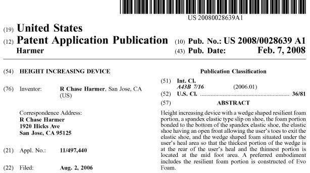
As I was going through the Make Me Taller forums yesterday I came across a discussion thread which again was looking at any alternatives of height increase beyond the limb lengthening surgery. The thread lead to a patent from 2008 which seems to be a real application. To get the link, just go to Google and type in “Height Increase Device Patent” and it should be one of the results on the first page. The original link is from FreePatentsOnline.com.

Screen Shot 2013-01-23 at 8.21.04 PMWhat is claimed is: 

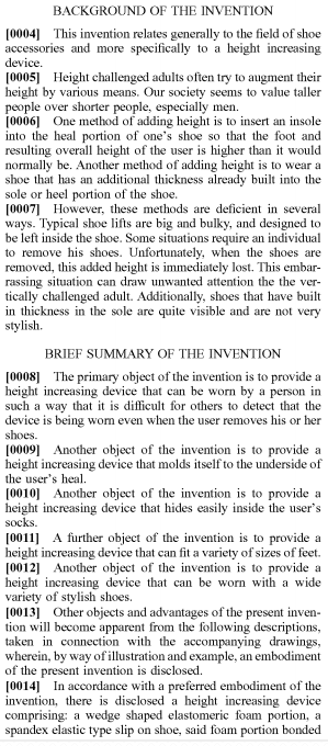
1. Height increasing device comprising: a wedge shaped resilient foam portion; a spandex elastic type slip on shoe; said foam portion bonded to the bottom of said spandex elastic shoe; said elastic shoe having an open front allowing the user’s toes to exit said elastic shoe; and said wedge shaped foam situated under the user’s heal area so that the thickest portion of said wedge is at the rear of the user’s heal and the thinnest portion is located at the mid foot area. 

2. Height increasing device as claimed in claim 1 wherein said resilient foam portion is constructed of Evo Foam. 

3. Height increasing device as claimed in claim 1 wherein said wedge shape can be as thick as one and one half inches at its thickest point at the rear of the heal. 

4. Height increasing device as claimed in claim 1 wherein said spandex shoe can fit a variety of foot sizes. 

5. Height increasing device as claimed in claim 1 wherein said spandex shoe and attached wedge shaped portion can be worn under a person’s standard sock so that said person can remove his or her shoes and still have the benefit of extra height because said spandex shoe can not easily be seen through said user’s sock.

Analysis Of The Patent:

There is a link HERE to the actual patent form filed with pictures of the device. The pictures are very crude and makes me wonder why a person would ever try to patent this type of idea. It is not very inforceable since I am quite certain that many shoe sellers and repair shops in Singapore, Indonesia, and China are already doing something similar in fashion to give some of their buyers a height boost from their shoes.

The shoe design seems to be able to remove 3 main problems that has been traditionally associated with any type of height increase shoes, which is that….

  1. The shoes inserts are big and bulky designed to be left inside the shoes.
  2. There will be some situations where the shoes do have to be removed which reveals the height difference and cause public embarrassment.
  3. The soles of the shoes are clearly visible to be thick and not very stylish.

After looking over this device for 10 minutes and looking at the really crudely drawn pictures of how the feet and shoe accessory is supposed to fit over the feet, I am still a little confused. On the one hand I think that this device is a sock like thing where the heel region is a relatively thick, strong, elastic part which causes the feet’s heel to thicken up at a sloped angle, which would give around 1 inch of concealed height. On the other hand, the device may be something that is placed into the shoe from the start and for the feet to go into and stay wearing until the shoes need to be removed for some reason. The device most likely will be staying on the feet while the shoes keep a normal thickness in the heel region.

I have clipped and posted the first page of the patent below.

Screen Shot 2013-01-23 at 7.58.56 PM

A Proposed Height Increase Method Using Microfracture Surgery Techniques With Fibrocartilage Formation

In a recent post “Increase Height And Grow Taller Using Microfracture Surgery, Part III” I had begun to propose a new height increase minimal invasive technique using the most general idea from microfracture surgery.

Here is how I interpret the entire idea behind microfracture surgery. You cut a section of the cartilage to get to the underlayer of bone. You use a type of thin drill or pick to poke very small holes, the microfractures, into the bone so that the device reaches deep enough to hit the inner of the epiphysis of the long bone. This means that the inner bone marrow, blood, and stem cells can seep out, filling up the hole made, and the clot eventually turns into into fibrocartilage.

I had stated at the end of the previous post…”What could work is that if a series of microfractures in a specific distribution design is created from drills on the side of the epiphysis to completely go around the bone in a closed path. This means that after a few days, the path of drilled microfractures would fill up with stem cells which will eventually turn into fibrocartilage. The fibrocartilage will not be that strong, but before they calcify into bone from vascularization, it would be possible to drill another set of microfractures around the same path to fill up the remaining bone bridges.”

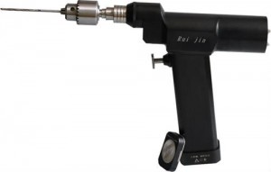
url-3I wanted to explain a little further into this idea so that the reader can understand from a 3 dimensional perspective what I am graphically proposing. The microfracture surgery used an awl (surgical device that punctures) to get the holes in the bones. What I would proposed for the height increase method is to use a similar device, the surgical drill, which would still have the same thickness as the awl.

The drill would go through the outer tissues like the skin and the muscle, just like what would be seen when the traditional ilizarov external method does when the drill is used to add the spokes and wires which are supposed to hold the two bone sides apart and in place when they are being distracted slowly by the long screws.

url-1The path from looking from a downward z direction would show that the drilled micro holes goes completely around the leg bone. Where the original external fixator method used only 3 drills to put metal spokes or wires into the bones to hole them into place, the drilling for this method would be in the dozens, but will be done under anesthesia. The drilling will go through the bone multiple times to make a path. After the drilling is done, the entire bone will leak out the marrow and stem cells. Something to hold the leaked marrow will keep the clot. After a few days, the outer surface of the bones would be clotted with initial fibrocartilage formation. When the cartilage is formed after 1 week, The rest of the undrilled areas of the bone is done. This would eventually cause the entire cortical area of an entire strip of length in the long bone to be drilled through. The other drilled fractures will also be filled with clots, which turns into cartilage. After the cartilage is formed around the entire bone, covering where the cortical bone is supposed to be, the tensile strength of the long bone will be reduced dramatically. Any type of method like LSJL or weight pulling would lead to cartilage width expansion, thus longitudinal growth.

Body Hack XIX: Changing Eye Color From Brown To Blue Permanently Using Quick Laser Treatment Without Surgery

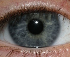
6a00d8341bf67c53ef015392e527f7970b-800wiI was reading through the science articles recently when I came through an article which talks about innovative medical innovations on the science of the eye. The BBC, Discovery Magazine, Washington Post, and other sources all have been able to pick up this story of the fact that a optic physician from California, a Dr. Gregg Homer, says that he has managed to create a type of laser that  can in only 20 seconds remove the pigmentation from a person’s iris.

From our high school biology books, we remember that the pigmentation from our eyes comes from some type of compound or protein called melanin. The melanin is what gives over 50% of the human world the color brown in their iris. Apparently only a small percentage of the world has what is traditionally blue eyes. However the blue hue or color in the iris is really just behind the brown pigment. All that the laser he has developed does is zap away the outer brown coloring to reveal the blue color underneath.

All the articles I have found say that the procedure will take either 20 seconds or 1 minute. The procedure is supposed to be painless, and the transformation from brown to blue eyes takes about 2-3 weeks, after the brown pigment is blasted away and degenerated. The blue color will apparently naturally remove the pigment. The cost of this cosmetic procedure will take about $5000 for each person. Currently, there is only one such process, from brown to blue. For black, green, hazel, and other colors of the eyes, there is no treatment found yet.

The article I decided to copy and post below is from the Discovery Magazine link…

New Procedure Would Turn Your Brown Eyes Blue

NOV 8, 2011 11:03 AM ET // BY MARIANNE ENGLISH

If you had the option to change your eye color — would you?

Now think about the question again, this time under two conditions: you’d have to pay $5,000 and undergo brief laser surgery. Would you still do it?

It’s obvious many people’s answer would flip. But one doctor thinks his method will attract a following. Gregg Homer, a California doctor, says he’s worked 10 years to perfect a laser treatment that can irreversibly turn brown eyes blue.

Homer believes that the eyes are the “window to the soul,” with blue being a preferred hue that looks less opaque than darker colors. In essence, he says, blue eyes allow others to look more deeply into them.

Here’s how the “20-second long” laser procedure works: A computer scans the iris and uses a laser to disrupt the brown pigment on the surface of the eye. Since blue pigment lies beneath the brown pigment, removing the outer layer of melanin can reveal a bluer look behind it. Since the brown pigment is damaged and will not regenerate, Homer says, the eye removes it naturally, according to one BBC article. Between two and three weeks later, the pigment lightens up, eventually transforming brown eyes to blue.

While addressing questions about the risks associated with the procedure, Homer told KTLA, a news station in Los Angeles, that he uses 15 “sophisticated” tests to ensure that no tissues are damaged. But in the very next sentence, he says, “Is it possible that something comes down the road? It’s possible.”

Unless the video was edited completely out of context, the previous statement suggests that Homer does not know the long-term effects of his laser treatment — a glaring problem other doctors point out in one ABC News article.

One NYU eye specialist, Robert Cykiert, told ABC News that the procedure is “probably risky.” He also said:

“When you burn the brown pigment away with a laser, the debris that is created in the front of the eye — think of it as ashes resulting from burning anything — is likely to clog up the microscopic channels in the front of the eye, known as trabecular meshwork,” said Cykiert. “ is very likely to cause a high pressure in the eye, known as glaucoma.”

Ironically, one of the treatments for glaucoma is more surgery.

So far, Homer says he has worked on the technique in animals, cadavers and human participants in Mexico. The method, called Lumineyes has been launched under Homer’s business, Stroma Medical Corporation (as of November 2011, neither site lists anything other than an email address).

He estimates the procedure will be available outside the United States in as few as 18 months, while it may take longer to gain approval in the states.

Also, it appears the technology only works to change from brown to blue, not other colors. Homer writes in one of his first patents that color eye contacts as well as implants aren’t permanent enough for people desiring pretty blues.

Photo by Dottie Mae/Flickr.com

The Connection Between The Wnt Beta Catenin Signaling Pathway And Growth, Part I

For the longest time I have been hearing from other height increase researchers talk about the Wnt/Beta-Catenin Signaling Pathway and how it effects so many different protein signaling pathways so I wanted to try to explain in my own words in this post what I have come to understand from reading from PubMed articles, Wikipedia articles, Medical journals, and other scientific and biological resources why this pathway is so important and how it is involved in the process of human growth and height.

Since my degree was in chemistry and engineering, I never formally took a class on molecular biology or on genetics before so this will be a slow process. The one class I ever took for Biochemistry I remember almost nothing from it so this study on the Wnt/Beta-Catenin Signaling Pathway will be absolutely necessary for any serious researcher on protein pathways and intracellular and extracellular pathways.

Here is what I remember from high school and college courses.

  • The purpose of all genes is to create proteins.
  • Genes are found either in the nucleus or the mitochondria of a cell.
  • A gene is a molecular unit of heredity of a living organism. It is a name given to some stretches of DNA and RNA that code for a polypeptide or for an RNA chain that has a function in the organism. (Wikipedia)

Since I remember that a lot of biology, biochemistry, and molecular biology, was about remembering names and terms, I think it was important to make a few terms and the terminology clear.

The definitions were mainly taken from the Wikipedia articles on these terms.


  • Intracellular – In cell biology, molecular biology and related fields, the word intracellular means “inside the cell”. It is used in contrast to extracellular (outside the cell)….This terms also means existing within the cells.
  • Extracellular – In cell biology, molecular biology and related fields, the word extracellular (or sometimes extracellular space) means “outside the cell”. This space is usually taken to be outside the plasma membranes, and occupied by fluid. The term is used in contrast to intracellular (inside the cell).
  • Ligand – In biochemistry and pharmacology, a ligand (from the Latin ligandumbinding) is a substance (usually a small molecule), that forms a complex with a biomolecule to serve a biological purpose. In a narrower sense, it is a signal triggering molecule, binding to a site on a target protein.
  • Receptor – In the field of biochemistry, a receptor is a molecule most often found on the surface of a cell, which receives chemical signals originating externally from the cell. Through binding to a receptor, these signals direct a cell to do something—for example to divide or die, or to allow certain molecules to enter or exit.Receptors are protein molecules, embedded in either the plasma membrane (cell surface receptors) or the cytoplasm or nucleus (nuclear receptors) of a cell, to which one or more specific kinds of signaling molecules may attach. A molecule which binds (attaches) to a receptor is called a ligand, and may be a peptide (short protein) or other small molecule, such as a neurotransmitter, a hormone, a pharmaceutical drug, or a toxin.
  • Cell surface receptors ( aka membrane receptors or transmembrane receptors) – are specialized integral membrane proteins that take part in communication between the cell and the outside world. Extracellular signaling molecules (usually hormones, neurotransmitters, cytokines, growth factors or cell recognition molecules) attach to the receptor, triggering changes in the function of the cell. This process is called signal transduction: The binding initiates a chemical change on the intracellular side of the membrane. In this way the receptors play a unique and important role in cellular communications and signal transduction.
  • Kinase – In biochemistry, a kinase is a type of enzyme that transfers phosphate groups from high-energy donor molecules, such as ATP,[2] to specific substrates, a process referred to as phosphorylation.
  • Protein – are large biological molecules consisting of one or more chains of amino acids. Proteins perform a vast array of functions within living organisms, including catalyzing metabolic reactions, replicating DNA, responding to stimuli, and transporting molecules from one location to another. Proteins differ from one another primarily in their sequence of amino acids, which is dictated by the nucleotide sequence of their genes, and which usually results infolding of the protein into a specific three-dimensional structure that determines its activity
  • Like other biological macromolecules such as polysaccharides and nucleic acids, proteins are essential parts of organisms and participate in virtually every process within cells. Many proteins are enzymes that catalyze biochemical reactions and are vital to metabolism. Proteins also have structural or mechanical functions, such as actin and myosin in muscle and the proteins in the cytoskeleton, which form a system of scaffolding that maintains cell shape. Other proteins are important in cell signaling, immune responses, cell adhesion, and the cell cycle.
  • Protein Kinase – is a kinase enzyme that modifies other proteins by chemically adding phosphate groups to them (phosphorylation). Phosphorylation usually results in a functional change of the target protein (substrate) by changing enzyme activity, cellular location, or association with other proteins. The human genome contains about 500 protein kinase genes and they constitute about 2% of all human genes.[1] Protein kinases are also found in bacteria and plants. Up to 30% of all human proteins may be modified by kinase activity, and kinases are known to regulate the majority of cellular pathways, especially those involved in signal transduction.
  • Phosphorylation – is the addition of a phosphate (PO43-) group to a protein or other organic molecule. Phosphorylation turns many protein enzymes on and off, thereby altering their function and activity. Protein phosphorylation is one type of post-translational modification.
  • Signaling Molecule – is a chemical involved in transmitting information between cells. Such molecules are released from the cell sending the signal, cross over the gap between cells by diffusion, and interact with specific receptors in another cell, triggering a response in that cell by activating a series of enzyme controlled reactions which lead to changes inside the cell.
  • Signaling Pathway (aka Signal transduction) – occurs when an extracellular signaling molecule activates a cell surface receptor. In turn, this receptor alters intracellular molecules creating a response. There are two stages in this process:
    1. A signaling molecule activates a specific receptor protein on the cell membrane.
    2. A second messenger transmits the signal into the cell, eliciting a physiological response.

    In either step, the signal can be amplified. Thus, one signalling molecule can cause many responses. A signal transduction functions much like a switch.

  • Target gene – (A personal definition) – the gene that a particular protein signal pathway will eventually have an affect on. The pathway’s main or secondary goal was to affect this one type of gene specifically.
  • Transcription – Transcription is the first step of gene expression, in which a particular segment of DNA is copied into RNA by the enzyme RNA polymerase. Both RNA and DNA are nucleic acids, which use base pairs of nucleotides as a complementary language that can be converted back and forth from DNA to RNA by the action of the correct enzymes. During transcription, a DNA sequence is read by anRNA polymerase, which produces a complementary, antiparallel RNA strand.

From the Wikipedia article on the Wnt Signaling Pathway


Mechanism

  • The Wnt pathway involves a large number of proteins that can regulate the production of Wnt signaling molecules, their interactions with receptors on target cells and the physiological responses of target cells that result from the exposure of cells to the extracellular Wnt ligands.
  • The canonical Wnt pathway describes a series of events that occur when Wnt proteins bind to cell-surface receptors of the Frizzled family, causing the receptors to activate Dishevelled family proteins and ultimately resulting in a change in the amount of β-catenin that reaches the nucleus

From the Wikipedia article on Beta-Catenin


Beta-catenin (or β-catenin) is a protein that in humans is encoded by the CTNNB1 gene. In Drosophila, the homologous protein is called armadillo. β-catenin is a subunit of the cadherin protein complex and acts as an intracellular signal transducer in the Wnt signaling pathway.

Function

β-Catenin is part of a complex of proteins that constitute adherens junctions (AJs). AJs are necessary for the creation and maintenance of epithelial cell layers by regulating cell growth and adhesion between cells. β-Catenin also anchors the actin cytoskeleton and may be responsible for transmitting the contact inhibition signal that causes cells to stop dividing once the epithelial sheet is complete.[6]

Recent evidence suggests that β-catenin plays an important role in various aspects of liver biology including liver development (both embryonic and postnatal), liver regeneration following partial hepatectomy, HGF-induced hepatomegaly, liver zonation, and pathogenesis of liver cancer.[7]

Role in the Wnt signaling pathway

When Wnt is not present, GSK-3 (a kinase) constitutively phosphorylates the β-catenin protein. β-catenin is associated with axin (scaffolding protein) complexed with GSK3 and APC (adenomatous polyposis coli). The creation of said complex acts to substantially increase the phosphorylation of β-catenin by facilitating the action of GSK3. When β-catenin is phosphorylated, it is degraded and, thus, will not build up in the cell to a significant level. When Wnt binds to frizzled (Fz), its receptor, dishevelled (Dsh) is recruited to the membrane. GSK3 is inhibited by the activation of Dsh by Fz. Because of this, β-catenin is permitted to build up in the cytosol and can be subsequently translocated into the nucleus to perform a variety of functions. It can act in conjunction with TCF and LEF to activate specific target genes involved in different processes

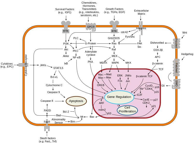
The picture below represents a the overview of the major signal transduction pathways. It is taken from the Wikipedia article for signal tranduction pathway .

Note: The post is continued below the picture of the signal transduction pathway diagram. Yes, there is more. Much, MUCH more in this post for me to study and learn. Hope you guys are reading and learning too.
Screen Shot 2013-01-22 at 8.45.20 PM


What we are seeing is that the Wnt/Beta-Catenin Signaling Pathway is one of the major signal transduction pathways which goes from the extracellular layer and area through the cell outer membrance inward, into the intracellular area, and then into the Nucleus, through the nuclear membrance.

The first question I would be asking myself is…’What is Wnt? What does the term refer to?

From what I can gather from the Wikipedia artilce on the word “Wnt” itself, it seems that the term “Wnt” is the combined word and meaning from two words and terms, Int and Wg.

Wg represents Wingless.

Int represents Integrated.

From PubMed study “Wnt signal transduction pathways“…”The name Wnt is resultant from a fusion of the name of the Drosophila segment polarity gene wingless and the name of the vertebrate homolog, integrated or int-1.”

The term Wnt is used in two cases. It refers to both the genes that make a certain type of protein, and the proteins that are made. So there are….

  • Wnt genes – The wnt genes make the wnt proteins. (I know, confusing). There is more than one type of Wnt gene, but an entire group (or family) of these genes.
  • Wnt proteins – these proteins act almost exclusively as signal transduction proteins, which give signals as a form of communication between individual cells.

From Wikipedia….

  • The Wnt proteins are a group of secreted lipid-modified (palmitoylation) signaling proteins of 350-400 amino acids in length
  • The WNT gene family consists of structurally related genes that encode secreted signaling proteins. These proteins have been implicated in oncogenesis and in several developmental processes, including regulation of cell fate and patterning during embryogenesis.
  • Wnt proteins are a major class of secreted morphogenic ligands of profound importance in establishing the pattern of development in the bodies of all multicellular organisms studied.

From looking at the diagram above, we can assume that the names used in the Wnt/Beta-Catenin Pathway are the key players. There is…

  1. Wnt – 
  2. Frizzled
  3. Dihevelled
  4. GSK-3Beta
  5. APC
  6. Beta-Catenin
  7. TCF

It would be difficult for me to explain the pathway well right now so I take a paragraph from a page from Stanford

Wnt proteins form a family of highly conserved secreted signaling molecules that regulate cell-to-cell interactions during embryogenesis. Insights into the mechanisms of Wnt action have emerged from several systems: genetics in Drosophila and Caenorhabditis elegans; biochemistry in cell culture and ectopic gene expression in Xenopus embryos. Mutations in Wnt genes or Wnt pathway components lead to specific developmental defects, while various human diseases, including cancer, are caused by abnormal Wnt signaling. As currently understood, Wnt proteins bind to receptors of the Frizzled and LRP families on the cell surface. Through several cytoplasmic relay components, the signal is transduced to beta-catenin, which enters the nucleus and forms a complex with TCF to activate transcription of Wnt target genes

Me: From the Stanford website, there is 19 documented Wnt genes from the mice genome. The reason why we can say that the study of mice to find elementary protein pathways and that information can be eventually applied to humans is because of this. It would appear that the Wnt pathway is a type of signal transduction, extracellularly pathway that almost all animals seem to have, derived from some billion years ago evolved process. If there is 19 currently found Wnt genes, we could be reasonably confident in saying that there is probably 19 Wnt genes for humans as well, and that the genes for humans have the same function as the genes in the mice.

From the looks fo the diagram, it seems that the Wnt gene will create a Wnt protein, which is and acts as a signal transduction from one cell to another cell. It is also a ligand. When it reaches the outer surface of the cell, it interacts with a certain type of receptor. This process is a where the Wnt ligand/protein and attaches itself to a specific type of protein, known as the target protein. The surface of animal cells is called the plasma membrane.

From a first glance, it would seem that the Wnt protein first interacts with something called a Frizzled, which is a type of protein already on the plasma membrane. This send a signal to something called a Dihevelled.

From the webpage from Stanford….

  1. Wnt – 
  2. Frizzled – Frizzled proteins are seven-transmembrane receptors. There is biochemical and genetic evidence that Frizzleds act as receptors for Wnt proteins, in particular in the case of Frizzled and Dfrizzeld2 interacting with Wingless in Drosophila. Wnts can bind the the CRD (cysteine-rich domain) of Frizzled, an extracellular part of the receptor. The structure of the CRD has been solved by Dann (2001) FRP/FrzB molecules consist of the CRD only and can act as secreted antagonists of Wnt signaling. Little is known about the mechanism of Frizzled signaling. Some but not all Frizzleds stimulate Ca release and PKC activity.
  3. Dihevelled – Dishevelled is one of the multi-module proteins working in the Wnt pathway. The DIX domain in Axin is similar to a domain in Dishevelled, and may promote interacions between these two domains. The protein has been found at multiple locations in cells, including the nucleus
  4. GSK-3 – GSK-3 (zw3 or shaggy in Drosophila) is a key enzyme in Wnt signaling. It phosphorylates b-catenin leading to the subsequent degradation of this molecule. The phosphorylation is primed by CK1alpha.
  5. APC – APC is an enormous protein that has muliple roles in the cell. In the Wnt pathway, it binds to b-catenin and is necessary for its down-regulation. APC also interacts directly with Axin.
  6. Beta-Catenin – β-catenin (armadillo in Drosophila) is the key mediator of the Wnt signal. In cells not exposed to the signal, β-catenin levels are kept low through interactions with the protein kinase zw3/GSK-3, CK1a, APC and Axin. β-catenin is degraded, after phosphorylation by GSK-3 and CK1 alpha, through the ubiquitin pathway.
  7. TCF – In the nucleus, in the absence of the Wnt signal,TCF acts as a repressor of Wnt/Wg target genes. TCF can form a complex with Groucho. The repressing effect of Groucho is mediated by interactions with Histone Deacetylases. b-catenin can convert TCF into a transcriptional activator of the same genes that are repressed by TCF alone

Axin – Axin associates directly with β-catenin, GSK-3β and APC and is implicated in down- regulating Wnt signaling. A gene related to Axin, called Conductin or Axil was cloned by virtue of interacting with β-catenin. Overexpressed Axin or Conductin destabilize β-catenin

So the pathway from the diagram goes Wnt –> Frizzled –> Dihevelled –> GSK-3Beta –> APC –> Beta-Catenin –> TCF –> Gene regulation.

From the information I have gathered through Wikipedia and Stanford, this is what I can piece together at this point on the entire pathway.

  • The Frizzled is a big receptor that goes across the lipid bilayer which is the plasma membrane. It is big, long, and has ends sticking out to the outside and side the cell. The Frizzeled act as a receptor for the Wnt proteins.
  • The GSK-3 is an enzyme in the pathway. It adds a phosphate group to beta-catenin which causes the beta-catenin to degrade. Before it can add the phosphate group, there is another element called CK1alpha which needs to do something on the GSK-3.
  • The APC seems to have multiple roles in the cell. For the Wnt pathway, it binds to the beta-catenin to down regulate the beta-catenin. It also interacts directly with Axin.
  • The Beta-Catenin itself does not directly interact with the Wnt signal from the beginning of the pathway. The levels of this element is kept low from binding and interacting with other elementts, the GSK-3 (aka zw3), the CK1a, the APC, and the Axin. The Beta will be phosphorylated by the GSK-3 and the Ck1 alpha which will cause it to degrade.
  • For the TCF, its function is to repress Wnt/Wg target genes if there is to Wnt signal.
  • The Axin is used to down-regulate Wnt signaling.

From this quick analysis on why the Wnt/Beta-Catenin signaling pathway is important for growth and overall height, it would seem that the pathway is one of the most important in controlling the life cycle of cells during embryo formation.

From PubMed study “Wnt signal transduction pathways“…

“The extra-cellular Wnt signal stimulates several intra-cellular signal transduction cascades, including the canonical or Wnt/β-catenin dependent pathway and the non-canonical or β-catenin-independent pathway which can be divided into the Planar Cell Polarity pathway and the Wnt/Ca2+ pathway”

Interpretation: This sentence implies that there Wnt/Beta-Catenin signal pathway is one of several pathways all started by the extracellular Wnt signal. The canonical version has the Beta-Catenin and the non-canonical does not have the beta-catenin. The study would go on to explain why the Wnt family of genes and protein and the pathway named after them are so important in growth and development of the human body….

Conclusion: I wanted to end this post here due to the fact that the Wnt/Beta-Catenin signaling pathway is one of most important and most studied intracellular signaling pathways. There is much more to learn about this pathway than what I have learn here and I think that I should come back to the learning of this pathways in another time. There are quite a few PubMed studies and articles which show how the target genes and the eventually expressed (or not expressed) proteins result in regulating some part of a the chondrocyte proliferation and/or chondrocyte differentiation processes. As for now, there is clear connections on how the Wnt/Beta-Catenin signaling pathways effects growth, but I just haven’t had the chance or have more time to go further into studying and researchers its effects.