Increase Height And Grow Taller Using Microfracture Surgery, Part III

So far we have looked at the idea of using microfractures surgery principles and techniques as a starting point for ideas to cause potential height increase. In the first post, we looked at where the origin of the idea came from. The 2nd post was to go explain what is the knee microfracture surgery is, which is a arthroscopy procedure.

This last post is to see whether we can from an integration of the ideas behind the micrafracture surgery can come up with something real.

Analysis And Interpretation:

The reason why I thought we might be able to use the ideas of microfracture surgery to develop an height increase minimal surgical method was because of how minimally invasive the approach was, and how easily cartilage can be formed, at least for articular cartilage repair or decreased degeneration and damage. Microfractures have been mentioned extensively within this community and this medical procedure, specifically on the knee and leg region seemed to have some similar and maybe even applicable ideas.

The main issue that the Wikipedia source seems to reveal is that the cartilage that is formed from the bone marrow stem cell clots is that it is fibrocartilage, not hyaline cartilage. From the Wikipedia article on cartilage, we find out that there is apparently 3 types of cartilage. (I had stated before that there was only 2, so I was wrong about that point.) From the article…

Cartilage is composed of specialized cells called chondroblasts that produce a large amount of extracellular matrix composed of collagen fibers, abundant ground substance rich in proteoglycan, and elastin fibers. Cartilage is classified in three types, elastic cartilage, hyaline cartilage and fibrocartilage, which differ in the relative amounts of these three main components. Chondroblasts that get caught in the matrix are called chondrocytes. They lie in spaces called lacunae with up to eight chondrocytes per lacuna.

So they say that the difference between the hyaline cartilage and the fibrocartilage is the relative amounts of the 3 main components…

  1. collagen fibers
  2. ground substance composed highly of proteoglycans
  3. elastin fibers.

I remember when I was doing research that the chondrocytes do leave a type of waste, that the waste is two main components that make up the cartilage extracellular matrix, the collagen type II, and the proteoglycans.

Continued….

Unlike other connective tissues, cartilage does not contain blood vessels. The chondrocytes are supplied by diffusion, helped by the pumping action generated by compression of the articular cartilage or flexion of the elastic cartilage. Thus, compared to other connective tissues, cartilage grows and repairs more slowly.

We do remember that the perichondrium, which is a layer of cells that surrounds cartilage that is in DEVELOPING bones. This makes me wonder whether after the bones have stopped growing, is the perichondrium still there, at least in the articular cartilage layer? From it’s Wiki page…

The perichondrium is a layer of dense irregular connective tissue which surrounds the cartilage of developing bone. It consists of two separate layers: an outer fibrous layer and inner chondrogenic layer. The fibrous layer contains fibroblasts, which produce collagenous fibers. The chondrogenic layer remains undifferentiated and can form chondroblasts or chondrocytes. Perichondrium can be found around the perimeter of elastic cartilage and hyaline cartilage. Fibrocartilage and articular cartilage both lack perichondrium

Perichondrium is a type of Irregular Collagenous Ordinary Connective Tissue, and also functions in the growth and repair of cartilage

Once vascularized, the perichondrium becomes the periosteum.

So when we combine the two wiki articles together, we get a better image of how the development of cartilage to bone and cartilage and bone separately work. Cartilage seems to always have some tendency to turn into bone. The cartilage does not contain blood vessels, but get their food or energy by the process of diffusion. In the developing bone at least, when there is still an epiphyseal plate to talk about, the growth plate cartilage has a surrounding perichondrium. There is two layers, an outer fibrous layer (which I would guess has the function of structure and protection) and an inner chondrogenic layer (for proliferation and acts as a source for new chondrocyte formation). The chondrogenic layer doesn’t get differentiated but can change into either the chondorcytes or chondroblasts.

One of the most most important phrases in the wiki article on perichondrium is this “Once vascularized, the perichondrium becomes the periosteum.I remember from other studies on the periosteum that the periosteum around the long bones also have two layers like their predecessor, the perichondrium. The outer layer of the periosteum is something I haven’t looked at extensively but I did write a post looking at the possible idea for using the inner layer of the periosteum as a way to increase height before.

From the Wikipedia article on cartilage continued….

Repair

Cartilage has limited repair capabilities: Because chondrocytes are bound in lacunae, they cannot migrate to damaged areas. Therefore cartilage damage is difficult to heal. Also, because hyaline cartilage does not have a blood supply, the deposition of new matrix is slow. Damaged hyaline cartilage is usually replaced by fibrocartilage scar tissue. Over the last years, surgeons and scientists have elaborated a series of cartilage repair procedures that help to postpone the need for joint replacement.

Bioengineering techniques are being developed to generate new cartilage, using a cellular “scaffolding” material and cultured cells to grow artificial cartilage.

This shows the same issues that is talked about in the microfracture surgery issue, which is that hyaline cartilage is replaced with fibrocartilage.

From source (McGrawHill)…

Hyaline Cartilage

The type of protein fiber embedded within the matrix of cartilage determines the cartilage type.  In hyaline cartilage protein fibers are large and predominantly collagen.  The optical density of these fibers is the same as the ground substance surrounding them and as a result, they are not visible within the extracellular matrix.  Hyaline cartilage subsequently appears as a very uniform, glossy type tissue with evenly dispersed chondrocytes in lacunae.  Typically, perichondreum is found around hyaline cartilage.

  • Chondroblasts would be found as flattened, elongate cells between the perichondreum and cartilage.

Elastic Cartilage

Elastic cartilage has a preponderance of dark-staining elastic fibers embedded in ground substance.  These fibers are clearly visible and this trait is the single, best identifier to be used for differentiating elastic cartilage from hyaline.  Perichondreum is also typically found around elastic cartilage.  Elastic cartilage is found in the pharyngotympanic(eusatachian) tubes, epiglottis, and ear lobes where needs dictate supportive tissues possess elasticity.

Fibrocartilage

Fibrocartilage(fibrous) is a type of cartilage that contains fine collagen fibers arranged in layered arrays.  In contrast to the very uniform appearance of hyaline cartilage, fibrocartilage possesses a more open or spongey architecture with gaps between lacunae and collagen fiber bundles.  It is this open spongey structure that makes fibrocartilage a good shock-absorbing material in the pubic symphysis and intervertebral disks.  It can appear quite different in these two locales.  Most textbooks show images of fibrocartilage from the intervertebral disks where it is very open and loose.  In the pubic symphysis, it can be much tighter in construction, appearing like a dense connective tissue with lacunae.  

What we are seeing for the difference between the fibrocartilage and the hyaline cartilage is that the collagen fibers are arranged differently. The hyaline cartilage is a very uniform, glossy type tissue. The fibrocartilage is different because it has a more open and spongey architecture of the gaps between the lacunae and collagen fiber bundles. The hyaline cartilage in contrast has a uniform, evenly dispersed chondrocytes in lacunae.

Implications for Height Increase:

If we decided to try to drill microfractures into the long bone ends, what would happen is that the stem cells that seeps out would be disorganized and not be distributed in uniform which would be what determines hyaline cartilage from the other types. Fibrocartilage formation from the clotting process would be a given. It might be possible that through adding a type of scaffold that the cartilage type can be changed to be hyaline. What could work is that if a series of microfractures in a specific distribution design is created from drills on the side of the epiphysis to completely go around the bone in a closed path. This means that after a few days, the path of drilled microfractures would fill up with stem cells which will eventually turn into fibrocartilage. The fibrocartilage will not be that strong, but before they calcify into bone from vascularization, it would be possible to drill another set of microfractures around the same path to fill up the remaining bone bridges. The idea that we are using is to get a punctured hole to the subchondral, subcortical bone layer so that the stem cells inside would come out and turn into cartilage.
Increase Height And Grow Taller Using Microfracture Surgery, Part II

Screen Shot 2013-01-22 at 3.53.04 PMIn the previous post, I wanted to establish the basis for the origin of the idea for this question to see if we can somehow use the microfracture surgery technology to increase height. From a post, Tyler had said…

Scientists have utilized the power of microscopic fractures in stem cells with knee microfracture surgery to stimulate cartilage growth.  You can also cause microscopic fractures in the ends of the long bones to release these powerful red bone marrow stem cells.  Laterally pressing against the ends of your bones also increases fluid flow within the bone.  This increase in fluid flow sends the stem cells to your now opened growth plate!

The idea is theoretically sound and might work from a quick analysis. I would do a little more research on microfracture surgery. Let’s first see what Wikipedia says with it’s article on Microfracture Surgery. From Wikipedia…


Microfracture surgery is an articular cartilage repair surgical technique that works by creating tiny fractures in the underlying bone. This causes new cartilage to develop from a so-called super-clot. Microfracture surgery has gained popularity in sports in recent years; numerous professional athletes including …. have undergone the procedure.

The surgery is quick (typically lasting between 30-90 minutes), minimally invasive, and can have a significantly shorter recovery time than an arthroplasty (knee replacement).

Background

Chronic articular cartilage defects do not heal spontaneously. However, acute traumatic osteochondral lesions or surgically created lesions extending into subchondral bone, e.g. by Pridie drilling, spongialization abrasion or microfracture causing the release of pluripotent mesenchymal stem cells from the bone marrow, may heal with repair tissue consisting of fibrous tissue, fibrocartilage or hyaline-like cartilage. The quality of the repair tissue after these “bone marrow stimulating techniques” depends on various factors including the species and age of the individual, the size and localization of the articular cartilage defect, the surgical technique, e.g., how the subchondral bone plate is treated, and the postoperative rehabilitation protocol.

Procedure

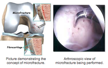
The surgery is performed by arthroscopy, after the joint is cleaned of calcified cartilage. Through use of an awl, the surgeon creates tiny fractures in the subchondral bone plate. Blood and bone marrow (which contains stem cells) seep out of the fractures, creating a blood clot that releases cartilage-building cells. The microfractures are treated as an injury by the body, which is why the surgery results in new, replacement cartilage. The procedure is less effective in treating older patients, overweight patients, or a cartilage lesion larger than 2.5 cm. Further on, chances are high that after only 1 or 2 years of the surgery symptoms start to return as the fibrocartilage wears away, forcing the patient to reengage in articular cartilage repair.

The effectiveness of cartilage growth after microfracture surgery is thought to be dependent on the patient’s bone marrow stem cell population and some think increasing the number of stem cells increases the chances of success. A couple of physicians are promoting an alternative treatment implanting autologous mesenchymal stem cells directly into the cartilage defect, without having to penetrate the subchondral bone.

Microfracture Reports

Studies have shown that microfracture techniques do not fill in the chondral defect fully, forming fibrocartilage rather than hyaline cartilage. Fibrocartilage is not as mechanically sound as hyaline cartilage; it is much denser and unable to withstand the demands of everyday activities as well as the original cartilage and is thus at higher risk of breaking down. The blood clot is very delicate after surgery and needs to be protected. In terms of time, the clot takes about 8 weeks to 15 weeks convert to fibrous tissue and is usually fibrocartilage by about four months post surgery, holding implications for the rehabilitation.

Chondrocyte Implantation procedures (CCI), a cell based articular cartilage repair procedure that aims to provide complete hyaline repair tissues for articular cartilage repair, have been posed by some as an alternative to microfracture surgery. In February 2008, Saris et. al published a large-scale study claiming that CCI results in better structural repair for symptomatic cartilage defects of the knee than microfracture surgery. According to the study, one year after treatment, the tissue regenerate associated with CCI is of better quality than that of microfracture surgery.


So we know a few things just from a Wikipedia article. This idea of purposely creating microfractures in the knee subchondral bone plate area in the shape of small circular holes will cause blood and bone marrow to flow out, cause some clotting, which will cause cartilage formation. It seems that the clotting action of the bone marrow with the stem cells inside which seeps out is what causes cartilage formation. The problem seems to be that the cartilage formed is fibrocartilage, NOT hyaline cartilage. We must remember that there are different kinds of cartilage. The growth plates and the articular cartilage are hyaline cartilage, which is what we are looking for and to create. However the idea of making very small microfractures and punctures through the bone into the subchondral layer is enticing. Cartilage is formed so that is a start.

From a section of the website for the US National Library of Medicine, National Institutes of Health


Knee microfracture surgery

Knee microfracture surgery is a common procedure used to repair damaged knee cartilage. Cartilage is the material that helps cushion and cover the area where bones meet in the joints.

Description

Three different types of anesthesia may be used for knee arthroscopy surgery:

  • Medicine to relax you, and numbing the knee using shots of pain killers
  • Spinal (regional) anesthesia
  • General anesthesia, where you will be asleep and pain-free.

The surgeon will make a 1/4-inch surgical cut (incision) on your knee.

  • A long, thin device called an arthroscope is placed through this cut. It is like a camera. It is attached to a video monitor in the operating room. This tool allows the surgeon to look inside your knee area and work directly on the joint. See also: Knee arthroscopy
  • The surgeon makes another surgical cut and passes tools through this opening. A small pointed tool called an awl is used to make very small holes, called microfractures (tiny breaks), in the bone near the damaged cartilage.
  • These holes release cells in your bones that build new cartilage. Your body will build new cartilage to replace the damaged cartilage.

Why the Procedure is Performed
Your doctor may recommend this procedure if you have some damage to the cartilage in the knee joint and on the underside of the kneecap.

The goal of this surgery is to prevent or slow further damage to the cartilage, preventing knee arthritis. It can help you avoid the need for a partial or total knee replacement.

This procedure is also used to treat knee pain due to cartilage injuries.

Another surgery, autologous chondrocyte implantation, is done for similar reasons.


What we see from this resource is overall a repeat of the same information. From the website for the London Knee Clinic, it would seem that the procedure is similar in effect as the Autologous Chondrocyte Implantation (aka Transplantation). It says that in the beginning, there will be 1/4th of an inch in cut on the articular cartilage surface area. There is a 2nd cut and surgical tools are passed through this opening. Something called an awl (sharp pointy device??) is used to make the small holes, which is what the microfractures are. The hole are big enough to release the cells from inside the bones to the outside where the cartilage covering is but small enough that bone healing from the clotting effect will be easy and will be relatively simple and noninvasive.

This is Part II in a 3 post series where I look at the potential idea on using the techniques and theories from microfracture surgery to possibly find a way to increase height.

Increase Height And Grow Taller Using Microfracture Surgery, Part I

In the previous post I mentioned that an old post entitled “Grow Taller Basics” from HeightQuest.com made me realize that from something as simple as head and feet rubbing can possibly lead to height increase. Something else that caught my eye from the post was this segment he added in…

“Scientists have utilized the power of microscopic fractures in stem cells with knee microfracture surgery to stimulate cartilage growth.  You can also cause microscopic fractures in the ends of the long bones to release these powerful red bone marrow stem cells.  Laterally pressing against the ends of your bones also increases fluid flow within the bone.  This increase in fluid flow sends the stem cells to your now opened growth plate!”

I recently been looking at the idea of possibly using the current microfracture surgery technology to increase height after I went back to an old post about the Shinbone routine and using Microfractures to grow taller to add to these old posts which I edited and upgraded. The idea on growing taller using induced microfractures has been around a long time and there were even groups and teams of people who tried out their own created routines to see if the idea would work.

Tyler’s idea is interesting and has some points with…”Their is a myth that the growth plates completely fuse after puberty but that is untrue.  Their is actually a thin growth plate line that is simply inactive.

I would actually have to disagree with this idea, because I have read from sources that the epiphyseal line itself will also eventually go away. At this point, I can’t find those sources which state that the line itself will also at one point disappear too.

From WiseGeek.com“The epiphyseal line the part of the bone that replaces the epiphyseal growth plate in long bones once a person has reached their full adult height. An epiphyseal line is visible on a standard x-ray. It looks like a thin dark streak that stretches horizontally across the rounded ends of the bone.”

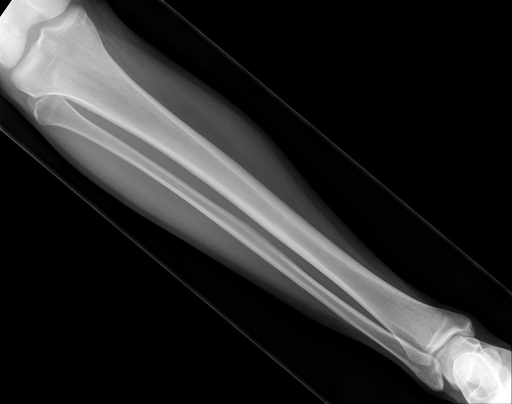
I would have to validate this type of claim from an pic of 1 X-Ray (out of 3 provided) of a tibia I received in the website email today I got where a person asked me whether I can see if their growth plates are fully closed and how to reopen them again. A guy named Felipe (last name not revealed over privacy issues) would give me this message…

“…here there are some pictures of my tibia distal and proximal growth plates,i had been using mens routine for 2 years and i hadnt any result, Actually i will be turning 20 in 2 months more,and i need a way to open my growth plates.”

[Note: As always, if this picture breaks any type of privacy or medical confidentiality laws, and I am informed by the person who gave me this picture that I am doing something wrong, I will of course take this picture down for privacy and legal reasons.]

Of the 3 X-rays he provided, this picture is probably the clearest. We can see that even a young 19 year old male already no longer has even an epiphyseal line. My claim is that the line also eventually goes away. There is a slight hint of some type of indention on the ends at the epiphysis but they are not where the epiphyseal plates are supposed to end up when they finally die out and calcify completely. I would guess that the line like indention is an indication of where the articular cartilage covering of the ends end off. If we use WiseGeek’s advice, I can’t find where the dark think streak is. Do you?

This is Part I of a 3 part series of posts where I look at whether we can use microfractures to cause some type of height increase.

18395516-0-RX DE PIERNA DERECHA-img0

Increase Height And Grow Taller From Head And Feet Rubbing And Tapping, A Simple Idea With High Feasibility

urlAs I was scouring through the usual sites that deals with height increase, I somehow came across a post on the GrowTallInfo.com website which made me realize that there may be a VERY easy and simple idea on how to possibly cause some bit of height increase. From the thread entitled “Grow Taller Basics” I realized that the thread was actually an old post that Tyler Made on HeightQuest.com HERE. After reading over the post, I was rather shocked to realize that a very simple and easy method for some height increase was possible that I completely forgot about which has a really good chance to work.

The post that was on HeightQuest.com mentions the fact that there are other types of bones in the human skeleton, not just the long bones. The skull for example is a clear example of a non-long bone but an irregular bone. The top of the skull is irregular in shape with a flat surface. As he states…

“The same tissue that is present on the width of long bones is present at the top bone of the skull.  Therefore, since you can increase the width of this tissue height increase after puberty is possible.”

“Now how do you increase the width of this outer sheet?  You rub against it.  Since it is the outer covering of the bone, you only need to target the surface of the bone.  So, a vigorous head massage may be one of the ways to get taller!”

When I thought over this simple and easy to do idea, I was very surprised at how simple it could be. All that one really has to do is do some vigorous top of head rubbing or massage. When I was little I would hear old wive’s tale that for a balding or bald man to regrow hair, he should just rub a raw potato on the top or back of his head, where the bald stop is. Maybe head and skull rubbing may cause more than just hair regeneration, but also some additional height that will come from the flat surface on the top of the head.

I remember looking over the idea of a dynamic bone loading method to widen the shoulder bones which I wrote a post about at “Review Of Claim To Widen Shoulder Bone, Lengthen Forearms, And Lengthen Lower Legs” and I had stated that the idea for shoulder widening really could work since the shoulder bone is irregular. Any possible loading on the surface of the shoulder could cause the bone sides to increase in width, thus leading to wider shoulder. The same idea is for the top skull flat bone.

I would maybe even suggest that instead of rubbing the head, it might be better to tap the entire upper head area, but get the loading to be of equal pressure distribution. If the tapping on the top of the head leads to just one small sized lump, like what we see in the cartoons when an anvil falls on a person’s head. However

If we remember, the way that real height is measure is from the top most point of a person’s top of head to their feet when they are standing completely erect and straight. The bump could mean that any bumps formed from appositional growth and bone remodeling from certain loading can lead to maybe a little bit of skull flat bone width increase and height increase.

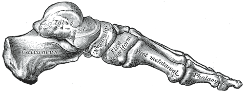
Gray290As for the feet…

Tyler would state…”There is also a bone in your feet.  Your heel bone.  This bone is completely covered with the sheet that can increase in width.  If you can increase the width of this bony sheath it will act sort of as height increasing shoes and you will be taller!

What he is referring to is known as the Calcaneus. From Wikipedia, “In humans, the calcaneus (from the Latin calcaneum, meaning heel[1]) or heel bone is a bone of the tarsus of the foot which constitute the heel. In some other animals, it is the point of the hock.”

Theoretically I would guess that the calcaneus may have the same bone remodeling effects as the flat surface of the top of the head, although I would suspect that in real life, the results would not be positive. We have to remember that in terms of the amount of weight and load any of the bones in the body gets most exerted on them, the heel bone probably has the most load on it. When a 200 lb human being jumps up, when they land back down, the heel bone and the ankles are the area of the body which gets the greatest amount of force per area and shock. Even if a person did manage to put enough load over the sheet of the heel bone, I would guess that the incredible load that the human body from the gravitational force exerted on it would experience would negate any real bone width increasing, but it might be possible to get 1-2 mm of height increase from applying pressure on the heel bone. I would rather suggest that it might be smarter to use a hammer or a 50-75 lb dumbbell for a stronger enough load on the bone.

Cfm1 and Cfm2

LSJL upregulates Cftr.  Although other than the name I can’t find a connection between this gene and Cfm1 and Cfm2.

According to Hierarchical fine mapping of the cystic fibrosis modifier locus on 19q13 identifies an association with two elements near the genes CEACAM3 and CEACAM6., Cfm1 is located on the same gene as Tgfb1 a height increase gene.  “seven microsatellite markers on 19q13 spanning a 4.8-Mb genomic area encompassing both, TGFB1 and CFM1”

“Cystic fibrosis (CF) is an autosomal-recessively inherited disease transmitted by two defective copies of the cystic fibrosis transmembrane conductance regulator gene (CFTR). The disease manifests as a generalized exocrinopathy, affecting all tissues that express the chloride and bicarbonate channel CFTR ”

Cystic fibrosis can result in shorter height.

Filamin-interacting proteins, Cfm1 and Cfm2, are essential for the formation of cartilaginous skeletal elements.

“Mutations of Filamin genes, which encode actin-binding proteins, cause skeletal abnormalities. The molecular mechanisms underlying Filamin functions in skeletal system formation remain elusive. In our screen to identify skeletal development molecules, we found that Cfm (Fam101) genes, Cfm1 (Fam101b) and Cfm2 (Fam101a), are predominantly co-expressed in developing cartilage and intervertebral discs (IVDs). To investigate the functional role of Cfm genes in skeletal development, we generated single knockout mice for Cfm1 and Cfm2, as well as Cfm1/Cfm2 double-knockout (Cfm DKO) mice, by targeted gene disruption. Mice with loss of a single Cfm gene displayed no overt phenotype, whereas Cfm DKO mice showed skeletal malformations including spinal curvatures, vertebral fusions, and impairment of bone growth, showing that the phenotypes of Cfm DKO mice resemble those of Filamin B (Flnb)-deficient mice. The number of cartilaginous cells in IVDs is remarkably reduced, and chondrocytes are moderately reduced in Cfm DKO mice. We observed increased apoptosis and decreased proliferation in Cfm DKO cartilaginous cells. In addition to direct interaction between Cfm and Filamin proteins in developing chondrocytes, Cfm is required for the interaction between Flnb and Smad3, which [regulates] Runx2 expression. Cfm DKO primary chondrocytes showed decreased cellular size and fewer actin bundles compared to those of wild-type chondrocytes. Cfms are essential partner molecules of Flnb in regulating differentiation and proliferation of chondryocytes and actin dynamics.”

“Inhibitors of actin polymerization stimulate chondrocyte differentiation in cultured mesenchymal cells and murine embryonic stem cells”

“mouse Cfm2 transcripts markedly increased in ATDC5 cells upon differentiation.”

“The CR length and tibial length of Cfm DKO mice were significantly shorter than those of wild-type, Cfm+/−/Cfm2−/−, and Cfm1−/− / Cfm2+/− mice at 4 weeks”

“Cfm1 and Cfm2 bind to Filamins (Flna, Flnb and Flnc)”<-LSJL upregulates Filamin C.

“Flna and Flnb are expressed in chondrocytes”

“Cfm1 and Cfm2 regulate chondrocyte survival and proliferation, and interact with Filamins. Cfms interacted with Filamins to organize perinuclear actin networks and regulated nuclear shape”

“Cfm is required for the interaction between Flnb and Smad3 in chondrocytes, and the Flnb-Cfm-Smad3 complex may play an important role in chondrogenesis.”

“Flnb normally prevents excessive Smad3 phosphorylation.”

“TGF-β1 stimulation up-regulated Cfm1 protein expression, but Cfm2 protein and mRNA were below detection level in an epithelial cell line”

The Multiple Difficulties And Challenges Involved In The Idea Of Going With Chondrocyte And Cartilage Implants And Transplants

As a response to the previous post I did about a member from the Make Me Taller forums named Mordred on a post HERE who talked about the idea of implanting chondrocytes into the closed growth plate area, I wanted to list the issues and challenges and problems which I can see at this point with this overall method.

Coming with an idea like implanting chondrocytes, progenitor cells like mesenchymal stem cells (MSCs), or completely in vitro epiphyseal cartilage plates into the original closed epiphyseal regions is easy and it makes sense.

However the hard part will always be…

  • Actually regrowing the epiphyseal hyaline cartilage plates and get the extracellular matrix of the cartilage to be the right compositions, with the collagen type II, the preoteoglycans, growth factors, cytokines, the gylcoaminoglycans, and the sulphated alkalines to be as close to what is found in vivo as possible.
  • You still have to cause some form of initial distraction or fracture or break in the bone so you can embedded the cartilage or chondrocyte material. The type of initial break would have to be uniform in a specific shape so that the shape of the completely regrown plate can slip in the distraction. The fact that the ilizarov method with its easy hammer and chisel quick fracture may seem it would be faster to go with the old idea. Even then, the process will be surgical where the outer muscle and skin tissue may have to be cut open to implant the cartilage.
  • You would have to check the immunological issues with this approach because of the fact that human bodies which detect foreign objects will try to attack it with its white blood cells and other resistance and defensive biological. This would cause the completely functioning body to REJECT the implant. Remember from talks with medical professionals about how hard it is to find the right blood donor, or organ donor? It is because of the human body’s natural defensive system, in rejecting foreign bodies.
  • You must remember that the human body when it was young had at least two dozen growth plate cartilage areas, not just the knee region with the distal femoral area and the proximal tibial area. – If you want to get a proportional body, you would have to open or embedded the foreign in vitro grown cartilage into the humerus at least so that your arms are as long as your legs, in proportion. We must remember that in general, the wingspan of a human is about the same length as their height.
  • There were growth plate cartilage in the hip joint area, in each other vertebrate of the torso, in both ends of the knee, both ends, wrist, elbow, metatarsals, metacarpals, collarbone, etc. – Would we spend possibly hundreds of thousands of dollars also embedding more cartilage into our body to get the entire body to be proportional?

Conclusion: What is clear is that the overal idea and method is reasonable and makes sense. There are just a few major challenges we would have to get around to make the idea reasonable and practical enough to get it working for the average person who hopes for an alternative to the limb lengthening solution.