Even More Evidence That Pregnancy Can Lead To Height Increase, This Must Be Considered!!

Me: So after the quick google search I thought the results and posting would end but they seem to only increase in number and the stories are vast in number. There is something going on here which I can’t explain. Tyler would try to say that the growth plates never fully disappeared because their senescence was slower from DNA methylation or something. I would say that the correlation is with whatever hormone increases were occurring during pregnancy.

My Theory: These women are now eating for two. Their appetite goes up, their food intake increases, and this is very similar to what happens during puberty where the young adolescent starts to eat at an extremely high rate. However it doesn’t explain why they would ever gain height. In terms of symptomes, their food intake resembles teenagers who are going though puberty eating everythiing. One women who is 41 gains 1 inch and another woman gains 3 inches through pregnancy.

From BabyCenter.Com… there is one post of a women who noticed her pants have gotten shorter and she doesn’t fit in them anymore.

From TheBump.Com community…

01-21-2011 at 10:57 PM
jennlynn53
Not Ranked
Joined on 07-21-2010
Louisiana
25 Posts
378 Points
jennlynn53 is not online. Last active: 09-01-2011, 12:45 PMNewbie

Growing taller because of pregnancy?

Is it possible for pregnancy to give you a growth spurt of some kind? I have been 5’3” since I was 12/13 and I had my FI measure me and I am now 5’5”. I tried searching on the web for an answer but I just kept coming to dead ends. So has this happened to anyone else? Or has anyone heard of such a thing? TIA

Me: This woman poster was measured 9-10 months ago and she was 5’3″. Now she is 5′ 5″ and she is 23. However a reply on the same board link is even crazier. Read below…

02-06-2011 at 7:25 PM
sfuqua1
Not Ranked
Joined on 11-16-2010
1 Posts
15 Points
sfuqua1 is not online. Last active: 02-06-2011, 7:25 PMNewbie

Even though my doctor said it’s impossible he can’t deny that I have grown.  I was 26 during my first pregnancy.  I started the pregnancy at 5’1 and ended it at 5’3.  I stayed at 5’3 for two years and then got pregnant again.  I am now 28 and started this pregnancy at 5’3 and already at 7 1/2 months pregnant I have grown to 5’4.  I seem to gain 1-2 inches with every pregnancy.  My doctor has no explanation for this and neither does the internet.  But it’s such a relief to know I’m not the only “freak” 🙂 out there who is doing this!

Me: So this mother grows 2 inches in her first pregnancy at the age of 26 and grows another  1 inch on her 2nd pregnancy at age 28. Her doctors can’t explain it.
April 17th, 2012, 11:19 PM
TuckrLeesMommy TuckrLeesMommy is offline

Mega Super Mommy

Join Date: Apr 2012
Location: Michigan
Posts: 1,289
Hey ladies,
so something interesting has just recently come to my attention.
I am almost 22 weeks pregnant now, i am due the end of august.
I am 22 years old and i have a younger sister who is 12.
now i am on the shorter side, i have been 5 foot even since i was 17. Just before my 3 month mark i was weighed and measured for my first WIC appointment at my local Health Department, and once again i was about 5 foot even.
Well my 12 year old younger sister has been about a 1/4 inch taller than me for several months now, well just a few days ago she was standing next to me and i realized that i was looking down to talk to her.
my mom laughed (she frequently ribs me about my height) and stood us back to back and proclaimed that i was indeed taller than my sister, when just a few months before we had done the same and my little sis was taller.
My mom immediately measured me and i’ll be ****** the tape measure read clearly…
5′ 2 1/2″
somehow i had gained over 2 inches of height!!!
now i have scoured the internet and i cant find ANY medical findings on this, but a few other accounts of women asking something similar.
has anyone else had this?
has pregnancy caused anyone else to grow?
is there a name for this kind of phenomenon?

Me: This girl who is 22 was first measured at 3 months of pregnancy (12 weeks) at 5 feet tall and in the 22 week she is measured at 5′ 2.5″ which means over a 2 inch increase over 10 weeks. I could say that she is unique at age 22 for having another growth spurt. Since she is rather short I could claim that catch up growth is what caused it.

From the website Mothering.Com

Getting taller during pregnancy?

post #1 of 9
10/22/08 at 3:55pm
THREAD STARTER
  • funkychunkymunky
  • offline
  • 118 Posts. Joined 3/2008
I’m out of the age range of height growth and yet I found myself growing about three inches during my pregnancy. Has this happened to anyone? My height never went back to normal.
10/22/08 at 6:12pm
  • 456 Posts. Joined 11/2007
  • Location: Roma, Italia

Hmmm…. Now you’ve got me thinking.

I have always been 5’2″, and I’m way after the age range for height growth. (I’ll be 41 in a few months.)

This past spring I had to measure myself for a costume and I was, very surprisingly: 5’3″! Now, that’s only one inch compared to your three inches, but it is possible that it was from the pregnancy… I honestly don’t know what else it could be. Other than maybe Yoga, but I’ve been doing yoga since ’93 and it had never caused any height increse before. And I’ve been very neglectful of my yoga practice these past few years.

Hmmmm……

I should mention that my feet remained their normal size through out the prenancy and although my butt and chest got bigger they’ve mostly gone back to Pre pregnancy size.

More Evidence That Pregnancy Can Increase Height, WOW!

Ever since I found the stories of women who have claimed to have increased in height during pregnancy I decided to do a quick search on google for stories, anecdotal evidence, and posts by women who claim they had gotten taller.

Me: If you read through the comments left by other moms you realize that this phenomena seems to be far more common than I ever expected. Of the 17 replies this women got, a good 2 of them said that they saw an increase in height beyond what would obviously be measurement error. The crazy anedotal story is the man’s wife who grew a “couple” inches when she was 30 and on her 3rd child. Wow, what is going on? Of course it is important to realize that a few of the replies stated that they actually lost height from the pregnancy. I don’t know what to take from this post and the replies except be more confused now.

From this post on an Australian Huggies web board HERE the poster says that she grew 1.5 cm during her pregnancy.

Here is one from CafeMom (source link HERE)

Height growth during pregnancy

Summer222009
Posted by

  • Summer222009

on Jul. 6, 2011 at 3:54 AM

  • 17 Replies

I know this may sound weird, but I was talking to someone else today about growing an inch or 2 taller while they were pregnant. I was 5’2″ pre pregnancy and now I’m 5’3 1/2″. This girl is also saying she got taller. I thought it was just the way they measured my height. But can you grow while pregnant?

Posted by

  • Summer222009

on Jul. 6, 2011 at 3:54 AM

  • Vipergirl22

Jessica on Jul. 6, 2011 at 5:38 AM

haha I would like to know that answer too…be interesting if it is true.

KaroGreenly

by

  • KaroGreenly

Karo on Jul. 6, 2011 at 8:30 AM

How old are you? Maybe you just weren’t finished growing to begin with.

Posted on CafeMom Mobile
.Angelica.

by

  • .Angelica.

Angie on Jul. 6, 2011 at 9:29 AM

I have heard of this happening. Consider yourself lucky if you wanted to be taller I guess. LOL.

.Angelica.

by

  • .Angelica.

Angie on Jul. 6, 2011 at 9:30 AM

A guy my DH works with, when his wife was pregnant with her 3rd child and I think right around 30 she grew a couple inches. So she should have for sure been done growing.

Quoting KaroGreenly:

How old are you? Maybe you just weren’t finished growing to begin with.


Summer222009

by

  • Summer222009

on Jul. 6, 2011 at 2:40 PM

I’m 19 but my growth plates have stopped when I was 12.

Quoting KaroGreenly:

How old are you? Maybe you just weren’t finished growing to begin with.

momoftwins_2

by

  • momoftwins_2

on Jul. 6, 2011 at 4:34 PM

My feet grew but I didn’t get taller

Posted on CafeMom Mobile
ATG499

by

  • ATG499

Wendy on Jul. 6, 2011 at 5:22 PM

I didn’t get any taller during my pregnancy.

Posted on CafeMom Mobile
alyciajoseph

by

  • alyciajoseph

on Jul. 6, 2011 at 7:42 PM

Mine too 🙁

Quoting momoftwins_2:

My feet grew but I didn’t get taller

Posted on CafeMom Mobile
remaining_me

by

  • remaining_me

Kay on Jul. 6, 2011 at 7:55 PM

 hmm i never heard of that, lol i didnt get any taller. wider? yes, lol.

remaining_me

by

  • remaining_me

Kay on Jul. 6, 2011 at 7:57 PM

 yeah, i permanently went up a half size with my second. I never knew they actually GREW, odd enough. i thought they just swell up.

Quoting momoftwins_2:

My feet grew but I didn’t get taller

11-17 of 17 replies
first previous 12

Jump to page:GO
Maddies-Mommy

by

  • Maddies-Mommy

Member on Jul. 7, 2011 at 2:24 AM

I didn’t get taller, however I went up a shoe size. It was weird, they didn’t swell they just got longer and three years later they are still that size.
lovealot0587

by

  • lovealot0587

on Jul. 7, 2011 at 2:32 AM

I’m not too sure

Posted on CafeMom Mobile
hayvensmommy07

by

  • hayvensmommy07

Bronze Member on Jul. 7, 2011 at 9:03 AM

I was 5’2 before my son. Now I’m 5 foot. Yea I shrank. How I have no idea so if I can shrink then I would think women could get taller too.

Posted on CafeMom Mobile
JayBearsmomma

by

  • JayBearsmomma

Rachael on Jul. 7, 2011 at 9:47 AM

lol I shrunk and1/2 inch ! I was 5’2 for the longest then at my 6 week check up I was 5’1 1/2

Posted on CafeMom Mobile
YouGotToLoveIt

by

  • YouGotToLoveIt

on Sep. 9, 2011 at 8:11 PM

Hi,

This is my first attempt to investigate my growth during pregnancy on the web.  Although I have a friend who grew 1 1/2 inches during her 3rd pregnancy and only daughter.

I am now 60 and had my only child when I was 36.  I grew 3/4 of an inch during pregnancy.  I tried to document this growth with any of my doctors and they all made excuses saying that they wrote down my original height incorrectly.  It was very frustrating because my original height was 5 feet 4 3/4 inches and I always tried to stand super tall when measured so that I could say I was 5 feet 5 inches even.  My final height is 5 feet 5 1/2 inches.

Thanks for this forum.  It is nice to know that I am not alone.

Girl_Charlie

by

  • Girl_Charlie

on Sep. 9, 2011 at 9:39 PM

And you thought that a “young moms” group was the place to go? Even 24 years ago, 36 was on the upper side of having children, not the young side.

Quoting YouGotToLoveIt:

Hi,

This is my first attempt to investigate my growth during pregnancy on the web.  Although I have a friend who grew 1 1/2 inches during her 3rd pregnancy and only daughter.

I am now 60 and had my only child when I was 36.  I grew 3/4 of an inch during pregnancy.  I tried to document this growth with any of my doctors and they all made excuses saying that they wrote down my original height incorrectly.  It was very frustrating because my original height was 5 feet 4 3/4 inches and I always tried to stand super tall when measured so that I could say I was 5 feet 5 inches even.  My final height is 5 feet 5 1/2 inches.

Thanks for this forum.  It is nice to know that I am not alone.


JayBearsmomma

by

  • JayBearsmomma

Rachael on Sep. 9, 2011 at 10:28 PM

Lol I shrunk half an inch
Posted on CafeMom Mobile

There’s a study that says pregnancy can increase bone turnover. This may be partially the cause of the height gain as bone turnover equals more plastically deformable bones.

The effect of pregnancy on bone density and bone turnover

“We studied 16 women longitudinally, with baseline measurements before pregnancy; then at 16, 26, and 36 weeks of pregnancy; and postpartum. We measured total-body BMD and biochemical markers of bone resorption (urinary pyridinium crosslinks and telopeptides of type I collagen) and bone formation (serum bone alkaline phosphatase, propeptides of type I procollagen [PINP] and osteocalcin). We also measured parathyroid hormone (PTH), insulin-like growth factor I (IGF-I), and human placental lactogen. Postpartum, BMD increased in the arms (2.8%, P < 0.01) and legs (1.9%, P < 0.01) but decreased in the pelvis (-3.2%, P < 0.05) and spine (-4.6%, P < 0.01) compared with prepregnancy values. All biochemical markers, with the exception of osteocalcin concentration, increased during pregnancy. The change in IGF-I at 36 weeks was related to the change in biochemical markers (e.g., PINP, r = 0.72, P = 0.002). Pregnancy is a high-bone-turnover state. IGF-I levels may be an important determinant of bone turnover during pregnancy. Elevated bone turnover may explain trabecular bone loss during pregnancy.”

“The involuting uterus is also a possible source of collagen degradation products. The uterus is predominantly composed of type I collagen; pyridinoline (Pyd) is the principal cross-link, and deoxypyridinoline (Dpd) is present in only trace amounts. The collagen content of the uterus increases approximately sevenfold during pregnancy. An increase in the Pyd/Dpd ratio during the puerperium would indicate a contribution to maternal cross-link excretion from uterine collagen.”

“Previous studies have reported decreased serum intact osteocalcin during pregnancy.”

“there appears to be a loss of bone density at trabecular bone sites and an increase at cortical bone sites during pregnancy. Bone turnover is increased with the increase in bone resorption preceding that of formation.”

Pregnancy Causes Hand, Feet, Extremity Increase In Length And Width And Height Increase (IMPORTANT)

When I was doing research for my post on the possiblity of injecting GH or BMPs into the feet to increase it’s size, I seemed to have come across a very interesting phenomena which might help a little in our search. It turns out that during and after pregnancy, the women often has her hands, feet, extremities enlarge in size, and that size increase never goes away.

You can see from all the comments that this is a very common phenomena. The comments by other mothers on the link at Baby Center continues for 5 more pages all telling their stories. There seems to be this hormone relaxin which loosens the joints in their feet, causing the foot bone sto spread. However, that doesn’t seem to explain why it is that even after pregnancy, the feet stays in that enlarged size. I wonder whether it might also be that the very bones also thickened and lengthened in size.

The stories show that the fingers get thicker. The feet gets longer, which could indicate either the feet being squeezed and spread out, or maybe the bones are indeed getting longer.

The most crazy comment I saw from the post was on the second page (link HERE) where the mother said this!!

Update: It seems that a 2nd mother posted that she had grown taller during pregnancy as well on the 4th page of comments!! (HERE)

Was this comment helpful?

I not only grew my feet from size 7 to 8, but I also grew 1 1/2 inches in height!!
posted 2/28/2009 by a BabyCenter Member
Was this comment helpful?

I too went up a size in shoes for each pregnancy (from a 7-9) but also grew 1 1/2″ in height at 24! Now that my third pregnancy is occurring at 28, I am fairly sure I won’t be getting any taller, but I also hope I don’t go to a 10…
posted 4/29/2012 by a BabyCenter Member
Analysis: Now I know that when females are going through pregnancy, they have all this hormone going through their body. I an accept the idea that their fingers and feet get enlarged and engorged. I am also proposed and accept the idea that maybe one of the hormones going through their system can cause even the long bones to thicken and get wider. This could mean that the metatarsals in their feet expanded radially making them even 3-4 millimeters taller. However this women who commented says that her pregnancy caused her to grow 1.5 inches in height at 24! What is going on?? I am very confused because that is not supposed to happen from what we currently understand. It reminds me again of the Tanya Angus and Kosen cases before. In a previous post I had created a theory that if you expand your stomach and torso out to stretch out your vertebrate it could theoretically cause some height increase. However a pregnant women’s expanded stomach should actually decrease her height because her lumbar region is pulled further downwards. If this story is accurate, it is another example of the idea that extreme hormones may cause some height increase even after physical maturity. Update: 2nd mother has commented that she also grew 1.5 inches in height with a shoe size increase from 7 to 8.

From BabyCenter.Com

Expert Answers

I think my feet have grown. Is this possible?

Jane Andersenpodiatrist

Yes. As you’re discovering, your belly isn’t the only thing that gets bigger during pregnancy! This is partly due to pregnancy weight gain and swelling from the extra fluid your body retains while you’re pregnant (called edema). To ease the swelling, soak your feet in cool water and prop them up as often as you can.

But there’s another reason that feet seem to grow during pregnancy. The aptly named pregnancy hormone relaxin, which loosens the joints around your pelvis so your baby can make his way down the birth canal, also loosens the ligaments in your feet, causing the foot bones to spread. Your foot bones aren’t actually growing — it’s just that the ligaments that hold these 26 bones together aren’t as tight as they were.

You may notice your shoes getting a little tight as early as the second trimester, and they may continue to increase in size until late in pregnancy. While the foot swelling generally subsides within a month after delivery, any foot spreading caused by looser ligaments is permanent. I estimate that half the moms out there wear shoes a half or a whole size bigger than they wore before having babies.

The bright side, if there is one, is that you get to buy new shoes! Buy a few pairs of comfortable, roomy shoes to wear during your pregnancy, but be aware that you may have to go back for more once the swelling settles down and your shoe size stabilizes about a month after delivery. Whatever you do, don’t try to make do with your old shoes! Wearing tight shoes can aggravate bunions and cause a host of painful foot problems, ranging from ingrown toenails to corns and calluses.

Finally, since feet tend to grow with each pregnancy, you may want to hold off on those pricey Jimmy Choos or Manolo Blahniks until you’re finished having babies.

Member Comments

Mine certianly did with my first. (Now 4) First they were swollen from a size 7-7.5 to a tight open on both ends size 10 sandal. After my son was born the swelling was gone, but my feet are now at a size 8-8.5
posted 2/05/2008 by a BabyCenter Member
39 out of 40 found this comment helpful
Was this comment helpful?
Before pregnancy I had big feet already because I am 6ft and wore size 11 1/2 or 12. After the pregnancy of my first daughter I could only wear a size 12 and sometimes they were a little snug. After the pregnancy of my 2nd daughter I can only wear size 13. I am now pregnant with our 3rd child and wondering what shoe size I will be in after this pregnancy. Talk about hard to find shoes that aren’t super expensive.
posted 7/29/2008 by a BabyCenter Member
28 out of 28 found this comment helpful
Was this comment helpful?

Mine did! I originally went from a 9… to a definite 9.5 after my first child. Basically depended on the brand. Now after having my second I’m now at a 10!Gawww! LOL It’s not so bad I guess if you start out being a 7 lol. But I miss being an 8.5-9!!
posted 2/21/2008 by lnewman2
22 out of 23 found this comment helpful
Was this comment helpful?

I am so glad I saw this, and I wish that some doctors would tell you this because it would have saved me a lot of pain! I couldn’t figure out why I was having terrible ingrown toenails. Being a nurse I finally figured out that it started when I stopped wearing flip flops. Definetly can’t wear ANY of my pre pregnancy shoes anymore. My beautiful wedding ring needs to be resized too, oh bother! Thank you for this post…i feel sooo much better that it wasn’t just me.
posted 10/25/2008 by a BabyCenter Member
19 out of 19 found this comment helpful
Was this comment helpful?

Trying to buy new shoes definitely isn’t a bright side when you were already a 5’11” woman who wore a size 11W before getting pregnant and now find yourself searching for the elusive 12W. I’d be better off trying to cobble my own shoes. It’s almost as if manufacturers think that the bigger your feet are, the less taste you have. I don’t want to wear shoes even my grandmother would be ashamed to wear.
posted 9/18/2010 by a BabyCenter Member
19 out of 19 found this comment helpful
Was this comment helpful?

I swear my fingers grew during pregnancy…my rings no longer fit at all! I have waited 9 mos and they still do not fit! Did this happen to anyone else?
posted 2/14/2008 by MommyStar24
15 out of 17 found this comment helpful
Was this comment helpful?

being the cheap-O that i am i tried to wear my pre-pregnancy shoes after the pregnancy and ended up with horribly painful ingrown toenails on a few of my toes that got severely infected. that lasted for several months to a year after she was born. by all means buy larger shoes!!
posted 10/18/2009 by a BabyCenter Member
11 out of 11 found this comment helpful
Was this comment helpful?

Yep, my feet grew a full size while I was pregnant and have settled to a half size bigger (gave birth 8 months ago). I’m also finally coming to the realization that my fingers have grown too. I’m just about at pre-pregnancy weight and I still can’t get my wedding rings on my finger.
posted 7/09/2010 by a BabyCenter Member
9 out of 9 found this comment helpful
Was this comment helpful?

Mine also swelled up with my pregnancy, and unfortunately never swelled back. That wouldn’t be so bad if I were a size 7 or 8 to start with, but I’m 6’1″ and already wore a 12 which was next to impossible to find off the rack. Now I wear a 13, and well, that’s even harder to find, unless I want to wear shoes that make me look like a granny or a lady of the night. *sigh*
posted 10/08/2008 by a BabyCenter Member
10 out of 11 found this comment helpful
Was this comment helpful?

My mom and my aunt told me about this happening but but all my friends were like…”I’ve never heard of that” And thought I was just cracking jokes or making it up. I started out at size 7-7.5. After my first daughter I could no longer fit a size 7. I brushed it off as “well that must be how the shoe is made” Now after my 2nd daughter my shoe is a size 8. I attributed my new shoes size to the extra weight I had put on with the baby #2. Now with my new found realization I must no longer hope to fit in those heels in the garage…. You can find them on ebay (someday). DAMN YOU HORMONES!!!!!!
posted 1/22/2009 by a BabyCenter Member

New Proposed Height Increase Method Using Feet Calcaneus Enlargement Through GH Injections And BMPs

I was sitting around writing up another post today when I realized that I currently have 1 technique to increase the length of the legs, another to increase the length of the torso, but what about the other areas? I thought about the head but there is not a lot I could think of at the time except wear taller hats like Abe Lincoln. I moved to the feet and I had a rather simple idea.

We know that from the condition of gigantism and acromegaly that the extremities and localized regions in a person can be enlarged and made bigger through a lot of growth hormones. Anthony Robbins and Shaq are known for having extremely large hands and feet. Obviously Robbins large hands and teeth are from his pituitary issue.

So why can’t we just buy some growth hormones, somatotropin, the real stuff and inject some into the blood vessels that reach out feet? given enough time, the irregular bones in the feet should expand in width. Since the layout of the long bones in the feet is flat on the ground, any increase in width of the long bones and irregular bones will only cause the distance between the bones and the ground to increase, which is effectively increasing height.

From this link HERE we find a male who states that his feet grew more than 1 shoe size when he is 28.

We know that the calcaneus is the heel bone at the back of the feet. Any increase of this bone will cause the entire body to be pushed up. I had in a previous post talked about feet heel implants to increase in height and that was implants at the calcaneus. I would say that a person who tries to inject growth hormones or BMPs into the blood stream that feed the mineralization and maintenance of the bones in the feet will lead to enlarged bones and cause some height increase, say 1-1.5 cm throughout a 1-5 year range time.

The height gain will be minimal and probably would lead to possible deformity and disfigurement but my point for this idea is a possible proof of concept.

Heel spurs may be another way to increase height:

The plantar calcaneal spur: a review of anatomy, histology, etiology and key associations

“The plantar calcaneal spurs (PCS) are a bony outgrowth from the calcaneal tuberosity “
 
“PCS are typically described as bony outgrowths arising just anterior to the medial process of the calcaneal tuberosity”
 
Here’s the tuberosity of the calcaneus:


So it specifically needs to be the lateral process of tuberosity.

Here’s some bone spurs:

” projections of larger than 1 or 2 mm”<-bone spur size.

“The prevalence of PCS increases with weight”

“The prevalence of PCS increases in older age groups”

“PCS consists of a core of mature lamellar bone and demonstrates evidence of degeneration and fibro-cartilaginous proliferation, along with one or more of intramembranous, chondroidal and endochondral ossification occurring at the surface”

“There is a statistically significant correlation between foot pronation and the development of PCS, with 62% of patients with a spur and 81% with a painful spur having a pronated foot radiographically”<-flat feet

“general tendency toward ossification of ligaments”

“over half of PCS are not within the PF or the intrinsic muscles of the foot but rather are surrounded by loose connective tissue”

“the epiphysis of the calcaneus extends down to the plantar aspect and that repetitive trauma results in PCS formation”

“repetitive trauma with low forces can cause histological changes and as the impact force is increased, fewer repetitions are required”

New Proposed Height Increase Method Using PEMF, BMP-7, BMP-6, And TGF-Beta3

This was an idea that I have been thinking about ever since I found and reviewed the study which showed that BMP-7 (aka OP-1) injections into a mice and sheep’s vertebrate lead to the regain of lost height and health of degenerating disks.

My idea is to create a type of padding or guase  which you would have BMP-7 and BMP-6 with TGF-Beta3 embedded in the padding. Dexamethasone might also help. Certain BMPs and growth factors have been shown to both lead to chondrogenesis and osteogenesis. BMP-6 is really good at osteogenesis. We are looking for those. The padding has these notches and bumps, with the bumps alligned with the location of the intervertebral disks. The padding is pressed agains tthe back skin, with small needles that can inject the growth factors  right next to the disks. Remember that the disks have an outer concentric layer (outer annulus fibrosus)  and the inner circular layer (nucleus pulposus (NP). The outer layer is what the growth factors have to both get around and into. There is still some cartilage in the vertebrate even after we have reached physical maturity.

The padding can be just even 1 cm thick and can be refilled like a syringe which the nurse replaces when she is collecting a lot of blood from you. The BMP will theoretically be soaked into the vertebral disks leading to first chondrogenesis, increased nucleus pulposus function (and maybe even size), and finally ossification leading to increased thickness in the disks causing torso and height lengthening. The sequence of body physiology can be controlled by manipulating the injected device to switch from one type of growth factor into another. The pads can be wrapped around the body with either velcro or an elastic strap.

The second part of the idea was to use PEMF technology on the sides of the disks and give short duration, low intensity sine wave electrically capacitive signals to stimulate the collagenous material… At this point I would like to test the PEMF idea on the collagenous fibers of the annulus fibrosus so see if they would get any larger.

Overall, this idea is rather simple to try out even at home. The hard part may be getting BMP-7 and the TGF-Beta 3 from a non-natural source.

From PubMed Study 1 HERE

Spine J. 2008 May-Jun;8(3):466-74. Epub 2007 Jun 26.

Bone morphogenetic protein-7 protects human intervertebral disc cells in vitro from apoptosis.

Wei A, Brisby H, Chung SA, Diwan AD.

Source

Orthopaedic Research Institute, The University of New South Wales, St George Hospital Campus, level 2, 4-10 South Street, Kogarah, NSW 2217, Australia.

Abstract

BACKGROUND CONTEXT:

Disc degeneration includes dysfunction and loss of disc cells leading to a decrease in extracellular matrix (ECM) components. Apoptosis has been identified in degenerated discs. Bone morphogenetic protein-7 (BMP-7) has been reported to stimulate ECM synthesis in the intervertebral disc (IVD), but its effect on disc cell viability is unknown.

PURPOSE:

To investigate whether BMP-7 can protect disc cells from programmed cell death while enhancing ECM production.

STUDY DESIGN:

An in vitro study to examine the effect of BMP-7 on apoptosis of IVD cells.

METHODS:

Human nucleus pulposus (NP) cells were cultured in monolayer, and human recombinant pure BMP-7 (rhBMP-7) was added to the medium when the cells were in the second passage. Thereafter, apoptosis was induced by either tumor necrosis factor-alpha (TNF-alpha) or hydrogen peroxide (H(2)O(2)). Cellular apoptosis was evaluated by terminal deoxynucleotidyl transferase-mediated dUTP nick end labeling assay and caspase-3 activity. ECM synthesis was assessed by immunofluorescence for collagen-2 and aggrecan. To study the possibility of bone induction by rhBMP-7 in disc cells, alkaline phosphatase activity and Alizarin red-S staining were evaluated.

RESULTS:

Apoptosis was induced by both TNF-alpha and H(2)O(2). Addition of rhBMP-7 resulted in inhibition of the apoptotic effects caused by both inducers. Further, BMP-7 decreased caspase-3 activity. In the presence of BMP-7, ECM production was maintained by the cells despite being in an apoptotic environment. No osteoblastic induction of the disc cells was seen.

CONCLUSIONS:

BMP-7 was demonstrated to prevent apoptosis of human disc cells in vitro. One of the antiapoptotic effects of BMP-7 on NP cells might be a result of its inactivation of caspase-3. Collagen production was maintained by addition of rhBMP-7 in an apoptotic environment.

PMID: 18082466   [PubMed – indexed for MEDLINE]

From PubMed study 2 HERE

Tissue Eng Part B Rev. 2010 Aug;16(4):435-44.

Comparative review of growth factors for induction of three-dimensional in vitro chondrogenesis in human mesenchymal stem cells isolated from bone marrow and adipose tissue.

Puetzer JL, Petitte JN, Loboa EG.

Source

Joint Department of Biomedical Engineering at UNC-Chapel Hill and North Carolina State University, Raleigh, North Carolina 27695, USA.

Abstract

The ability of bone-marrow-derived mesenchymal stem cells (MSCs) and adipose-derived stem cells (ASCs) to undergo chondrogenic differentiation has been studied extensively, and it has been suggested that the chondrogenic potential of these stem cells differ from each other. Here, we provide a comprehensive review and analysis of the various growth factor induction agents for MSC and ASC three-dimensional in vitro chondrogenic differentiation. In general, the most common growth factors for chondrogenic induction come from the transforming growth factor beta (TGFbeta) superfamily. To date, the most promising growth factors for chondrogenesis appear to be TGFbeta-3 and bone morphogenetic protein (BMP)-6. A thorough review of the literature indicates that human MSCs (hMSCs) appear to exhibit the highest chondrogenic potential in three-dimensional culture in the medium containing both dexamethasone and TGFbeta-3. Some reports indicate that the addition of BMP-6 to TFGbeta-3 and dexamethasone further increases hMSC chondrogenesis, but these results are still not consistently supported. Induction of human ASC (hASC) chondrogenesis appears most successful when dexamethasone, TGFbeta-3, and BMP-6 are used in combination. However, to date, current formulations do not always result in stable differentiation to the chondrocytic lineage by hMSCs and hASCs. Continued research must be performed to examine the expression cascades of the TFGbeta superfamily to further determine the effects of each growth factor alone and in combination on these stem cell lines.

PMID: 20196646    [PubMed – indexed for MEDLINE]

New Proposed Height Increase Method Using Knee Chondral Defects And BMPs, TGF-Beta, GDFs

Me: After going back and reading on my old posts on the power of possibly using BMPs to induce chondrocytes and also heal and regenerate articular cartilage, I have created a new idea on how it might be possible to induce longitudinal growth using a combination of at least 3 main ideas I have been exploring for the last 3 months. This a culmination of all of my learning and research so far. You can see from another post way back entitled” Increase Height Through Surgical Method By Cartilage Harvesting And Chondrocyte Implantation With Growth Factor Injections” that I tried also at that time to think up a possible height increase less invasive surgical path as well, but this new method is more likely to work out and the procedure is more detailed, due to new understanding and research. 
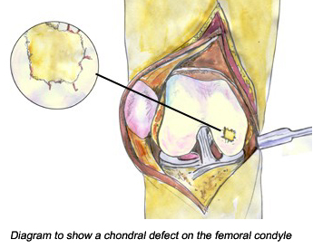
Note: One of the biggest issues we have to understand is what exactly are the differences between the articular cartilage found at the end of the long bones and the original growth plates we had. Remember that both of these cartilage ar hyaline to begin with. The major differences are…

1. The epiphyseal has a perichondrium, the articular does not.

2. The epiphyseal cartilage is created from two centers of ossification, a primary and secondary. The articular cartilage does not.  This means that the epiphyseal is directly connected to layers of trabecular bone which house progenitor cells on both sides. The articular does not.

3. The epiphyseal has a resting zone area, the articular does not.

One idea I originally thought about is try to get the articular cartilage to behave as much like the growth plates as possible.

Note: The other big thing we would have to get right is the right growth factor combination. At this point I am guessing to use BMP-7 (obviously), TGF-Beta2 & 3 (since there have been articles that showed that BMPs need it to work out), BMP-2 since it is the only other BMP that leads to chondorgenesis. We want to avoid BMPs which give better results for osteogenesis but have shown to result in chondrogenesis. The other big one I wanted to use was GDFs which are growth differentiation factors. 

So Step 1…

You create two chondral defects through the knee which connects the inner epiphysis trabecular bone with the articular cartilage. This can be done through two holes punctures through the cartilage of a bend knee (from diagram above). The puncture can be just 2-3 mm in diameter (from study 1). I would suggest using a diameter of 4-5 mm if that is possible. The exact location the punctures will go through will be the trochlea and the femoral condyle (from study 2). The penetration will be through the hard bone, into the epiphysis until it reaches the axis distance where the original epiphyseal plates will be (from X-ray).
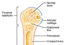
Note: From this diagram on the right it suggest that even after the epiphyseal line is gone, there seems to be still a band of trabecular bone which might be slightly thicker and denser in the area where the epiphysis and metaphysis are connect than in other areas in the inner bone. That is the line you want to reach, but right before you hit that thick band of dense bone. 

Step 2….

You implant a type of capsule which has a specific mixture of  growth factors that focuses on chondrogenesis, not osteogenesis. The growth factors can be encapsulated in a collagen and proteoglycan mixture. It is essentially cartilage matrix with growth factors inside but no chondrocytes. The mixture will have BMP-7 and BMP-2 which has shown to help differentiate the progenitor cells in the right way to give you the type of cell desired . You want to avoid types of growth factors which are mainly osteogenic.

We know that adult marrow is mostly yellow marrow of adiposytes and fatty acids. Study 5 suggest using instead BMP-6 and TGF-Beta 3 to convert them into chondrocytes. Remember that the bones are one of the tissues that regenerate and heal the fastest. That means that one must focus less on bone healing and more on how to get the cartilage to expand.

Step 3…

After the implant capsule of growth factors, you implant another capsule that has chondrocytes encased surrounded by a perichondrium layer. This is actual growth plate cartilage grown in vitro. I have researched one study that has shown that in vitro growth plates have been developed and can be reapplied back to function properly in vivo. Study 3 showed that chondorcytes can be differentiated from using perichondrium with BMP-7 so this step should be easy.

Step 4…

You make a cut around the outer edge of the long bone right at the distance where the initial penetration was done. You remove 3-4 mm in thickness axially of outer bone which will be the periosteum but also 4-5 cm radially inward. This is to remove the outer hard cortical bone. The other reason you want it to be at least this thick, is to prevent bone repair. If the created fracture was any thinner the bones will automatically heal over it. You implant resting zone progenitor cells in these locations which are surrounded in cartilage which is themselves surrounded by perichondrium.

Step 5…

A type of gause wrapping with BMP-7 and other growth factors in it which promote chondrogenesis is wrapped around the incision location and the cartilage implant. This has two functions. One is to prevent the cartilage inside the epiphysis to push out laterally. The other is to be a source of more growth factor to get the outer cartilage implant to push inward.

Step 6…

You fill up the original knee chondral defect first using a type of ostegenic BMP like 6 or 9 with TGF-Beta 3 (study 5), and later use a chondrogenic growth factor like BMP-7 which has shown to regrow the articular cartilage (from study 2).

Step 7…

What this method have effectly done is removed every single contraint the long bone has which would restrict longitudinal growth. We learned that the compressive strength of long bone is from the calcium hydroxylapatite in the inorganic matric which leads to the hard dense cortical bone to have a youngs modulus yield of upwards of 150 MPa. The tensile strength is from collagen fibrils arranged in lamellae (source is from previous post HERE). This is around 100 MPa. Since step 4 means the hardest material is fractured at a level which the body’s natural osteogenetic, most of the high value of young’s modulus tensile strength from the intact long bone will be gone. It will be just the trabecular bone which is holding the bone attached in that location and that can be stretched relatively easily by the chondrocytes if they go through hypertrophy.

If we remember, the way that the original ilizarov method works, and how distraction osteogenesis works in general is from callus distraction, which means you pull the trabecular bones apart over time. What is great about this method is that Since step 4 is done in the thicker area, you should not need the fixator to hold things stable unlike the ilizarove method, where the fracture created makes the long bone very weak and can break in half since the fracture was made around the middle (or diaphysis) of the long bone. This is one of the reasons you needed an external fixator to keep things stable and fixed so the inner bone which you had not separated wouldn’t move, causing deformities.

Step 8…

What you have done is basically created an entirely new growth plate similar to what the body’s original prenatal ossification development was like. Remember that the growth plate was formed from two primary ossification centers, My idea is to create two places which sort of goes through chondrocyte proliferation embedding into the bone matrix in the ossification process which will eventually come together fusing into another growth plate in the inside of the lone bone which is around the same location as the natural one.

If you have studied physics you might remember the calculations you have had to do for electrical fields or magnetic fields of a type of 3 dimensional body form called an annulus. I am treating the long bone as a concentric annulus. where the outer most circular region has cartilage implantation and the inner circle has cartilage implantation. The growth factors from both the inside implant and the outside implant  will diffuse into the surrounding bones turning any adult fatty acid marrow MCSs into chondrocytes. If that is sustained, the osteoclasts can clean up most of the dead bone lucanae. If the calcium crystals are hard to remove, we can swing the PTH/TH control so that calcium can be removed from the bones and goes back into the blood stream.

Any expansion the implanted cartilage will do have to move to the state or postion of lowest resistance. Since the lowest resistance is the area where you have made a fracture and implanted cartilage, the inner material will get pushed out. Since you are holding the outer edges intact, the force of pressure will push in the longitudinal direction. since the edges of the outer fracture will be moving away from each other.

Sorry if I have no pictures to explain what I mean.


From PubMed study 1 HERE

Br J Oral Maxillofac Surg. 2002 Jun;40(3):201-6.

Regeneration of defects in the articular cartilage in rabbit temporomandibular joints by bone morphogenetic protein-2.

Suzuki T, Bessho K, Fujimura K, Okubo Y, Segami N, Iizuka T.

Source

Department of Oral and Maxillofacial Surgery, Kanazawa Medical University, Ishikawa, Japan.

Abstract

The purpose of this study was to investigate the therapeutic use of recombinant human bone morphogenetic protein-2 (rhBMP-2) in internally deranged temporomandibular joints (TMJ). Defects (2 mm in diameter) were created in the surface of the condylar head. Lyophilized rhBMP-2 with collagen as the carrier was implanted in the defects in different doses: rhBMP-2 15 microg (n = 5); rhBMP-2 3 microg (n = 5); rhBMP-2 0.6 microg (n = 5). In the two control groups, the defects were either filled with collagen alone (n = 5) or left untreated (n = 5). Three weeks postoperatively the sites of defects were examined under light microscopy. In the 15 micromg and the 3 microg groups, new cartilage had filled the defects; endochondral ossification was also found deep within the defect. In the 0.6 microg group, fibrous tissue was proliferating in most areas of the defect, although cartilage was also found in some parts. In the two control groups, there was either soft tissue repair only or no evidence of tissue repair. These findings suggest that BMP-2 could stimulate the repair of defects in the articular cartilage of the mandibular condyle head during the 3 weeks postoperatively. To observe the progress of endochondral ossification in more detail, it may be necessary to extend the experiment for a longer period of time. However, this study supports the contention that BMP-2 may be useful in the regeneration of cartilage in TMJ disease.

Copyright 2002 The British Association of Oral and Maxillofacial Surgeons.

PMID:  12054709     [PubMed – indexed for MEDLINE]

From PubMed study HERE
Growth Factors. 2001;19(2):101-13.

Regeneration of articular cartilage chondral defects by osteogenic protein-1 (bone morphogenetic protein-7) in sheep.

Jelic M, Pecina M, Haspl M, Kos J, Taylor K, Maticic D, McCartney J, Yin S, Rueger D, Vukicevic S.

Source

Department of Anatomy, School of Medicine, University of Zagreb, Croatia.

Abstract

The efficacy of osteogenic protein-1 (OP-1; BMP-7) in regeneration of articular cartilage was examined by creating knee chondral defects in sheep. With a specially designed instrument in both knees, two 10 mm (diameter) chondral defects were created: one in the trochlea and the other on the femoral condyle. The recombinant BMP was delivered via an extra-articulary positioned mini-osmotic pump, which was fixed to the femoral diaphysis above the knee joint, and connected by a polyethylene tubing to the articular space. Prior to use, the compatibility of OP-1 with mini-osmotic pumps was tested in vitro by measuring aggregation/precipitation and modification of the released protein by size exclusion and reversed phase HPLC. The average amount of aggregation was 15% and about 5% of OP-1 was modified. However, the biological activity of OP-1 released from pumps over a period of 2 weeks at 37 degrees C was equal to ROS cell assay OP-1 standard. Following surgery, a total of 55 microg (low dose) or 170 microg (high dose) OP-1 in acetate buffer (pH 4.5) was slowly released from the pump over a period of 2 weeks. The pumps connected to control knees were filled with acetate buffer as a vehicle. Twelve animals were operated, six of which were treated with the low OP-1 dose, and six with the high OP-1 dose. Three sheep of each group were killed either at 3 or 6 months following surgery, based on arthroscopical evaluation. The chondral defects in the control knees remained empty during the observation period. At 3 months following surgery, defects treated with both OP-1 doses were filled with connective tissue and cartilage. At 6 months following surgery, both doses of OP-1 stimulated regeneration in treated knees. The boundaries between new and old cartilage were well fused and mechanically resisted animals’ weight bearing. The regenerated cartilage was rich in proteoglycans and type II collagen, as demonstrated by toluidine blue staining and immunohistochemistry. No signs of endochondral bone formation above the bony tidemark were observed. We suggest that a recombinant bone morphogenctic protein stimulates ingrowth of mesenchymal cells into the chondral defects which then transform into newly formed articular cartilage-like tissue.

PMID: 11769970      [PubMed – indexed for MEDLINE]

From PubMed study 3 HERE
Tissue Eng. 1998 Fall;4(3):305-13.

Stimulation of cartilage differentiation by osteogenic protein-1 in cultures of human perichondrium.

Klein-Nulend J, Semeins CM, Mulder JW, Winters HA, Goei SW, Ooms ME, Burger EH.

Source

Department of Oral Cell Biology, ACTA-Vrije Universiteit, 1081 BT Amsterdam, The Netherlands.

Abstract

Exposure of progenitor cells with chondrogenic potential to recombinant human osteogenic protein-1 [rhOP-1, or bone morphogenetic protein-7 (BMP-7] may be of therapeutic interest in the regeneration of articular cartilage. Therefore, in this study, we examined the influence of rhOP-1 on cartilage formation by human perichondrium tissue containing progenitor cells with chondrogenic potential in vitro. Fragments of outer ear perichondrium tissue were embedded in clotting autologous blood to which rhOP-1 had been added or not (controls), and the resulting explant was cultured for 3 weeks without further addition of rhOP-1. Cartilage formation was monitored biochemically by measuring [&sup35;S]sulfate incorporation into proteoglycans and histologically by monitoring the presence of metachromatic matrix with cells in nests. The presence of rhOP-1 in the explant at the beginning of culture stimulated [&sup35;S]sulfate incorporation into proteoglycans in a dose-dependent manner after 3 weeks of culture. Maximal stimulation was reached at 40 microgram/ml. Histology revealed that explants treated with 20-200 microgram/ml rhOP-1, but not untreated control explants, contained areas of metachromatic-staining matrix with chondrocytes in cell nests. These results suggest that rhOP-1 stimulates differentiation of cartilage from perichondrium tissue. The direct actions of rhOP-1 on perichondrium cells to stimulate chondrocytic differentiation and production of cartilage matrix in vitro provide a cellular mechanism for the induction of cartilage formation by rhOP-1 in vivo. Thus, rhOP-1 may promote early steps in the cascade of events leading to cartilage formation. Therefore, rhOP-1 could be an interesting factor for regeneration of cartilage in articular cartilage defects.

PMID: 9836793     [PubMed – indexed for MEDLINE]

From PubMed study 4 HERE 
Osteoarthritis Cartilage. 2002 May;10(5):394-401.

Cartilage-derived morphogenetic protein-1 and -2 are endogenously expressed in healthy and osteoarthritic human articular chondrocytes and stimulate matrix synthesis.

Bobacz K, Gruber R, Soleiman A, Graninger WB, Luyten FP, Erlacher L.

Source

Department of Rheumatology, Internal Medicine III, University of Vienna, Austria.

Abstract

OBJECTIVE:

We investigated whether chondrocytes derived from osteoarthritic cartilage may lose their responsiveness to cartilage-derived morphogenetic protein-1, -2 (CDMP-1, -2) and osteogenic protein-1 (OP-1) compared with healthy cells, thus leading to an impaired maintenance of matrix integrity.

DESIGN:

Chondrocytes were isolated from articular cartilage from patients with and without osteoarthritic lesions. Cells were grown as monolayer cultures for 7 days in a chemically defined serum-free basal medium (BM) in the presence of recombinant CDMP-1, -2, and OP-1. Glycosaminoglycan synthesis was measured by [35S]Sulfate incorporation into newly synthesized macromolecules. Cell proliferation was investigated by [3H]Thymidine incorporation. The endogenous gene expression of CDMPs/OP-1 and their respective type I and type II receptors was examined using RT-PCR. The presence of CDMP proteins in tissue and cultured cells was detected by Western immunoblots.

RESULTS:

mRNAs coding for CDMPs and their respective receptors are endogenously expressed not only in healthy, but also in osteoarthritic cartilage. CDMP proteins are present in both normal and osteoarthritic articular cartilage and cultured chondrocytes. CDMP-1, CDMP-2 and OP-1 markedly increased glycosaminoglycan synthesis in both healthy (P< 0.01) and osteoarthritic (P< 0.05) human articular chondrocytes. A comparison of the glycosaminoglycan biosynthetic activity between healthy and osteoarthritic samples revealed no detectable difference, neither in stimulated nor in unstimulated cultures. [(3)H]Thymidine incorporation showed that CDMPs/OP-1 did not affect cell proliferation in vitro.

CONCLUSION:

CDMPs and OP-1 exert their anabolic effects on both healthy and osteoarthritic chondrocytes indicating no loss in responsiveness to these growth factors in OA. The endogenous expression of CDMPs/OP-1 and their receptors suggest an important role in cartilage homeostasis.

Copyright 2002 OsteoArthritis Research Society International. Published by Elsevier Science Ltd. All rights reserved.

PMID: 12027540     [PubMed – indexed for MEDLINE]

From PubMed study 5 HERE
Tissue Eng Part B Rev. 2010 Aug;16(4):435-44.

Comparative review of growth factors for induction of three-dimensional in vitro chondrogenesis in human mesenchymal stem cells isolated from bone marrow and adipose tissue.

Puetzer JL, Petitte JN, Loboa EG.

Source

Joint Department of Biomedical Engineering at UNC-Chapel Hill and North Carolina State University, Raleigh, North Carolina 27695, USA.

Abstract

The ability of bone-marrow-derived mesenchymal stem cells (MSCs) and adipose-derived stem cells (ASCs) to undergo chondrogenic differentiation has been studied extensively, and it has been suggested that the chondrogenic potential of these stem cells differ from each other. Here, we provide a comprehensive review and analysis of the various growth factor induction agents for MSC and ASC three-dimensional in vitro chondrogenic differentiation. In general, the most common growth factors for chondrogenic induction come from the transforming growth factor beta (TGFbeta) superfamily. To date, the most promising growth factors for chondrogenesis appear to be TGFbeta-3 and bone morphogenetic protein (BMP)-6. A thorough review of the literature indicates that human MSCs (hMSCs) appear to exhibit the highest chondrogenic potential in three-dimensional culture in the medium containing both dexamethasone and TGFbeta-3. Some reports indicate that the addition of BMP-6 to TFGbeta-3 and dexamethasone further increases hMSC chondrogenesis, but these results are still not consistently supported. Induction of human ASC (hASC) chondrogenesis appears most successful when dexamethasone, TGFbeta-3, and BMP-6 are used in combination. However, to date, current formulations do not always result in stable differentiation to the chondrocytic lineage by hMSCs and hASCs. Continued research must be performed to examine the expression cascades of the TFGbeta superfamily to further determine the effects of each growth factor alone and in combination on these stem cell lines.

PMID: 20196646   [PubMed – indexed for MEDLINE]